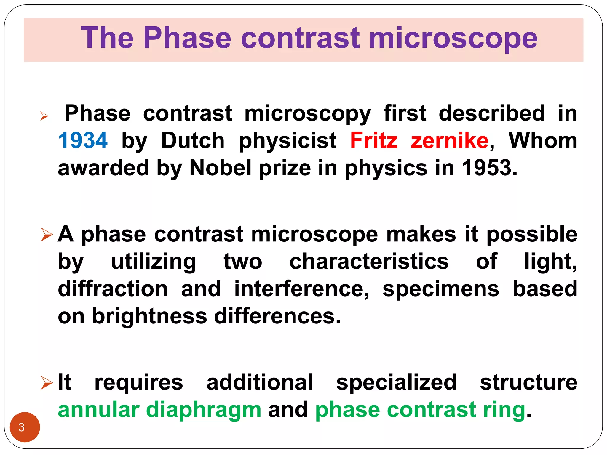
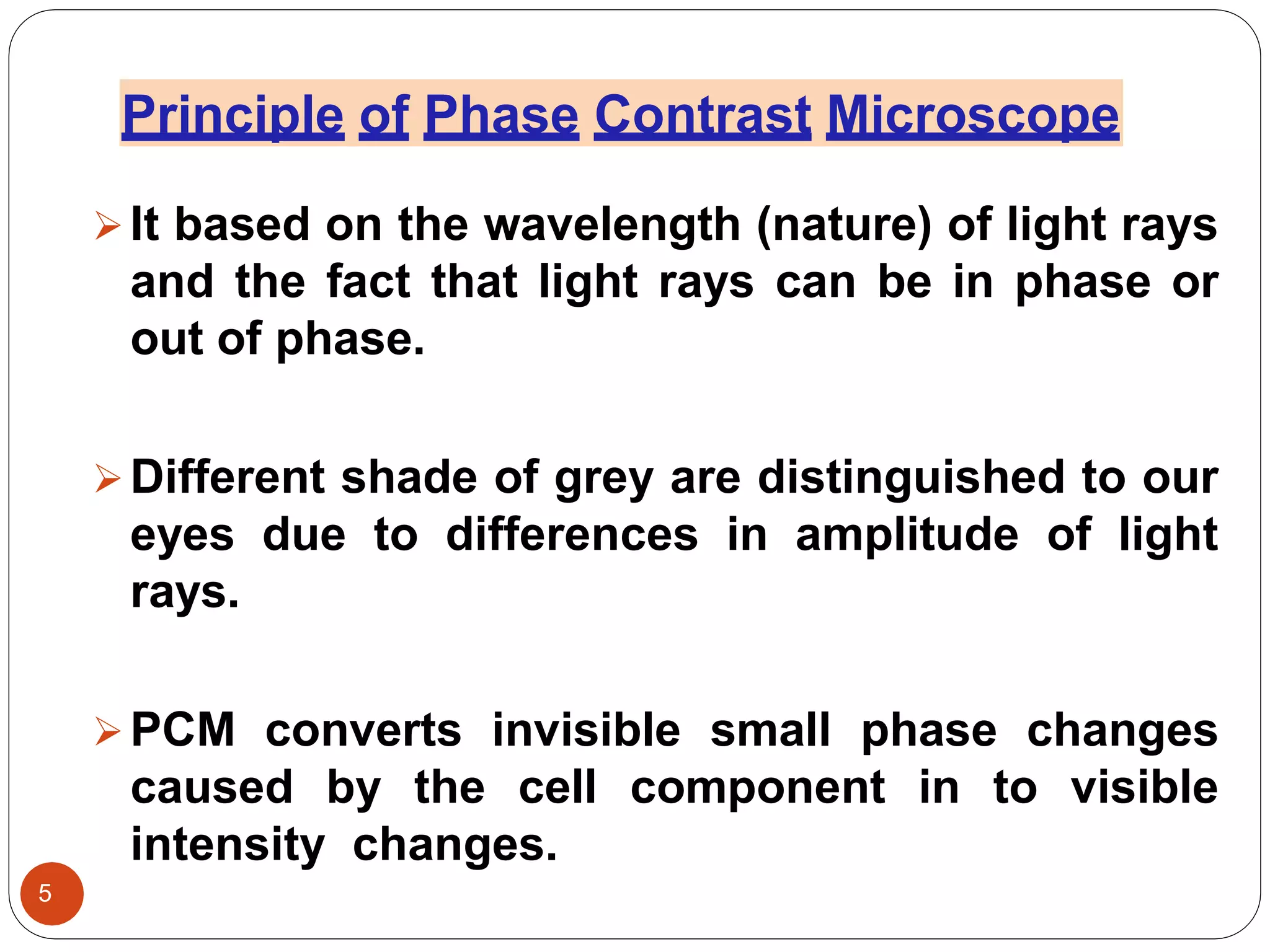
SUMIT
180040420016
M.Sc. (Evn.)
Department of
Environmental
science and Eng. ,
G.J.U.& S.T., Hisar.
Principles and application phase
contrast microsope
Presented by…
1
Contents
 Introduction
 Phase contrast microscope
◘ Principle
◘ working
◘ Applications
◘ Advantages
◘ Disadvantages
2
The Phase contrast microscope
3
 Phase contrast microscopy first described in
1934 by Dutch physicist Fritz zernike, Whom
awarded by Nobel prize in physics in 1953.
A phase contrast microscope makes it possible
by utilizing two characteristics of light,
diffraction and interference, specimens based
on brightness differences.
It requires additional specialized structure
annular diaphragm and phase contrast ring.
4 Sources: www.biologydiscussion
.com/microscope/phase contrast-
Principle of Phase Contrast Microscope
It based on the wavelength (nature) of light rays
and the fact that light rays can be in phase or
out of phase.
Different shade of grey are distinguished to our
eyes due to differences in amplitude of light
rays.
PCM converts invisible small phase changes
caused by the cell component in to visible
intensity changes.
5
In a Phase contrast microscope, one set of
light rays comes directly from the light
sources.
The other set comes from light that is reflected
or diffracted from a particular structure in the
specimen.
The images differences in refractive index of
cellular structure. Light passes through thicker
parts of cell is held up relative to the light that
passes through thinner parts of cytoplasm.
6
 How does it work ?
 Light that does not interact with the speciman
is collected by the objective passes through
the phase ring, and is regarded exactly ¼
wavelength.
The Phase shifted is not detectable by the eye
so the resulting image on the image plane in
the microscope appears as a normal bright
background.
7
Light passing through one material & into
another material of slightly different refractive
index or thickness will undergo a charge in
phase.
This charge in are translated into variations in
brightness of the structures.
 Natural light vibrates in many directions but
polarized light only one direction.
8
9 Sources :
https://www.bwoptics.com/newsend2asp
Application of Phase Contrast microscope
10
transparent specimens such as living cells
 Most commonly used to provide contrast of
or
small organisms.
 Useful in observing cells cultured in vitro during
mitosis.
 Phase contrast enables visualization of internal
cellular components.
 It’s used in examination of growth, dynamics, and
behaviour of a wide variety of living cells in cell
culture.
 It applied for equipment from the study of
the living biological specimens, medical
applications, study of live blood cells, and
other biological and science applications.
 It’s used in diagnosis of tumour cells.
11
ADVANTAGES
Unstained specimens can be viewed and imaged .
Live cells or organisms can be imaged in their natural
form without fixing or killing them.
Produces images that are information-rich and
detailed.
12
DISADVANTGES
 Not useful to see the natural colours of specimens
or if staining is involved.
Each objective has its separate phase contrast filter.
Accurate measurements of samples cannot be
acquired to technical. limitations where light cannot
penetrate beyond a particular sample thickness.
13
1) Biophysics
Author : Vasantha pattabhi ,
N. Gautham.
Edition : 2009 ( Second )
2) Basic Biophysics For Biologist
Author : M. Daniel
Edition : 2003
14
15

Phase contrast microscope

  • 1.
    SUMIT 180040420016 M.Sc. (Evn.) Department of Environmental scienceand Eng. , G.J.U.& S.T., Hisar. Principles and application phase contrast microsope Presented by… 1
  • 2.
    Contents  Introduction  Phasecontrast microscope ◘ Principle ◘ working ◘ Applications ◘ Advantages ◘ Disadvantages 2
  • 3.
    The Phase contrastmicroscope 3  Phase contrast microscopy first described in 1934 by Dutch physicist Fritz zernike, Whom awarded by Nobel prize in physics in 1953. A phase contrast microscope makes it possible by utilizing two characteristics of light, diffraction and interference, specimens based on brightness differences. It requires additional specialized structure annular diaphragm and phase contrast ring.
  • 4.
  • 5.
    Principle of PhaseContrast Microscope It based on the wavelength (nature) of light rays and the fact that light rays can be in phase or out of phase. Different shade of grey are distinguished to our eyes due to differences in amplitude of light rays. PCM converts invisible small phase changes caused by the cell component in to visible intensity changes. 5
  • 6.
    In a Phasecontrast microscope, one set of light rays comes directly from the light sources. The other set comes from light that is reflected or diffracted from a particular structure in the specimen. The images differences in refractive index of cellular structure. Light passes through thicker parts of cell is held up relative to the light that passes through thinner parts of cytoplasm. 6
  • 7.
     How doesit work ?  Light that does not interact with the speciman is collected by the objective passes through the phase ring, and is regarded exactly ¼ wavelength. The Phase shifted is not detectable by the eye so the resulting image on the image plane in the microscope appears as a normal bright background. 7
  • 8.
    Light passing throughone material & into another material of slightly different refractive index or thickness will undergo a charge in phase. This charge in are translated into variations in brightness of the structures.  Natural light vibrates in many directions but polarized light only one direction. 8
  • 9.
  • 10.
    Application of PhaseContrast microscope 10 transparent specimens such as living cells  Most commonly used to provide contrast of or small organisms.  Useful in observing cells cultured in vitro during mitosis.  Phase contrast enables visualization of internal cellular components.  It’s used in examination of growth, dynamics, and behaviour of a wide variety of living cells in cell culture.
  • 11.
     It appliedfor equipment from the study of the living biological specimens, medical applications, study of live blood cells, and other biological and science applications.  It’s used in diagnosis of tumour cells. 11
  • 12.
    ADVANTAGES Unstained specimens canbe viewed and imaged . Live cells or organisms can be imaged in their natural form without fixing or killing them. Produces images that are information-rich and detailed. 12
  • 13.
    DISADVANTGES  Not usefulto see the natural colours of specimens or if staining is involved. Each objective has its separate phase contrast filter. Accurate measurements of samples cannot be acquired to technical. limitations where light cannot penetrate beyond a particular sample thickness. 13
  • 14.
    1) Biophysics Author :Vasantha pattabhi , N. Gautham. Edition : 2009 ( Second ) 2) Basic Biophysics For Biologist Author : M. Daniel Edition : 2003 14
  • 15.