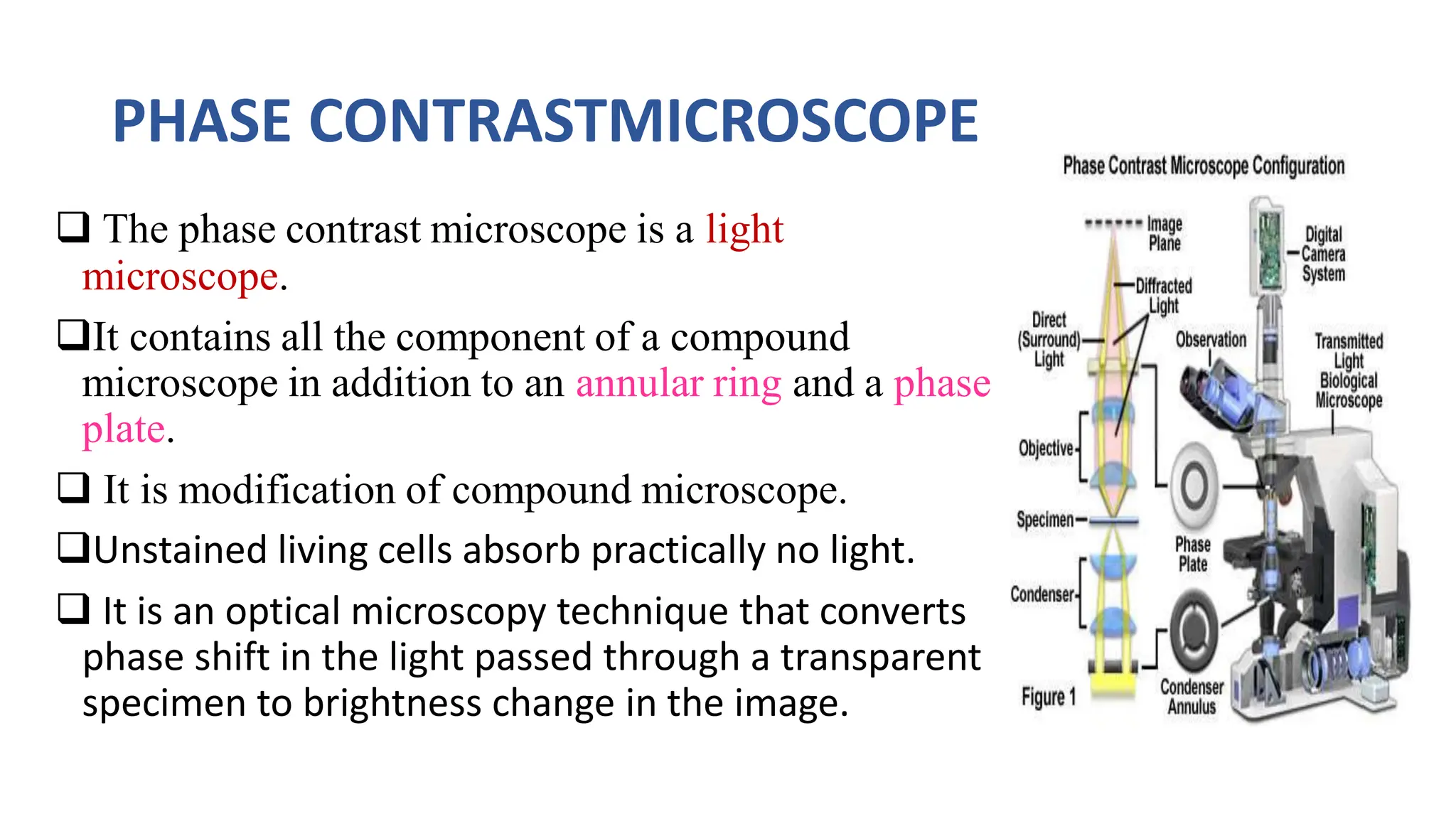
VIVEKANANDHA ARTS AND SCIENCECOLLEGE FOR WOMEN
VEERACHIPALAYAM,SANKAGIRI-6366007
DEPARTMENT OF MICROBIOLOGY
SUBJECT: GENERAL MICROBIOLOGY AND MICROBIAL DIVERISTY
TOPIC: PHASE CONTRAST MICROSCOPE
SUBJECT INCHARGE PRESENTED BY;
Dr.V.BHARATHI, M.sc.,M.Phil.,Ph.D. ANUJHA K.R
ASSISTANT PROFESSOR I MSc MICROBIOLOGY
DEPARTMENT OF MICROBIOLOGY DEPARTMENT OF
VIAAS,SANKAGIRI MICROBIOLOGY
VIAAS,SANKAGIRI
MICROSCOPE
A microscope is an instrument that magnifies an image of
a small object so that details can be seen that are too
small to be seen by the naked eye. Microscopes use at
least one lens to bend light towards the eye, making the
object appear larger than it actually is an instrument that
magnifies.
HISTORY OF MICROSCOPE
Antony van Leeuwenhoek (1632-
1723)is given credit as the
“FATHER OF MICROSCOPY”
 He developed simple microscopes
that used only one lens.
He was able to produce lenses with
magnifications of 270 diameters the
best at the time, and could see
objects not visible to the naked eye.
He observed the microscopic
organisms in pond water.
 Using his handcrafted
microscopes, he was the first to
observe and describe single-celled
organisms.
2.Early Microscopes
•The earliest microscope
was invented around
around 1590 by the
Zacharias Janssen .
•It had two lenses, one
magnifying the other.
a. Frits Zernike(1888-1966)
 It was first described in 1934
by Dutch physicist Frits
Zernike.
 It was invented by Frits
Zernike and was awarded noble
prize in 1953.
 Phase contrast microscope is
the first microscopic method
which allow the observation of
Living cell.
PHASE CONTRASTMICROSCOPE
 The phase contrast microscope is a light
microscope.
It contains all the component of a compound
microscope in addition to an annular ring and a phase
plate.
 It is modification of compound microscope.
Unstained living cells absorb practically no light.
 It is an optical microscopy technique that converts
phase shift in the light passed through a transparent
specimen to brightness change in the image.
PRINCIPLE OF PHASE CONTRAST MICROSCOPY
 The phase contrast microscope separates the illuminating background
light and the specimen scattering light.
 It is used to visualize transparent, colorless, living biological specimens.
These objects are called phase objects.
In a phase-contrast microscope, these phase shifts are converted into
changes in amplitude, which can be observed as differences in image
contrast.
The Working of Phase Contrast Microscopy
1. Partially coherent illumination by the tungsten-halogen lamp is directed
through a collector lens and focused on a specialized annulus(labeled
condenser annulus) positioned in the substage condenser front focal plane.
2. Wavefronts passing through the annulus illuminate the specimen and either
pass through underived or are diffracted and retarded in phase by structures
and phase gradients present in the specimen.
3. Undeviated and diffracted light collected by the objective is segregated at the
rear focal plane by a phase plate and focused at the intermediate image plane
to form the final phase-contrast image observed in the eyepieces.
Parts of phase contrast Microscopy
Phase-contrast microscopy is basically a specially designed light microscope
with all the basic parts in addition to which an annular phase plate and annular
phase plate and annular phase plate and annular diaphragm are fitted.
The annular diaphragm
• It is situated below the condenser.
• It is made up of a circular disc having a circular annular groove
• The light rays are allowed to pass through the annular groove.
• Through the annular groove of the annular diaphragm, the light rays fall on
the specimen or object to be studied.
• At the back focal plane of the objective develops an image.
• the annular phase plate is placed at this back focal plane.
The Phase Plate
• It is either a negative phase plate having a thick circular area or a
positive phase plate having a thin circular groove.
• This thick or thin area in the phase plate is called the conjugate
area.
• The phase plate is a transparent disc.
• With the help of the annular diaphragm and the phase plate, the
phase contrast is obtained in this microscope.
• This is obtained by separating the direct ray from the diffracted rays.
• The direct light rays pass through the annular groove whereas the
diffracted light ray pass through the region outside the groove.
• Depending upon the different refractive indices of different cell
components, the object to be studied show a different degree of
contrast in this microscope.
APPLICATIONS OF PHASE CONTRAST MICROSCOPY
To produce high-Contrast image of transparent specimens, such as
1. Living cells
2. Microorganisms,
3. Thin tissue slices,
4. Lithographic patterns,
5. Fibers,
6. Latex dispersions,
7. Glass fragments, and
8. Subcellular particles( Including nuclei and other organelles).
Advantages of Phase contrast Microscopy
• Living cell can be observed in their natural state without previous
fixation or labeling.
• It make a highly transparent object more visible.
• No special preparation of fixation or staining etc.is needed to study an
object under a phase contrast microscope which saves a lot of time.
• Examining intracellular components of living cells at relatively high
resolution.
Eg:The dynamic motility of mitochondria, mitotic chromosomes
&vacuoles.
• It made it possible for biologists to study living cells and how they
proliferate through cell division.
Limitations of phase contrast microscopy
• Phase-contrast codensers and objective lenses add considerable
cost to a microscope, and so phase contrast is often not used in
teaching labs except perhaps in the health professions.
• To use phase-contrast the light path must be aligned.
• Generally, more light is needed for phase contrast than for
corresponding bright-field viewing, since the technique is based on
the diminishment of the brightness of most objects.
Phase Contrast Microscopy Image
figure: Paramecium caudatum Figure: Bacteria in
phase contrast
Reference
1. https://en.wikipedia.org/wiki/Phase-contrast_microscopy
2. https://en.wikipedia.org/wiki/Phase-contrast_microscopy
3. https://www.microscopyu.com/techniques/phase-
contrast/introduction-to-phase-contrast-microscopy
4. https://www.microscopyu.com/techniques/phase-
contrast/phase-contrast-microscope-configuration

phase contrast microscope in general MB.

  • 1.
    VIVEKANANDHA ARTS ANDSCIENCECOLLEGE FOR WOMEN VEERACHIPALAYAM,SANKAGIRI-6366007 DEPARTMENT OF MICROBIOLOGY SUBJECT: GENERAL MICROBIOLOGY AND MICROBIAL DIVERISTY TOPIC: PHASE CONTRAST MICROSCOPE SUBJECT INCHARGE PRESENTED BY; Dr.V.BHARATHI, M.sc.,M.Phil.,Ph.D. ANUJHA K.R ASSISTANT PROFESSOR I MSc MICROBIOLOGY DEPARTMENT OF MICROBIOLOGY DEPARTMENT OF VIAAS,SANKAGIRI MICROBIOLOGY VIAAS,SANKAGIRI
  • 2.
    MICROSCOPE A microscope isan instrument that magnifies an image of a small object so that details can be seen that are too small to be seen by the naked eye. Microscopes use at least one lens to bend light towards the eye, making the object appear larger than it actually is an instrument that magnifies.
  • 3.
    HISTORY OF MICROSCOPE Antonyvan Leeuwenhoek (1632- 1723)is given credit as the “FATHER OF MICROSCOPY”  He developed simple microscopes that used only one lens. He was able to produce lenses with magnifications of 270 diameters the best at the time, and could see objects not visible to the naked eye. He observed the microscopic organisms in pond water.  Using his handcrafted microscopes, he was the first to observe and describe single-celled organisms.
  • 4.
    2.Early Microscopes •The earliestmicroscope was invented around around 1590 by the Zacharias Janssen . •It had two lenses, one magnifying the other.
  • 5.
    a. Frits Zernike(1888-1966) It was first described in 1934 by Dutch physicist Frits Zernike.  It was invented by Frits Zernike and was awarded noble prize in 1953.  Phase contrast microscope is the first microscopic method which allow the observation of Living cell.
  • 6.
    PHASE CONTRASTMICROSCOPE  Thephase contrast microscope is a light microscope. It contains all the component of a compound microscope in addition to an annular ring and a phase plate.  It is modification of compound microscope. Unstained living cells absorb practically no light.  It is an optical microscopy technique that converts phase shift in the light passed through a transparent specimen to brightness change in the image.
  • 7.
    PRINCIPLE OF PHASECONTRAST MICROSCOPY  The phase contrast microscope separates the illuminating background light and the specimen scattering light.  It is used to visualize transparent, colorless, living biological specimens. These objects are called phase objects. In a phase-contrast microscope, these phase shifts are converted into changes in amplitude, which can be observed as differences in image contrast.
  • 8.
    The Working ofPhase Contrast Microscopy 1. Partially coherent illumination by the tungsten-halogen lamp is directed through a collector lens and focused on a specialized annulus(labeled condenser annulus) positioned in the substage condenser front focal plane. 2. Wavefronts passing through the annulus illuminate the specimen and either pass through underived or are diffracted and retarded in phase by structures and phase gradients present in the specimen. 3. Undeviated and diffracted light collected by the objective is segregated at the rear focal plane by a phase plate and focused at the intermediate image plane to form the final phase-contrast image observed in the eyepieces.
  • 9.
    Parts of phasecontrast Microscopy Phase-contrast microscopy is basically a specially designed light microscope with all the basic parts in addition to which an annular phase plate and annular phase plate and annular phase plate and annular diaphragm are fitted. The annular diaphragm • It is situated below the condenser. • It is made up of a circular disc having a circular annular groove • The light rays are allowed to pass through the annular groove. • Through the annular groove of the annular diaphragm, the light rays fall on the specimen or object to be studied. • At the back focal plane of the objective develops an image. • the annular phase plate is placed at this back focal plane.
  • 10.
    The Phase Plate •It is either a negative phase plate having a thick circular area or a positive phase plate having a thin circular groove. • This thick or thin area in the phase plate is called the conjugate area. • The phase plate is a transparent disc. • With the help of the annular diaphragm and the phase plate, the phase contrast is obtained in this microscope. • This is obtained by separating the direct ray from the diffracted rays. • The direct light rays pass through the annular groove whereas the diffracted light ray pass through the region outside the groove. • Depending upon the different refractive indices of different cell components, the object to be studied show a different degree of contrast in this microscope.
  • 11.
    APPLICATIONS OF PHASECONTRAST MICROSCOPY To produce high-Contrast image of transparent specimens, such as 1. Living cells 2. Microorganisms, 3. Thin tissue slices, 4. Lithographic patterns, 5. Fibers, 6. Latex dispersions, 7. Glass fragments, and 8. Subcellular particles( Including nuclei and other organelles).
  • 12.
    Advantages of Phasecontrast Microscopy • Living cell can be observed in their natural state without previous fixation or labeling. • It make a highly transparent object more visible. • No special preparation of fixation or staining etc.is needed to study an object under a phase contrast microscope which saves a lot of time. • Examining intracellular components of living cells at relatively high resolution. Eg:The dynamic motility of mitochondria, mitotic chromosomes &vacuoles. • It made it possible for biologists to study living cells and how they proliferate through cell division.
  • 13.
    Limitations of phasecontrast microscopy • Phase-contrast codensers and objective lenses add considerable cost to a microscope, and so phase contrast is often not used in teaching labs except perhaps in the health professions. • To use phase-contrast the light path must be aligned. • Generally, more light is needed for phase contrast than for corresponding bright-field viewing, since the technique is based on the diminishment of the brightness of most objects.
  • 14.
    Phase Contrast MicroscopyImage figure: Paramecium caudatum Figure: Bacteria in phase contrast
  • 15.
    Reference 1. https://en.wikipedia.org/wiki/Phase-contrast_microscopy 2. https://en.wikipedia.org/wiki/Phase-contrast_microscopy 3.https://www.microscopyu.com/techniques/phase- contrast/introduction-to-phase-contrast-microscopy 4. https://www.microscopyu.com/techniques/phase- contrast/phase-contrast-microscope-configuration