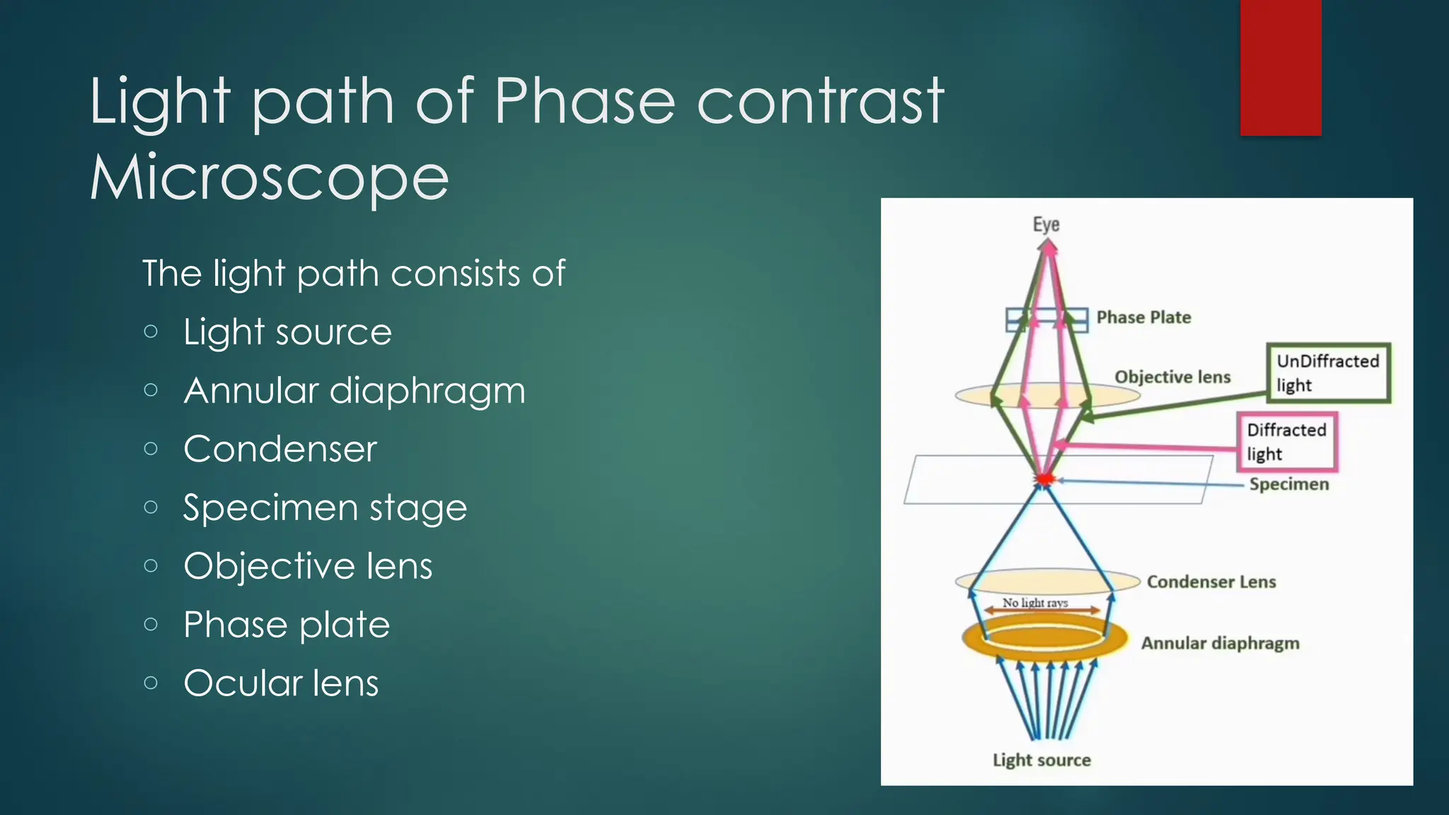
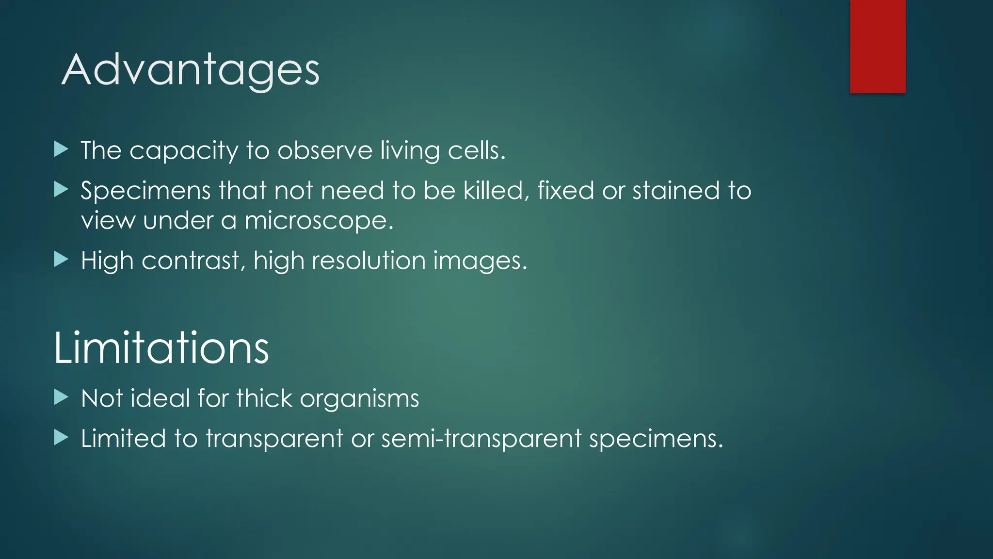
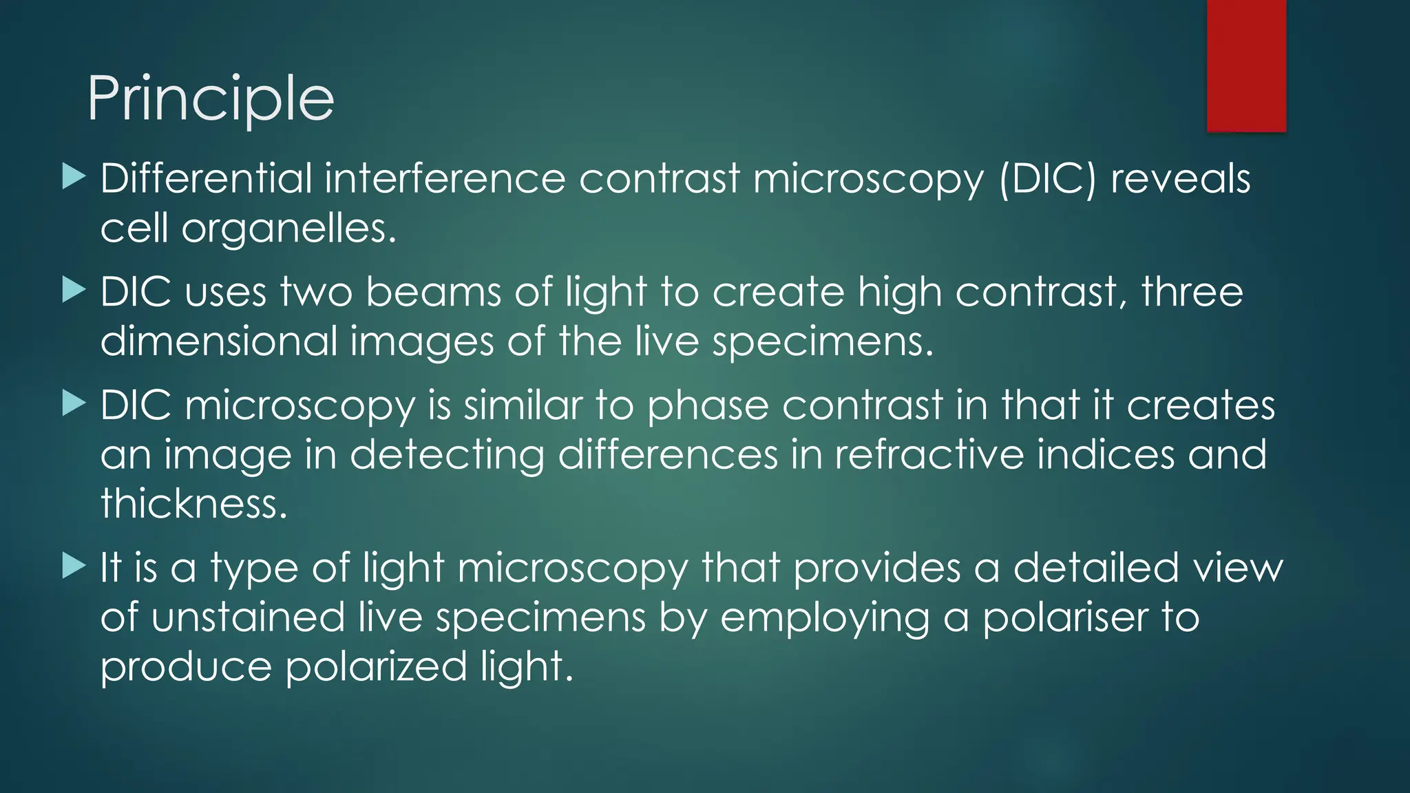
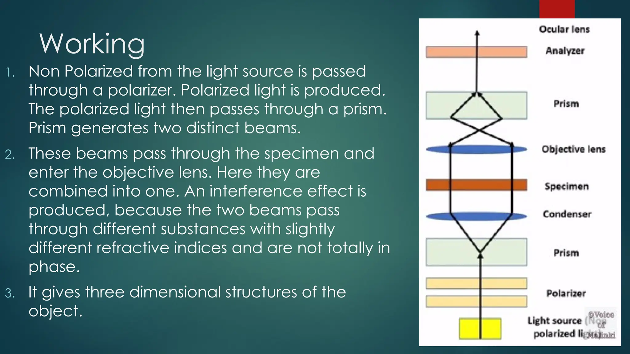
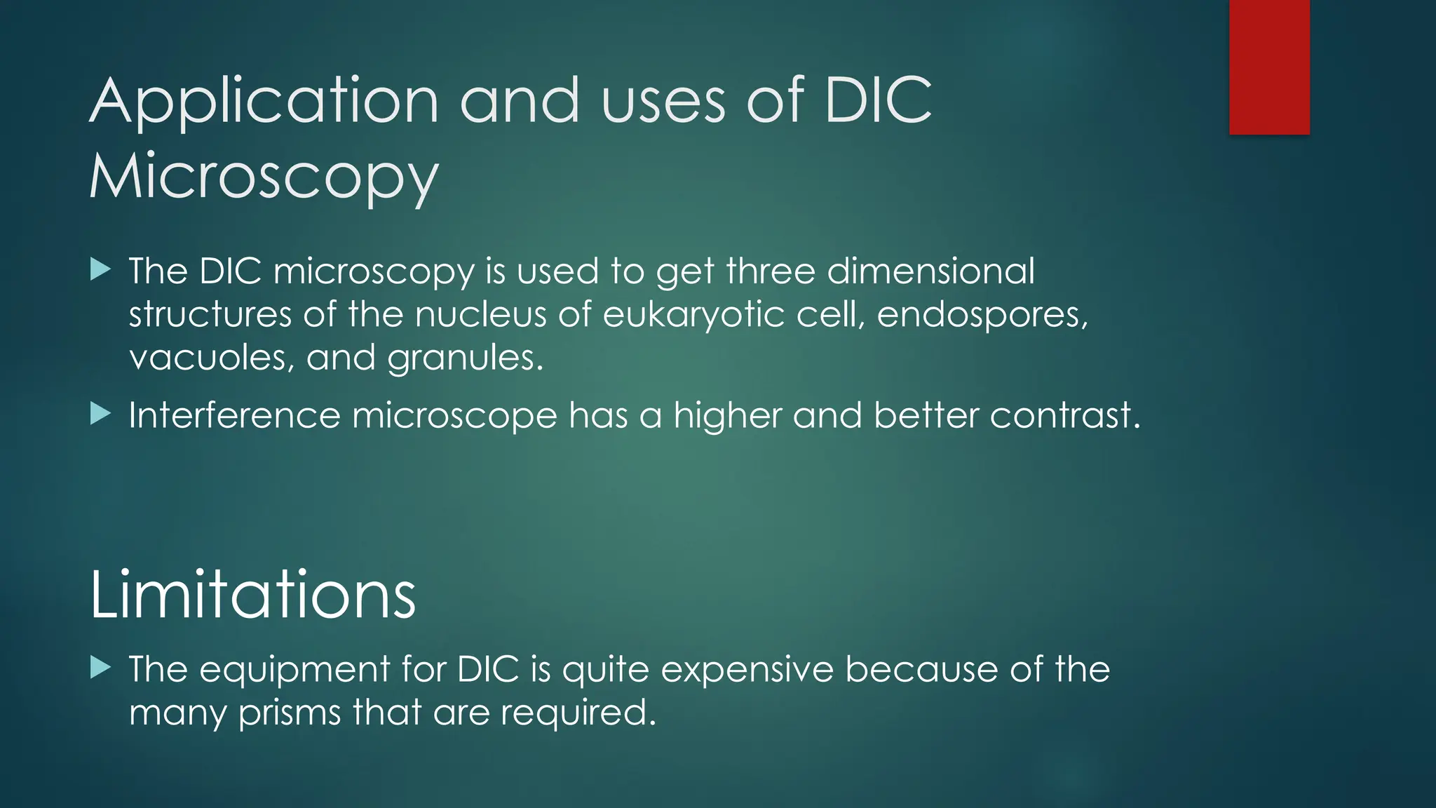
Phase Contrast
Microscopy
 This microscopic technique is slightly different from the
bright and dark field techniques because in this technique
the light rays of different phases are combined and used
to generate an image.
 Different components of cell (cell organelles) have
different refractive indices (power to turn the light from its
path). Due to this property, when the light passes through
the cell, each cell part change the path of light
according to its refractive indices, this create phase
difference in light rays.
 This microscope is used for visualization of cell culture and
live cells. Living cells can be observed without any
staining.
Principle
 Phase contrast microscopy is based on the
principle that small phase changes in the
light rays, induced by differences in the
thickness and refractive index of the
different parts of an object, can be
transformed into differences in brightness or
light intensity
Light path of Phase contrast
Microscope
The light path consists of
o Light source
o Annular diaphragm
o Condenser
o Specimen stage
o Objective lens
o Phase plate
o Ocular lens
 The light rays enter the annular diaphragm
from its source
 This annular diaphragm blocks most of the
light from the illuminator. Hence it produces a
hollow cone of light.
 Hollow cone of light is focused on specimen
before reaching the objective lens.
 Light travelling directly from the illuminator
(undiffracted wave) passes through one part
of the phase plate whereas light refracted or
scattered by the specimen (diffracted wave)
passes through another part of the phase
plate.
 This causes undiffracted wave to be out of
phase with that of diffracted wave. As a
result, diffracted and undiffracted rays
formed different images.
Applications of Phase-contrast
Microscopy
 To produce high-contrast images of transparent
specimens, such as
 Living cells (usually in culture),
 Microorganims,
 This tissues slices,
 Subcellular particles (including nuclei and other
organelles)
 Used in the study of dynamic cellular processes like
bacterial spore formation and germination. It avoids the
need to examine killed and fixed specimens.
Advantages
 The capacity to observe living cells.
 Specimens that not need to be killed, fixed or stained to
view under a microscope.
 High contrast, high resolution images.
Limitations
 Not ideal for thick organisms
 Limited to transparent or semi-transparent specimens.
Differential Interference
Contrast Microscopy
(DIC)
Principle
 Differential interference contrast microscopy (DIC) reveals
cell organelles.
 DIC uses two beams of light to create high contrast, three
dimensional images of the live specimens.
 DIC microscopy is similar to phase contrast in that it creates
an image in detecting differences in refractive indices and
thickness.
 It is a type of light microscopy that provides a detailed view
of unstained live specimens by employing a polariser to
produce polarized light.
Working
1. Non Polarized from the light source is passed
through a polarizer. Polarized light is produced.
The polarized light then passes through a prism.
Prism generates two distinct beams.
2. These beams pass through the specimen and
enter the objective lens. Here they are
combined into one. An interference effect is
produced, because the two beams pass
through different substances with slightly
different refractive indices and are not totally in
phase.
3. It gives three dimensional structures of the
object.
Application and uses of DIC
Microscopy
 The DIC microscopy is used to get three dimensional
structures of the nucleus of eukaryotic cell, endospores,
vacuoles, and granules.
 Interference microscope has a higher and better contrast.
Limitations
 The equipment for DIC is quite expensive because of the
many prisms that are required.

Phase Contrast Microscopy & Differential Interference Contrast Microscopy (DIC).pptx

  • 1.
  • 2.
     This microscopictechnique is slightly different from the bright and dark field techniques because in this technique the light rays of different phases are combined and used to generate an image.  Different components of cell (cell organelles) have different refractive indices (power to turn the light from its path). Due to this property, when the light passes through the cell, each cell part change the path of light according to its refractive indices, this create phase difference in light rays.  This microscope is used for visualization of cell culture and live cells. Living cells can be observed without any staining.
  • 3.
    Principle  Phase contrastmicroscopy is based on the principle that small phase changes in the light rays, induced by differences in the thickness and refractive index of the different parts of an object, can be transformed into differences in brightness or light intensity
  • 4.
    Light path ofPhase contrast Microscope The light path consists of o Light source o Annular diaphragm o Condenser o Specimen stage o Objective lens o Phase plate o Ocular lens
  • 5.
     The lightrays enter the annular diaphragm from its source  This annular diaphragm blocks most of the light from the illuminator. Hence it produces a hollow cone of light.  Hollow cone of light is focused on specimen before reaching the objective lens.  Light travelling directly from the illuminator (undiffracted wave) passes through one part of the phase plate whereas light refracted or scattered by the specimen (diffracted wave) passes through another part of the phase plate.  This causes undiffracted wave to be out of phase with that of diffracted wave. As a result, diffracted and undiffracted rays formed different images.
  • 6.
    Applications of Phase-contrast Microscopy To produce high-contrast images of transparent specimens, such as  Living cells (usually in culture),  Microorganims,  This tissues slices,  Subcellular particles (including nuclei and other organelles)  Used in the study of dynamic cellular processes like bacterial spore formation and germination. It avoids the need to examine killed and fixed specimens.
  • 7.
    Advantages  The capacityto observe living cells.  Specimens that not need to be killed, fixed or stained to view under a microscope.  High contrast, high resolution images. Limitations  Not ideal for thick organisms  Limited to transparent or semi-transparent specimens.
  • 9.
  • 10.
    Principle  Differential interferencecontrast microscopy (DIC) reveals cell organelles.  DIC uses two beams of light to create high contrast, three dimensional images of the live specimens.  DIC microscopy is similar to phase contrast in that it creates an image in detecting differences in refractive indices and thickness.  It is a type of light microscopy that provides a detailed view of unstained live specimens by employing a polariser to produce polarized light.
  • 11.
    Working 1. Non Polarizedfrom the light source is passed through a polarizer. Polarized light is produced. The polarized light then passes through a prism. Prism generates two distinct beams. 2. These beams pass through the specimen and enter the objective lens. Here they are combined into one. An interference effect is produced, because the two beams pass through different substances with slightly different refractive indices and are not totally in phase. 3. It gives three dimensional structures of the object.
  • 12.
    Application and usesof DIC Microscopy  The DIC microscopy is used to get three dimensional structures of the nucleus of eukaryotic cell, endospores, vacuoles, and granules.  Interference microscope has a higher and better contrast. Limitations  The equipment for DIC is quite expensive because of the many prisms that are required.