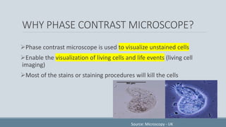
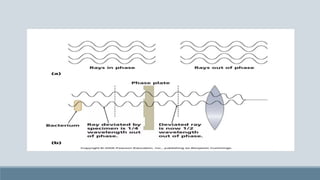
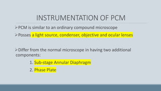
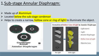
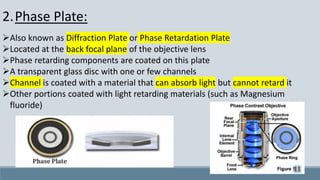
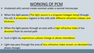
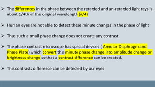
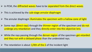
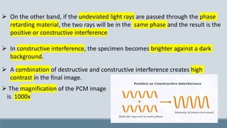
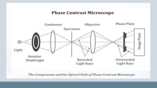
PHASE CONTRAST
MICROSCOPY
SOUSAN
A274153121006
HG&MM
“The eye of a human being is a microscope, which
makes the world seem bigger than it really is…”
-Khalil Gibran
CONTENT
➢ Introduction
➢ Background on microscopy
➢ Important terminologies
➢ Phase Contrast Microscopy
➢ Types of PCM
➢ History and Discovery
➢ Instrumentation
➢ Working principle
➢ Applications of PCM
➢ Advantages
➢ Limitations/Disadvantages
➢ References
INTRODUCTION
➢ A microscope is an instrument used to see objects that are too small for the
naked eyes
➢ The science of investigating small objects using such an instrument is called
microscopy
➢ There are different types of microscopes as follows:
1. Optical microscope: Uses light to image the sample
2. Electron microscope: Uses electrons to image the sample
2.1. Scanning Electron Microscope (SEM)
2.2. Transmission Electron Microscope (TEM)
3. Ultra Microscope
4. Scanning Probe Microscope
5. Stereomicroscope
LIGHT MICROSCOPE
➢It uses visible light to detect small objects
➢It is a good use or well-known research tool in biology
➢We can use a light microscope to enlarge cytoplasm, nucleus, cell
membrane, chloroplast, vacuoles, cell wall, etc.
TYPES OF LIGHT MICROSCOPE
1. Bright-field Microscope: Produce a dark image against a brighter
background
2. Darkfield Microscope: Produces a bright image of the object
against a dark background
3. Phase Contrast Microscope: Enhances the contrast between
intracellular structures having slight differences in refractive index
COMPARISON OF LIGHT MICROSCOPY
SOURCE: BOSHIDA
MAGNIFICATION AND RESOLUTION
Magnification:
➢Degree of enlargement
➢Decrease the number of times in length, breadth, and diameter of the object
are multiplied
Resolution:
➢The minimum distance between two visible bodies
at which they can be seen as separate and distinct
objects and not in contact with each other
HISTORY OF PCM
➢Developed by Zernike in the early 1930s
➢Won Nobel prize in physics-1953
PHASE CONTRAST MICROSCOPY
(PCM)
➢Made it possible to study living cells and how they proliferate through cell
division
➢Used in examining living organisms or specimens that would be damaged or
altered by attaching them to slides or staining
➢Uses a conventional light microscope fitted with a phase contrast objective and
phase contrast condenser
➢Light passing through one material and into another material of slightly different
refractive index or thickness will undergo a change in phase. This change is
translated into variations in the brightness of the structure.
TYPES OF PCM
1. Positive Phase Contrast Microscope:
➢The specimen is dark and the background is bright
➢The specimen is surrounded by a bright halo
2. Negative Phase Contrast Microscope:
➢The specimen is bright and the background is dark
➢The specimen is surrounded by a dark halo
Source: https://www.researchgate.net/figure/Comparison-of-
positive-A-and-negative-B-phase-contrast-images-of-the-MCF-
10A_fig14_281434136
WORKING WITH AN ORDINARY MICROSCOPE
➢ In ordinary microscopy, the object is viewed due to differences in the intensities
of the specimen
➢ To create color intensities, the specimen is stained with the suitable dyes
➢ Contrast is obtained when the light rays pass through a stained specimen
because different stains absorb different amounts of light
➢ The differential absorption properties of stained specimens modify the intensity
or amplitude of the light waves transmitted by different regions of the cells and
this ultimately creates contrast in the image
➢ Thus, staining is essential to create contrast in ordinary microscopy
➢ Unstained specimens cannot be observed through an ordinary microscope
WHY PHASE CONTRAST MICROSCOPE?
➢Phase contrast microscope is used to visualize unstained cells
➢Enable the visualization of living cells and life events (living cell
imaging)
➢Most of the stains or staining procedures will kill the cells
Source: Microscopy - UK
WORKING PRINCIPLE OF PCM
➢Phase contrast microscopy is based on the principle that small
phase changes in the light rays, induced by differences in the
thickness and refractive index of the different parts of an object, can
be transformed into differences in brightness or light intensity.
➢It is the translation of visible phase shifts into visible differences in
intensities
➢Phase changes are not detectable to the human eye whereas the
brightness or light intensity can be detected by the human eyes.
INSTRUMENTATION OF PCM
➢PCM is similar to an ordinary compound microscope
➢Posses a light source, condenser, objective and ocular lenses
➢Differ from the normal microscope in having two additional
components:
1. Sub-stage Annular Diaphragm
2. Phase Plate
Source: Phase Contrast Microscopy - Wikipedia
1.Sub-stage Annular Diaphragm:
➢ Made up of Aluminium
➢ Located below the sub-stage condenser
➢ Helps to create a narrow, hollow cone or ring of light to illuminate the object.
2.Phase Plate:
➢Also known as Diffraction Plate or Phase Retardation Plate
➢Located at the back focal plane of the objective lens
➢Phase retarding components are coated on this plate
➢A transparent glass disc with one or few channels
➢Channel is coated with a material that can absorb light but cannot retard it
➢Other portions coated with light retarding materials (such as Magnesium
fluoride)
WORKING OF PCM
➢ Unstained cells cannot create contrast under a normal microscope
➢ When the light passes (The light source is a tungsten-halogen lamp) through
the cell, it encounters regions in the cells with different refractive indexes and
thickness
➢ When the light passes through an area with a high refractive index it has
deviated from its normal path
➢ Such a light ray experience a phase change or phase retardation
➢ Light rays pass through the area of less refractive index remain un-deviated (no
phase change)
➢ The differences in the phase between the retarded and un-retarded light rays is
about 1/4th of the original wavelength (λ/4)
➢ Human eyes are not able to detect these minute changes in the phase of light
➢ Thus such a small phase change does not create any contrast
➢ The phase contrast microscope has special devices ( Annular Diaphragm and
Phase Plate) which convert this minute phase change into amplitude change or
brightness change so that a contrast difference can be created.
➢ This contrasts difference can be detected by our eyes
➢ In PCM, the diffracted waves have to be separated from the direct waves
➢ This is achieved by the sub-stage annular diaphragm
➢ The annular diaphragm illuminates the specimen with a hallow cone of light
➢ Some rays (direct rays) through the thinner region of the specimen and do not
undergo any retardation and they directly enter into the objective lens
➢ While the rays passing through the denser region of the specimen get retarded
and they run with a delayed phase than the undeviated rays.
➢ The retardation is about 1/4th of the λ of the incident light
➢ Both the retarded and unretarded light have to pass through the phase plate
kept on the back focal plane of the objective.
➢ The phase plate is designed and positioned in such a way that the retarded
light rays will pass through the area of the phase plate where light retarding
materials are coated
➢ When the 1/4 (λ/4) retarded light passes through the plate, it is further
retarded by 1/4 (λ/4)
➢ Thus the final change or retardation will be:
¼ λ + ¼ λ = ½ λ Retardation of phase
➢ The unretarded rays will pass through the channels of the phase plate and their
phase is not altered by the phase plate
➢ When the unretarded and ½ λ retarded light are combined (at the focal point) a
negative or destructive interference is created because the crest and trough
cancel each other
➢ With the destructive interference, the specimen appears darker against a bright
background.
➢ On the other band, if the undeviated light rays are passed through the phase
retarding material, the two rays will be in the same phase and the result is the
positive or constructive interference
➢ In constructive interference, the specimen becomes brighter against a dark
background.
➢ A combination of destructive and constructive interference creates high
contrast in the final image.
➢ The magnification of the PCM image
is 1000x
APPLICATIONS OF PCM
Magnification and resolution PCM is similar to an ordinary microscope. Still,
PCM has many applications in biological sciences, such as:
➢ Enable visualization of living cells
➢ Enable visualization of unstained cells
➢ Can be used to view various cell organelles
➢ Helps to study cellular events such as cell division, phagocytosis, cyclosis,
etc.
➢ Visualize all types of cellular movements (chromosomal & flagellar)
➢ Enable the study of membrane permeability (phagocytosis)
➢ Extensively used to observe living cells in tissue culture to monitor their
growth
ADVANTAGES OF PCM
➢ Provide a clear image of unstained cells
➢ Avoid damage to the cells due to chemical preparation and staining
➢ Provide high-contrast images highlighting the fine details of the cells
➢ The optical construction is relatively simple
➢ A compound microscope can be elevated to PCM with minor additions
➢ Enable prolonged observation of living cells
➢ Living cell imaging is possible
➢ Affordable cost
LIMITATIONS OF PCM
➢ Produce a bright halo around the images
➢ The formation of a halo is due to the partial or incomplete separation of direct
and deviated rays
➢ Only useful for viewing individual cells or a thin layer of cells.
THANK YOU
REFERENCES
https://www.med.unc.edu/microscopy/wp-content/uploads/sites/742/2018/06/lm-ch-10-phase-
contrast.pdf
https://www.cn-microscope.com/zh_cn/biological-microscope2/what-is-bright-field-phase-contrast-
dark-field-dic-microscope.html
http://www.microscopy-uk.org.uk/mag/artmar06/go-phase.html
https://www.researchgate.net/figure/Comparison-of-positive-A-and-negative-B-phase-contrast-
images-of-the-MCF-10A_fig14_281434136
https://www.microscopyu.com/techniques/phase-contrast/introduction-to-phase-contrast-
microscopy
https://www.scientifica.uk.com/learning-zone/a-guide-to-phase-contrast
https://www.youtube.com/watch?v=YsHUqfxNj9M
**********

PHASE CONTRAST MICROSCOPY.pdf

  • 1.
  • 2.
    “The eye ofa human being is a microscope, which makes the world seem bigger than it really is…” -Khalil Gibran
  • 3.
    CONTENT ➢ Introduction ➢ Backgroundon microscopy ➢ Important terminologies ➢ Phase Contrast Microscopy ➢ Types of PCM ➢ History and Discovery ➢ Instrumentation ➢ Working principle ➢ Applications of PCM ➢ Advantages ➢ Limitations/Disadvantages ➢ References
  • 4.
    INTRODUCTION ➢ A microscopeis an instrument used to see objects that are too small for the naked eyes ➢ The science of investigating small objects using such an instrument is called microscopy ➢ There are different types of microscopes as follows: 1. Optical microscope: Uses light to image the sample 2. Electron microscope: Uses electrons to image the sample 2.1. Scanning Electron Microscope (SEM) 2.2. Transmission Electron Microscope (TEM) 3. Ultra Microscope 4. Scanning Probe Microscope 5. Stereomicroscope
  • 5.
    LIGHT MICROSCOPE ➢It usesvisible light to detect small objects ➢It is a good use or well-known research tool in biology ➢We can use a light microscope to enlarge cytoplasm, nucleus, cell membrane, chloroplast, vacuoles, cell wall, etc.
  • 6.
    TYPES OF LIGHTMICROSCOPE 1. Bright-field Microscope: Produce a dark image against a brighter background 2. Darkfield Microscope: Produces a bright image of the object against a dark background 3. Phase Contrast Microscope: Enhances the contrast between intracellular structures having slight differences in refractive index
  • 7.
    COMPARISON OF LIGHTMICROSCOPY SOURCE: BOSHIDA
  • 8.
    MAGNIFICATION AND RESOLUTION Magnification: ➢Degreeof enlargement ➢Decrease the number of times in length, breadth, and diameter of the object are multiplied Resolution: ➢The minimum distance between two visible bodies at which they can be seen as separate and distinct objects and not in contact with each other
  • 9.
    HISTORY OF PCM ➢Developedby Zernike in the early 1930s ➢Won Nobel prize in physics-1953
  • 10.
    PHASE CONTRAST MICROSCOPY (PCM) ➢Madeit possible to study living cells and how they proliferate through cell division ➢Used in examining living organisms or specimens that would be damaged or altered by attaching them to slides or staining ➢Uses a conventional light microscope fitted with a phase contrast objective and phase contrast condenser ➢Light passing through one material and into another material of slightly different refractive index or thickness will undergo a change in phase. This change is translated into variations in the brightness of the structure.
  • 11.
    TYPES OF PCM 1.Positive Phase Contrast Microscope: ➢The specimen is dark and the background is bright ➢The specimen is surrounded by a bright halo 2. Negative Phase Contrast Microscope: ➢The specimen is bright and the background is dark ➢The specimen is surrounded by a dark halo Source: https://www.researchgate.net/figure/Comparison-of- positive-A-and-negative-B-phase-contrast-images-of-the-MCF- 10A_fig14_281434136
  • 12.
    WORKING WITH ANORDINARY MICROSCOPE ➢ In ordinary microscopy, the object is viewed due to differences in the intensities of the specimen ➢ To create color intensities, the specimen is stained with the suitable dyes ➢ Contrast is obtained when the light rays pass through a stained specimen because different stains absorb different amounts of light ➢ The differential absorption properties of stained specimens modify the intensity or amplitude of the light waves transmitted by different regions of the cells and this ultimately creates contrast in the image ➢ Thus, staining is essential to create contrast in ordinary microscopy ➢ Unstained specimens cannot be observed through an ordinary microscope
  • 13.
    WHY PHASE CONTRASTMICROSCOPE? ➢Phase contrast microscope is used to visualize unstained cells ➢Enable the visualization of living cells and life events (living cell imaging) ➢Most of the stains or staining procedures will kill the cells Source: Microscopy - UK
  • 14.
    WORKING PRINCIPLE OFPCM ➢Phase contrast microscopy is based on the principle that small phase changes in the light rays, induced by differences in the thickness and refractive index of the different parts of an object, can be transformed into differences in brightness or light intensity. ➢It is the translation of visible phase shifts into visible differences in intensities ➢Phase changes are not detectable to the human eye whereas the brightness or light intensity can be detected by the human eyes.
  • 16.
    INSTRUMENTATION OF PCM ➢PCMis similar to an ordinary compound microscope ➢Posses a light source, condenser, objective and ocular lenses ➢Differ from the normal microscope in having two additional components: 1. Sub-stage Annular Diaphragm 2. Phase Plate
  • 17.
    Source: Phase ContrastMicroscopy - Wikipedia
  • 18.
    1.Sub-stage Annular Diaphragm: ➢Made up of Aluminium ➢ Located below the sub-stage condenser ➢ Helps to create a narrow, hollow cone or ring of light to illuminate the object.
  • 19.
    2.Phase Plate: ➢Also knownas Diffraction Plate or Phase Retardation Plate ➢Located at the back focal plane of the objective lens ➢Phase retarding components are coated on this plate ➢A transparent glass disc with one or few channels ➢Channel is coated with a material that can absorb light but cannot retard it ➢Other portions coated with light retarding materials (such as Magnesium fluoride)
  • 20.
    WORKING OF PCM ➢Unstained cells cannot create contrast under a normal microscope ➢ When the light passes (The light source is a tungsten-halogen lamp) through the cell, it encounters regions in the cells with different refractive indexes and thickness ➢ When the light passes through an area with a high refractive index it has deviated from its normal path ➢ Such a light ray experience a phase change or phase retardation ➢ Light rays pass through the area of less refractive index remain un-deviated (no phase change)
  • 21.
    ➢ The differencesin the phase between the retarded and un-retarded light rays is about 1/4th of the original wavelength (λ/4) ➢ Human eyes are not able to detect these minute changes in the phase of light ➢ Thus such a small phase change does not create any contrast ➢ The phase contrast microscope has special devices ( Annular Diaphragm and Phase Plate) which convert this minute phase change into amplitude change or brightness change so that a contrast difference can be created. ➢ This contrasts difference can be detected by our eyes
  • 22.
    ➢ In PCM,the diffracted waves have to be separated from the direct waves ➢ This is achieved by the sub-stage annular diaphragm ➢ The annular diaphragm illuminates the specimen with a hallow cone of light ➢ Some rays (direct rays) through the thinner region of the specimen and do not undergo any retardation and they directly enter into the objective lens ➢ While the rays passing through the denser region of the specimen get retarded and they run with a delayed phase than the undeviated rays. ➢ The retardation is about 1/4th of the λ of the incident light
  • 23.
    ➢ Both theretarded and unretarded light have to pass through the phase plate kept on the back focal plane of the objective. ➢ The phase plate is designed and positioned in such a way that the retarded light rays will pass through the area of the phase plate where light retarding materials are coated ➢ When the 1/4 (λ/4) retarded light passes through the plate, it is further retarded by 1/4 (λ/4) ➢ Thus the final change or retardation will be: ¼ λ + ¼ λ = ½ λ Retardation of phase
  • 24.
    ➢ The unretardedrays will pass through the channels of the phase plate and their phase is not altered by the phase plate ➢ When the unretarded and ½ λ retarded light are combined (at the focal point) a negative or destructive interference is created because the crest and trough cancel each other ➢ With the destructive interference, the specimen appears darker against a bright background.
  • 25.
    ➢ On theother band, if the undeviated light rays are passed through the phase retarding material, the two rays will be in the same phase and the result is the positive or constructive interference ➢ In constructive interference, the specimen becomes brighter against a dark background. ➢ A combination of destructive and constructive interference creates high contrast in the final image. ➢ The magnification of the PCM image is 1000x
  • 28.
    APPLICATIONS OF PCM Magnificationand resolution PCM is similar to an ordinary microscope. Still, PCM has many applications in biological sciences, such as: ➢ Enable visualization of living cells ➢ Enable visualization of unstained cells ➢ Can be used to view various cell organelles ➢ Helps to study cellular events such as cell division, phagocytosis, cyclosis, etc. ➢ Visualize all types of cellular movements (chromosomal & flagellar) ➢ Enable the study of membrane permeability (phagocytosis) ➢ Extensively used to observe living cells in tissue culture to monitor their growth
  • 30.
    ADVANTAGES OF PCM ➢Provide a clear image of unstained cells ➢ Avoid damage to the cells due to chemical preparation and staining ➢ Provide high-contrast images highlighting the fine details of the cells ➢ The optical construction is relatively simple ➢ A compound microscope can be elevated to PCM with minor additions ➢ Enable prolonged observation of living cells ➢ Living cell imaging is possible ➢ Affordable cost
  • 31.
    LIMITATIONS OF PCM ➢Produce a bright halo around the images ➢ The formation of a halo is due to the partial or incomplete separation of direct and deviated rays ➢ Only useful for viewing individual cells or a thin layer of cells.
  • 32.
  • 33.