SlideShare a Scribd company logo
Dr. P. Suganya
Assistant Professor
Department of Biotechnology
Sri Kaliswari College (Autonomous)
 The concept of confocal microscopy was initially
developed by Marvin Minsky in the 1950s, at Harvard
University with an aim of viewing the neural network
without staining the tissues but it did not bear fruit
due to lack of enough light source and a
computerized system to store the large data.
 The work was later adapted by David Egger M. and
Mojmir Petran, forming a multiple-beam confocal
microscope in the late 1960s. they used a spinning
disk known as Nipkow which they used to examine
brain tissues and ganglion cells that were unstained.
The technique was later modified and published by
Egger forming a mechanical scanned confocal laser
microscope, that was able to visualize images of
cells.
 Later developments in science including
development of computers and laser technology
and digital manipulations of images using
algorithms, grew the advances in confocal
microscopy, forming practically usable confocal
microscopes by a series of scientists
including G. Fred Brakenhoff (1979), Colin
Sheppard, Tony Wilson, Brad Amos, and John
White (1980s).
 The first commercial confocal microscope was
developed in 1987 with improved optics and
electronics, powerful lasers with high scanning
efficiency.
 The modern confocal Microscope has all the
possible integration of technology and
mechanical components including optical
components, which perform the primary function
of the configuration by use of electronic
detectors, a computer and laser systems.
 The functioning of the confocal microscope is a
collective role of all its components to produce
an electronic image. To date, these microscopes
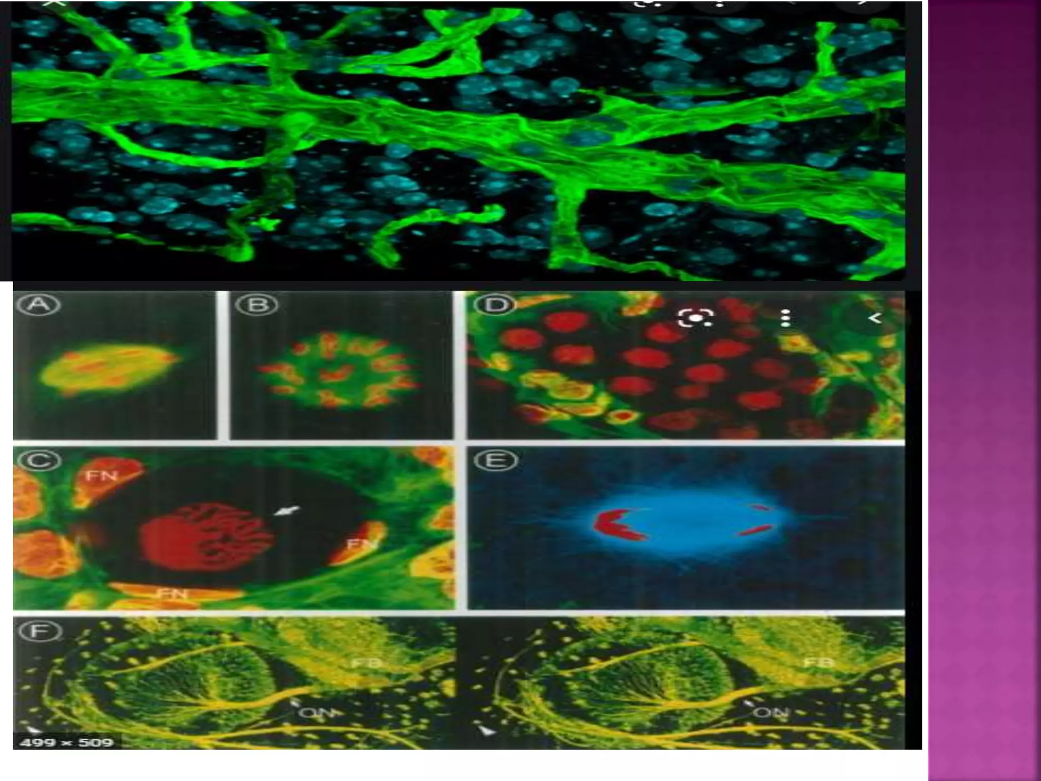
have been used to investigate molecules,
microbial cells, and tissues.
Image Source: DOI: 10.1186/1475-925X-
13-52
 Normally a conventional (wide-field) Microscope uses
different wavelengths from a light source, to visualize
and illuminate a large area of a specimen, forming
fuzzy, murky and crowded images, because cell sample
images are captured from all directions, without a
focal point.
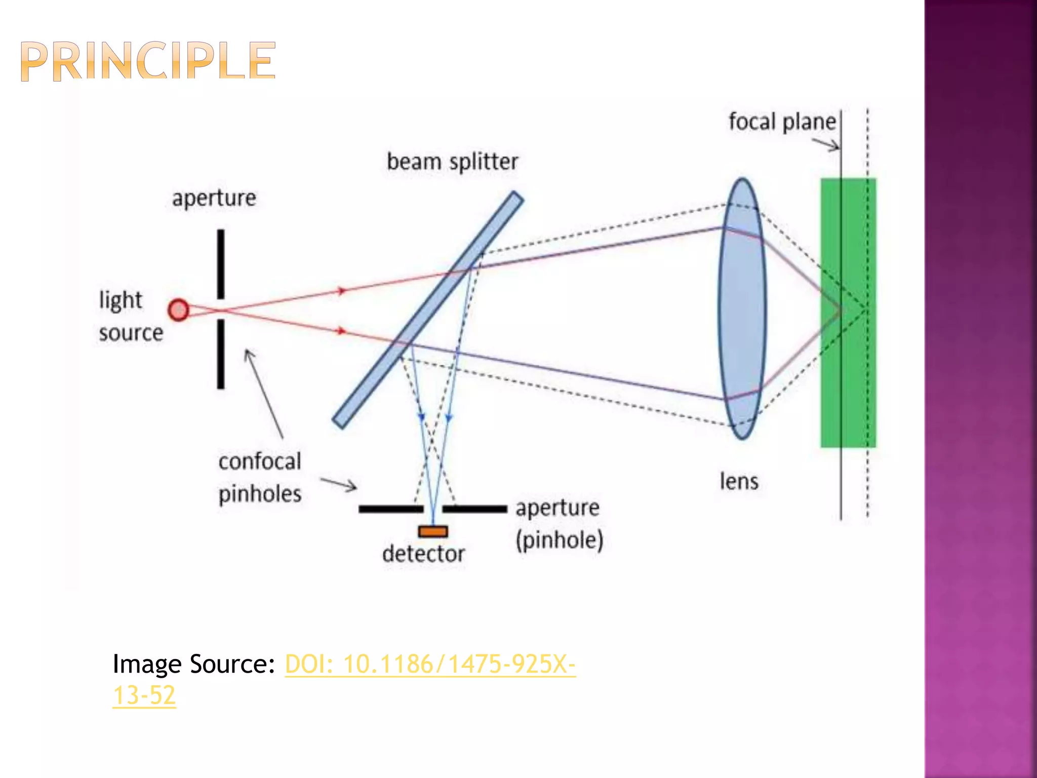
 To avoid these issues, a Confocal Microscope is used. In
wide-field or Fluorescent microscopes, the whole
specimen receives light, receiving complete
excitement and emitting light which is detected by a
photodetector on the microscope. However, with the
confocal microscope, point illumination is the principle
working mechanism.
 A specimen is stained with fluorochrome is examined.
When a beam of light is focused at a particular point of
the fluoro-chromatic specimen, it produces an
illumination that is focused by the objective lens to a
plane above the objectives. The objective has an
aperture on the focal plane located above it, which
primarily functions to block any stray light from
reaching the specimen.
 A measure of the illumination point is about
0.25 to 0.8 um in diameter, determined by
the objective numerical aperture and 0.5 to
1.5 um deep, with the brightest intensity.
 The specimen normally lies between the
camera lens and the perfect point of focus,
known as the plane of focus. Using the laser
from the microscope, the laser scans over a
plane on the specimen (beam scanning0 or
by moving the stage (stage scanning). A
detector then will measure the illumination
producing an image of the optical section.
scanning several optical sections, they are
collected in a computerized system as data,
forming a 3D image. The image can be
measured and quantified.
 Its outcome is also favored by the aperture
found above the objective which blocks stray
light.
 Images produced by the confocal microscope has
a very good contract and resolution capacity
despite the thickness of the specimen. Images
are stored in the high-resolution 3D image of the
cell complexes including its structures.
 The main characteristic of the Confocal
Microscope is that it only detects what is focused
and anything outside the focus point, appears
black.
 The image of the specimen is formed when
the microscope scanner, scans the focused
beam across a selected area with the control
of two high-speed oscillating mirrors. Their
movement is facilitated by galvanometer
motors. One mirror moves the beam from
left to right on the lateral X-axis while the
second mirror translates the beam along
the Y-axis. After a scan on the X-axis, the
beam moves rapidly back to the starting
point to start a new scan, a process known as
flyback. No information is collected during
the flyback process, therefore the point of
focus, which is the area of interest is what is
illuminated by the laser scanner.
The Confocal Laser Scanning Microscope is
made up a few components:
 Objective lens
 Out-of-focus plane
 In-focus plane
 Beam splitters
 Detector
 Confocal pinhole (aperture)
 Laser
 Oscillator Mirrors
 Confocal laser scanning Microscope – It uses
several mirrors that scan along the X and Y axes on
the specimen, by scanning and descanning, and the
image passes through a pinhole into the detector.
 Spinning disk, also known as the Nipkow disk, is a
type of confocal microscope that uses several
movable apertures (pinholes) on a disc to scan for
spots of light in a parallel manner over a specified
plane, over a long period. The longer the time the
less the excitation energy required for
illumination, as compared to the Confocal laser
scanning microscope. Lessened excitation energy
reduces phototoxicity and photobleaching, hence
its mainly used to imaging live cells.
 Dual spinning Disk or Microlens enhanced
confocal Microscope -, it was invented by
Yokogawa electric; it works similarly to the
spinning disk, the only difference is, it has a
second spinning-disk with micro-lenses that is
found before the spinning disk that contains the
pinholes. The micro-lenses capture broadband of
light focusing it into each pinhole, thus
increasing the amount of light that is directed
into each pinhole, reducing the amount of light
that is blocked by the spinning disk. This
Confocal Microscopes with enhanced Microlenses
are much more sensitive than the spinning disks.
 Programmable array Microscope (PAM) – this
type of confocal microscope uses a spatial
light modulator (SLM – an object that
imposes some form of spatially-varying
modulation on a beam of light). The SLM has
a set of movable apertures (pinholes), with
arrays of pixels of opacity, reflectivity or
optical rotation. The SLM also has
microelectrochemical mirrors that capture
the image by a charge-coupled device (CCD)
camera.
 Each of the confocal microscopes has its
advantages and disadvantages, but they all
capture the images by recording the images
and sometimes they can be programmed to
get high-density images, especially the
Programmed array Microscope and the
Spinning disk confocal Microscope.
 The Confocal Microscope is used in a wide range of
fields including Biomedical sciences, Cells Biology,
genetics, Microbiology, Developmental Biology,
Spectroscopy, Nanoscience (nanoimaging) and
Quantum Optics.
 In Biomedical sciences, it is used in the analysis of eye
corneal infections, by quantifying and qualitatively
analyzing the endothelial cells of the cornea.
 Used to identify the presence of fungal elements in
the corneal stroma, during keratomycosis infection, or
rapid diagnosis and quick therapeutic response.
 It is used in pharmaceutical industries, to ensure the
maintenance of thin-film pharmaceuticals, allowing
control of the quality and uniformity of drug
distribution.
 It is used to retrieve data from some 3D optical
storage systems. This has helped in quantifying the
age of Magdalen papyrus.
 The advantage of the Confocal microscope is that it
improves the outcome of the image because it
analyses the image from one optical point to
another, therefore there is no interference with
scattered light from other parts of the specimen.
 They have better resolution and each point of
interest is visualized and captured.
 It can be used to study live and fixed cells
 It can be used to collect serial optical sections.
 It illuminates uniformly across the focus points.
 The adjust their magnification electronically,
without changing the objectives, by a factor known
as the zoom factor.
 It generates 3D sets of images.
 They have a limited number of excitation
wavelengths, with very narrow bands.
 They are expensive to produce the
ultraviolet rays used by the Confocal
Microscopes
 They are also expensive to manufacture and
to purchase.
 https://microbenotes.com/confocal-
microscope/
Confocal microscope

More Related Content

What's hot

Electron microscopy
Electron microscopyElectron microscopy
Electron microscopy
Najam ul Sehar Aiman
 
5. Microsocope ELECTRON MICROSCOPE (TEM & SEM ) - Basics
5. Microsocope   ELECTRON MICROSCOPE (TEM & SEM ) - Basics5. Microsocope   ELECTRON MICROSCOPE (TEM & SEM ) - Basics
5. Microsocope ELECTRON MICROSCOPE (TEM & SEM ) - Basics
Nethravathi Siri
 
Principles and application of light, phase constrast and fluorescence microscope
Principles and application of light, phase constrast and fluorescence microscopePrinciples and application of light, phase constrast and fluorescence microscope
Principles and application of light, phase constrast and fluorescence microscope
MaitriThakor
 
phase contrast microscope
phase contrast microscopephase contrast microscope
phase contrast microscope
Manjunatha Sanka
 
Fluorescence Microscopy
Fluorescence MicroscopyFluorescence Microscopy
Fluorescence Microscopy
VIVEK KUMAR SINGH
 
Scanning electron microscopy (sem)
Scanning electron microscopy (sem)Scanning electron microscopy (sem)
Scanning electron microscopy (sem)
Sivasangari Shanmugam
 
Lecture 3 Unit-I Bright-field & Dark-field Microscopy.pdf
Lecture 3 Unit-I Bright-field & Dark-field Microscopy.pdfLecture 3 Unit-I Bright-field & Dark-field Microscopy.pdf
Lecture 3 Unit-I Bright-field & Dark-field Microscopy.pdf
SushmitaChoudhary16
 
Electron microscope, principle and application
Electron microscope, principle and applicationElectron microscope, principle and application
Electron microscope, principle and application
KAUSHAL SAHU
 
PHASE MICROSCOPY
PHASE MICROSCOPYPHASE MICROSCOPY
PHASE MICROSCOPY
Rajpal Choudhary
 
Electron microscope ppt
Electron microscope pptElectron microscope ppt
Electron microscope ppt
NithyaNandapal
 
Radioisotope technique and methods
Radioisotope technique and methodsRadioisotope technique and methods
Radioisotope technique and methods
University of Mumbai
 
Microscopy
 Microscopy  Microscopy
Microscopy
Baljinder Gill
 
transmission Electron Microscopy (Tem)
transmission Electron Microscopy (Tem)transmission Electron Microscopy (Tem)
transmission Electron Microscopy (Tem)
Sivasangari Shanmugam
 
Cofocal microscope
Cofocal microscopeCofocal microscope
Cofocal microscope
kishore gupta
 
Phase contrast microscope
Phase contrast microscopePhase contrast microscope
Phase contrast microscope
Ravi Kumar
 
microscopy
 microscopy microscopy
microscopy
sanchu yadav
 
Transmission electron microscope
Transmission electron microscopeTransmission electron microscope
Transmission electron microscope
Senthil Arasan
 
Confocal laser scanning microscopy (clsm)
Confocal laser scanning microscopy (clsm)Confocal laser scanning microscopy (clsm)
Confocal laser scanning microscopy (clsm)
Nethravathi Siri
 
Fluorescence microscope
Fluorescence microscopeFluorescence microscope
Fluorescence microscope
Leyon Raj
 
Electron Microscopy (SEM & TEM)
Electron Microscopy (SEM & TEM)Electron Microscopy (SEM & TEM)
Electron Microscopy (SEM & TEM)
Kalyan Acharjya
 

What's hot (20)

Electron microscopy
Electron microscopyElectron microscopy
Electron microscopy
 
5. Microsocope ELECTRON MICROSCOPE (TEM & SEM ) - Basics
5. Microsocope   ELECTRON MICROSCOPE (TEM & SEM ) - Basics5. Microsocope   ELECTRON MICROSCOPE (TEM & SEM ) - Basics
5. Microsocope ELECTRON MICROSCOPE (TEM & SEM ) - Basics
 
Principles and application of light, phase constrast and fluorescence microscope
Principles and application of light, phase constrast and fluorescence microscopePrinciples and application of light, phase constrast and fluorescence microscope
Principles and application of light, phase constrast and fluorescence microscope
 
phase contrast microscope
phase contrast microscopephase contrast microscope
phase contrast microscope
 
Fluorescence Microscopy
Fluorescence MicroscopyFluorescence Microscopy
Fluorescence Microscopy
 
Scanning electron microscopy (sem)
Scanning electron microscopy (sem)Scanning electron microscopy (sem)
Scanning electron microscopy (sem)
 
Lecture 3 Unit-I Bright-field & Dark-field Microscopy.pdf
Lecture 3 Unit-I Bright-field & Dark-field Microscopy.pdfLecture 3 Unit-I Bright-field & Dark-field Microscopy.pdf
Lecture 3 Unit-I Bright-field & Dark-field Microscopy.pdf
 
Electron microscope, principle and application
Electron microscope, principle and applicationElectron microscope, principle and application
Electron microscope, principle and application
 
PHASE MICROSCOPY
PHASE MICROSCOPYPHASE MICROSCOPY
PHASE MICROSCOPY
 
Electron microscope ppt
Electron microscope pptElectron microscope ppt
Electron microscope ppt
 
Radioisotope technique and methods
Radioisotope technique and methodsRadioisotope technique and methods
Radioisotope technique and methods
 
Microscopy
 Microscopy  Microscopy
Microscopy
 
transmission Electron Microscopy (Tem)
transmission Electron Microscopy (Tem)transmission Electron Microscopy (Tem)
transmission Electron Microscopy (Tem)
 
Cofocal microscope
Cofocal microscopeCofocal microscope
Cofocal microscope
 
Phase contrast microscope
Phase contrast microscopePhase contrast microscope
Phase contrast microscope
 
microscopy
 microscopy microscopy
microscopy
 
Transmission electron microscope
Transmission electron microscopeTransmission electron microscope
Transmission electron microscope
 
Confocal laser scanning microscopy (clsm)
Confocal laser scanning microscopy (clsm)Confocal laser scanning microscopy (clsm)
Confocal laser scanning microscopy (clsm)
 
Fluorescence microscope
Fluorescence microscopeFluorescence microscope
Fluorescence microscope
 
Electron Microscopy (SEM & TEM)
Electron Microscopy (SEM & TEM)Electron Microscopy (SEM & TEM)
Electron Microscopy (SEM & TEM)
 

Similar to Confocal microscope

Confocal Microscopy.pptx MSC BIOTECHNOLOGY
Confocal Microscopy.pptx MSC BIOTECHNOLOGYConfocal Microscopy.pptx MSC BIOTECHNOLOGY
Confocal Microscopy.pptx MSC BIOTECHNOLOGY
AnjaliKannaujiya1
 
Types of Light Microscopes used in Histological Studies.pptx
Types of Light Microscopes used in Histological Studies.pptxTypes of Light Microscopes used in Histological Studies.pptx
Types of Light Microscopes used in Histological Studies.pptx
ssuserab552f
 
Microscopy - Magnification, Resolving power, Principles, Types and Applications
Microscopy - Magnification, Resolving power, Principles, Types and ApplicationsMicroscopy - Magnification, Resolving power, Principles, Types and Applications
Microscopy - Magnification, Resolving power, Principles, Types and Applications
Nethravathi Siri
 
Confocal Microscopy.pptx
Confocal Microscopy.pptxConfocal Microscopy.pptx
Confocal Microscopy.pptx
Rajadr
 
2. Microscopy (Biochemistry)
2. Microscopy (Biochemistry)2. Microscopy (Biochemistry)
2. Microscopy (Biochemistry)
Jay Khaniya
 
MICROSCOPY.pptx
MICROSCOPY.pptxMICROSCOPY.pptx
MICROSCOPY.pptx
ArchanaBawari1
 
Histo tool.pptx
Histo tool.pptxHisto tool.pptx
Histo tool.pptx
DeanParamedics
 
Histological tools
Histological toolsHistological tools
Histological tools
Navneet Randhawa
 
To Study Principles of Microscopy: Light Microscope, Phase Contrast Microsco...
To Study Principles of Microscopy: Light Microscope, Phase Contrast  Microsco...To Study Principles of Microscopy: Light Microscope, Phase Contrast  Microsco...
To Study Principles of Microscopy: Light Microscope, Phase Contrast Microsco...
Om Prakash
 
3. Microscope simple, compound & stereo - Basics
3. Microscope   simple, compound & stereo - Basics3. Microscope   simple, compound & stereo - Basics
3. Microscope simple, compound & stereo - Basics
Nethravathi Siri
 
Confocal microscopy Likhith K
Confocal microscopy Likhith KConfocal microscopy Likhith K
Confocal microscopy Likhith K
LIKHITHK1
 
Optical microscope
Optical microscopeOptical microscope
Optical microscope
SaurabhShankar7
 
Techniques in cell biology by vinitha
Techniques in cell biology by vinithaTechniques in cell biology by vinitha
Techniques in cell biology by vinitha
VinithaVinitha24
 
Microscopy
MicroscopyMicroscopy
Microscopy
Mistarlal Singh
 
Principles of microscopy: A microscope is an instrument that produces an accu...
Principles of microscopy: A microscope is an instrument that produces an accu...Principles of microscopy: A microscope is an instrument that produces an accu...
Principles of microscopy: A microscope is an instrument that produces an accu...
Smitha Vijayan
 
ELECTRON MICROSCOPE-1.pptx
ELECTRON MICROSCOPE-1.pptxELECTRON MICROSCOPE-1.pptx
ELECTRON MICROSCOPE-1.pptx
OpenPhone
 
Microscope
MicroscopeMicroscope
Microscope
MerlinDayana2
 
Introducation to Microscopy
Introducation to MicroscopyIntroducation to Microscopy
Introducation to Microscopy
Narottam Priyadarsahi
 
Microscope of ppt for botany major this is a project
Microscope of ppt for botany major this is a projectMicroscope of ppt for botany major this is a project
Microscope of ppt for botany major this is a project
arpitakhairwar123
 

Similar to Confocal microscope (20)

Confocal Microscopy.pptx MSC BIOTECHNOLOGY
Confocal Microscopy.pptx MSC BIOTECHNOLOGYConfocal Microscopy.pptx MSC BIOTECHNOLOGY
Confocal Microscopy.pptx MSC BIOTECHNOLOGY
 
Types of Light Microscopes used in Histological Studies.pptx
Types of Light Microscopes used in Histological Studies.pptxTypes of Light Microscopes used in Histological Studies.pptx
Types of Light Microscopes used in Histological Studies.pptx
 
Microscopy - Magnification, Resolving power, Principles, Types and Applications
Microscopy - Magnification, Resolving power, Principles, Types and ApplicationsMicroscopy - Magnification, Resolving power, Principles, Types and Applications
Microscopy - Magnification, Resolving power, Principles, Types and Applications
 
Confocal Microscopy.pptx
Confocal Microscopy.pptxConfocal Microscopy.pptx
Confocal Microscopy.pptx
 
2. Microscopy (Biochemistry)
2. Microscopy (Biochemistry)2. Microscopy (Biochemistry)
2. Microscopy (Biochemistry)
 
MICROSCOPY.pptx
MICROSCOPY.pptxMICROSCOPY.pptx
MICROSCOPY.pptx
 
Histo tool.pptx
Histo tool.pptxHisto tool.pptx
Histo tool.pptx
 
Histological tools
Histological toolsHistological tools
Histological tools
 
To Study Principles of Microscopy: Light Microscope, Phase Contrast Microsco...
To Study Principles of Microscopy: Light Microscope, Phase Contrast  Microsco...To Study Principles of Microscopy: Light Microscope, Phase Contrast  Microsco...
To Study Principles of Microscopy: Light Microscope, Phase Contrast Microsco...
 
3. Microscope simple, compound & stereo - Basics
3. Microscope   simple, compound & stereo - Basics3. Microscope   simple, compound & stereo - Basics
3. Microscope simple, compound & stereo - Basics
 
Confocal microscopy Likhith K
Confocal microscopy Likhith KConfocal microscopy Likhith K
Confocal microscopy Likhith K
 
Optical microscope
Optical microscopeOptical microscope
Optical microscope
 
Techniques in cell biology by vinitha
Techniques in cell biology by vinithaTechniques in cell biology by vinitha
Techniques in cell biology by vinitha
 
Microscopy
MicroscopyMicroscopy
Microscopy
 
Principles of microscopy: A microscope is an instrument that produces an accu...
Principles of microscopy: A microscope is an instrument that produces an accu...Principles of microscopy: A microscope is an instrument that produces an accu...
Principles of microscopy: A microscope is an instrument that produces an accu...
 
ELECTRON MICROSCOPE-1.pptx
ELECTRON MICROSCOPE-1.pptxELECTRON MICROSCOPE-1.pptx
ELECTRON MICROSCOPE-1.pptx
 
Microscope
MicroscopeMicroscope
Microscope
 
Introducation to Microscopy
Introducation to MicroscopyIntroducation to Microscopy
Introducation to Microscopy
 
Microscope PPT.pdf
Microscope PPT.pdfMicroscope PPT.pdf
Microscope PPT.pdf
 
Microscope of ppt for botany major this is a project
Microscope of ppt for botany major this is a projectMicroscope of ppt for botany major this is a project
Microscope of ppt for botany major this is a project
 

More from SuganyaPaulraj

FOOD ADULTERATION.pptx
FOOD ADULTERATION.pptxFOOD ADULTERATION.pptx
FOOD ADULTERATION.pptx
SuganyaPaulraj
 
AMF.pptx
AMF.pptxAMF.pptx
AMF.pptx
SuganyaPaulraj
 
TRANSLATION.pptx
TRANSLATION.pptxTRANSLATION.pptx
TRANSLATION.pptx
SuganyaPaulraj
 
TCA CYCLE (KREBS).pptx
TCA CYCLE (KREBS).pptxTCA CYCLE (KREBS).pptx
TCA CYCLE (KREBS).pptx
SuganyaPaulraj
 
Structure of amino acids.pptx
Structure of amino acids.pptxStructure of amino acids.pptx
Structure of amino acids.pptx
SuganyaPaulraj
 
REGULATION OF GENE EXPRESSION IN PROKARYOTES.pptx
REGULATION OF GENE EXPRESSION IN PROKARYOTES.pptxREGULATION OF GENE EXPRESSION IN PROKARYOTES.pptx
REGULATION OF GENE EXPRESSION IN PROKARYOTES.pptx
SuganyaPaulraj
 
PROPERTIES OF AMINOACIDS.pptx
PROPERTIES OF AMINOACIDS.pptxPROPERTIES OF AMINOACIDS.pptx
PROPERTIES OF AMINOACIDS.pptx
SuganyaPaulraj
 
POST TRANSLATIONAL MODIFICATIONS.pptx
POST TRANSLATIONAL MODIFICATIONS.pptxPOST TRANSLATIONAL MODIFICATIONS.pptx
POST TRANSLATIONAL MODIFICATIONS.pptx
SuganyaPaulraj
 
POST TRANSCRIPTIONAL MODOFICATION.pptx
POST TRANSCRIPTIONAL MODOFICATION.pptxPOST TRANSCRIPTIONAL MODOFICATION.pptx
POST TRANSCRIPTIONAL MODOFICATION.pptx
SuganyaPaulraj
 
PENTOSE PHOSPHATE PATHWAY.pptx
PENTOSE PHOSPHATE PATHWAY.pptxPENTOSE PHOSPHATE PATHWAY.pptx
PENTOSE PHOSPHATE PATHWAY.pptx
SuganyaPaulraj
 
OXIDATIVE PHOSPHORYLATION.pptx
OXIDATIVE PHOSPHORYLATION.pptxOXIDATIVE PHOSPHORYLATION.pptx
OXIDATIVE PHOSPHORYLATION.pptx
SuganyaPaulraj
 
PROKAYOTIC TRANSCRIPTION.pptx
PROKAYOTIC TRANSCRIPTION.pptxPROKAYOTIC TRANSCRIPTION.pptx
PROKAYOTIC TRANSCRIPTION.pptx
SuganyaPaulraj
 
EUKARYOTIC TRANSCRIPTION.pptx
EUKARYOTIC TRANSCRIPTION.pptxEUKARYOTIC TRANSCRIPTION.pptx
EUKARYOTIC TRANSCRIPTION.pptx
SuganyaPaulraj
 
Rna
RnaRna
Dna
DnaDna
Genome organization of prokaryotes and eukaryotes
Genome organization of prokaryotes and eukaryotesGenome organization of prokaryotes and eukaryotes
Genome organization of prokaryotes and eukaryotes
SuganyaPaulraj
 
Carbohydrates
CarbohydratesCarbohydrates
Carbohydrates
SuganyaPaulraj
 
Classification of microorganisms
Classification of microorganismsClassification of microorganisms
Classification of microorganisms
SuganyaPaulraj
 
Fluoresence microscope
Fluoresence microscopeFluoresence microscope
Fluoresence microscope
SuganyaPaulraj
 
Microbial flora of soil
Microbial flora of soilMicrobial flora of soil
Microbial flora of soil
SuganyaPaulraj
 

More from SuganyaPaulraj (20)

FOOD ADULTERATION.pptx
FOOD ADULTERATION.pptxFOOD ADULTERATION.pptx
FOOD ADULTERATION.pptx
 
AMF.pptx
AMF.pptxAMF.pptx
AMF.pptx
 
TRANSLATION.pptx
TRANSLATION.pptxTRANSLATION.pptx
TRANSLATION.pptx
 
TCA CYCLE (KREBS).pptx
TCA CYCLE (KREBS).pptxTCA CYCLE (KREBS).pptx
TCA CYCLE (KREBS).pptx
 
Structure of amino acids.pptx
Structure of amino acids.pptxStructure of amino acids.pptx
Structure of amino acids.pptx
 
REGULATION OF GENE EXPRESSION IN PROKARYOTES.pptx
REGULATION OF GENE EXPRESSION IN PROKARYOTES.pptxREGULATION OF GENE EXPRESSION IN PROKARYOTES.pptx
REGULATION OF GENE EXPRESSION IN PROKARYOTES.pptx
 
PROPERTIES OF AMINOACIDS.pptx
PROPERTIES OF AMINOACIDS.pptxPROPERTIES OF AMINOACIDS.pptx
PROPERTIES OF AMINOACIDS.pptx
 
POST TRANSLATIONAL MODIFICATIONS.pptx
POST TRANSLATIONAL MODIFICATIONS.pptxPOST TRANSLATIONAL MODIFICATIONS.pptx
POST TRANSLATIONAL MODIFICATIONS.pptx
 
POST TRANSCRIPTIONAL MODOFICATION.pptx
POST TRANSCRIPTIONAL MODOFICATION.pptxPOST TRANSCRIPTIONAL MODOFICATION.pptx
POST TRANSCRIPTIONAL MODOFICATION.pptx
 
PENTOSE PHOSPHATE PATHWAY.pptx
PENTOSE PHOSPHATE PATHWAY.pptxPENTOSE PHOSPHATE PATHWAY.pptx
PENTOSE PHOSPHATE PATHWAY.pptx
 
OXIDATIVE PHOSPHORYLATION.pptx
OXIDATIVE PHOSPHORYLATION.pptxOXIDATIVE PHOSPHORYLATION.pptx
OXIDATIVE PHOSPHORYLATION.pptx
 
PROKAYOTIC TRANSCRIPTION.pptx
PROKAYOTIC TRANSCRIPTION.pptxPROKAYOTIC TRANSCRIPTION.pptx
PROKAYOTIC TRANSCRIPTION.pptx
 
EUKARYOTIC TRANSCRIPTION.pptx
EUKARYOTIC TRANSCRIPTION.pptxEUKARYOTIC TRANSCRIPTION.pptx
EUKARYOTIC TRANSCRIPTION.pptx
 
Rna
RnaRna
Rna
 
Dna
DnaDna
Dna
 
Genome organization of prokaryotes and eukaryotes
Genome organization of prokaryotes and eukaryotesGenome organization of prokaryotes and eukaryotes
Genome organization of prokaryotes and eukaryotes
 
Carbohydrates
CarbohydratesCarbohydrates
Carbohydrates
 
Classification of microorganisms
Classification of microorganismsClassification of microorganisms
Classification of microorganisms
 
Fluoresence microscope
Fluoresence microscopeFluoresence microscope
Fluoresence microscope
 
Microbial flora of soil
Microbial flora of soilMicrobial flora of soil
Microbial flora of soil
 

Recently uploaded

Nutraceutical market, scope and growth: Herbal drug technology
Nutraceutical market, scope and growth: Herbal drug technologyNutraceutical market, scope and growth: Herbal drug technology
Nutraceutical market, scope and growth: Herbal drug technology
Lokesh Patil
 
Seminar of U.V. Spectroscopy by SAMIR PANDA
 Seminar of U.V. Spectroscopy by SAMIR PANDA Seminar of U.V. Spectroscopy by SAMIR PANDA
Seminar of U.V. Spectroscopy by SAMIR PANDA
SAMIR PANDA
 
In silico drugs analogue design: novobiocin analogues.pptx
In silico drugs analogue design: novobiocin analogues.pptxIn silico drugs analogue design: novobiocin analogues.pptx
In silico drugs analogue design: novobiocin analogues.pptx
AlaminAfendy1
 
insect taxonomy importance systematics and classification
insect taxonomy importance systematics and classificationinsect taxonomy importance systematics and classification
insect taxonomy importance systematics and classification
anitaento25
 
EY - Supply Chain Services 2018_template.pptx
EY - Supply Chain Services 2018_template.pptxEY - Supply Chain Services 2018_template.pptx
EY - Supply Chain Services 2018_template.pptx
AlguinaldoKong
 
Structural Classification Of Protein (SCOP)
Structural Classification Of Protein  (SCOP)Structural Classification Of Protein  (SCOP)
Structural Classification Of Protein (SCOP)
aishnasrivastava
 
THE IMPORTANCE OF MARTIAN ATMOSPHERE SAMPLE RETURN.
THE IMPORTANCE OF MARTIAN ATMOSPHERE SAMPLE RETURN.THE IMPORTANCE OF MARTIAN ATMOSPHERE SAMPLE RETURN.
THE IMPORTANCE OF MARTIAN ATMOSPHERE SAMPLE RETURN.
Sérgio Sacani
 
The ASGCT Annual Meeting was packed with exciting progress in the field advan...
The ASGCT Annual Meeting was packed with exciting progress in the field advan...The ASGCT Annual Meeting was packed with exciting progress in the field advan...
The ASGCT Annual Meeting was packed with exciting progress in the field advan...
Health Advances
 
Richard's entangled aventures in wonderland
Richard's entangled aventures in wonderlandRichard's entangled aventures in wonderland
Richard's entangled aventures in wonderland
Richard Gill
 
Multi-source connectivity as the driver of solar wind variability in the heli...
Multi-source connectivity as the driver of solar wind variability in the heli...Multi-source connectivity as the driver of solar wind variability in the heli...
Multi-source connectivity as the driver of solar wind variability in the heli...
Sérgio Sacani
 
Predicting property prices with machine learning algorithms.pdf
Predicting property prices with machine learning algorithms.pdfPredicting property prices with machine learning algorithms.pdf
Predicting property prices with machine learning algorithms.pdf
binhminhvu04
 
Comparative structure of adrenal gland in vertebrates
Comparative structure of adrenal gland in vertebratesComparative structure of adrenal gland in vertebrates
Comparative structure of adrenal gland in vertebrates
sachin783648
 
Earliest Galaxies in the JADES Origins Field: Luminosity Function and Cosmic ...
Earliest Galaxies in the JADES Origins Field: Luminosity Function and Cosmic ...Earliest Galaxies in the JADES Origins Field: Luminosity Function and Cosmic ...
Earliest Galaxies in the JADES Origins Field: Luminosity Function and Cosmic ...
Sérgio Sacani
 
justice-and-fairness-ethics with example
justice-and-fairness-ethics with examplejustice-and-fairness-ethics with example
justice-and-fairness-ethics with example
azzyixes
 
Large scale production of streptomycin.pptx
Large scale production of streptomycin.pptxLarge scale production of streptomycin.pptx
Large scale production of streptomycin.pptx
Cherry
 
(May 29th, 2024) Advancements in Intravital Microscopy- Insights for Preclini...
(May 29th, 2024) Advancements in Intravital Microscopy- Insights for Preclini...(May 29th, 2024) Advancements in Intravital Microscopy- Insights for Preclini...
(May 29th, 2024) Advancements in Intravital Microscopy- Insights for Preclini...
Scintica Instrumentation
 
ESR_factors_affect-clinic significance-Pathysiology.pptx
ESR_factors_affect-clinic significance-Pathysiology.pptxESR_factors_affect-clinic significance-Pathysiology.pptx
ESR_factors_affect-clinic significance-Pathysiology.pptx
muralinath2
 
Orion Air Quality Monitoring Systems - CWS
Orion Air Quality Monitoring Systems - CWSOrion Air Quality Monitoring Systems - CWS
Orion Air Quality Monitoring Systems - CWS
Columbia Weather Systems
 
RNA INTERFERENCE: UNRAVELING GENETIC SILENCING
RNA INTERFERENCE: UNRAVELING GENETIC SILENCINGRNA INTERFERENCE: UNRAVELING GENETIC SILENCING
RNA INTERFERENCE: UNRAVELING GENETIC SILENCING
AADYARAJPANDEY1
 
GBSN- Microbiology (Lab 3) Gram Staining
GBSN- Microbiology (Lab 3) Gram StainingGBSN- Microbiology (Lab 3) Gram Staining
GBSN- Microbiology (Lab 3) Gram Staining
Areesha Ahmad
 

Recently uploaded (20)

Nutraceutical market, scope and growth: Herbal drug technology
Nutraceutical market, scope and growth: Herbal drug technologyNutraceutical market, scope and growth: Herbal drug technology
Nutraceutical market, scope and growth: Herbal drug technology
 
Seminar of U.V. Spectroscopy by SAMIR PANDA
 Seminar of U.V. Spectroscopy by SAMIR PANDA Seminar of U.V. Spectroscopy by SAMIR PANDA
Seminar of U.V. Spectroscopy by SAMIR PANDA
 
In silico drugs analogue design: novobiocin analogues.pptx
In silico drugs analogue design: novobiocin analogues.pptxIn silico drugs analogue design: novobiocin analogues.pptx
In silico drugs analogue design: novobiocin analogues.pptx
 
insect taxonomy importance systematics and classification
insect taxonomy importance systematics and classificationinsect taxonomy importance systematics and classification
insect taxonomy importance systematics and classification
 
EY - Supply Chain Services 2018_template.pptx
EY - Supply Chain Services 2018_template.pptxEY - Supply Chain Services 2018_template.pptx
EY - Supply Chain Services 2018_template.pptx
 
Structural Classification Of Protein (SCOP)
Structural Classification Of Protein  (SCOP)Structural Classification Of Protein  (SCOP)
Structural Classification Of Protein (SCOP)
 
THE IMPORTANCE OF MARTIAN ATMOSPHERE SAMPLE RETURN.
THE IMPORTANCE OF MARTIAN ATMOSPHERE SAMPLE RETURN.THE IMPORTANCE OF MARTIAN ATMOSPHERE SAMPLE RETURN.
THE IMPORTANCE OF MARTIAN ATMOSPHERE SAMPLE RETURN.
 
The ASGCT Annual Meeting was packed with exciting progress in the field advan...
The ASGCT Annual Meeting was packed with exciting progress in the field advan...The ASGCT Annual Meeting was packed with exciting progress in the field advan...
The ASGCT Annual Meeting was packed with exciting progress in the field advan...
 
Richard's entangled aventures in wonderland
Richard's entangled aventures in wonderlandRichard's entangled aventures in wonderland
Richard's entangled aventures in wonderland
 
Multi-source connectivity as the driver of solar wind variability in the heli...
Multi-source connectivity as the driver of solar wind variability in the heli...Multi-source connectivity as the driver of solar wind variability in the heli...
Multi-source connectivity as the driver of solar wind variability in the heli...
 
Predicting property prices with machine learning algorithms.pdf
Predicting property prices with machine learning algorithms.pdfPredicting property prices with machine learning algorithms.pdf
Predicting property prices with machine learning algorithms.pdf
 
Comparative structure of adrenal gland in vertebrates
Comparative structure of adrenal gland in vertebratesComparative structure of adrenal gland in vertebrates
Comparative structure of adrenal gland in vertebrates
 
Earliest Galaxies in the JADES Origins Field: Luminosity Function and Cosmic ...
Earliest Galaxies in the JADES Origins Field: Luminosity Function and Cosmic ...Earliest Galaxies in the JADES Origins Field: Luminosity Function and Cosmic ...
Earliest Galaxies in the JADES Origins Field: Luminosity Function and Cosmic ...
 
justice-and-fairness-ethics with example
justice-and-fairness-ethics with examplejustice-and-fairness-ethics with example
justice-and-fairness-ethics with example
 
Large scale production of streptomycin.pptx
Large scale production of streptomycin.pptxLarge scale production of streptomycin.pptx
Large scale production of streptomycin.pptx
 
(May 29th, 2024) Advancements in Intravital Microscopy- Insights for Preclini...
(May 29th, 2024) Advancements in Intravital Microscopy- Insights for Preclini...(May 29th, 2024) Advancements in Intravital Microscopy- Insights for Preclini...
(May 29th, 2024) Advancements in Intravital Microscopy- Insights for Preclini...
 
ESR_factors_affect-clinic significance-Pathysiology.pptx
ESR_factors_affect-clinic significance-Pathysiology.pptxESR_factors_affect-clinic significance-Pathysiology.pptx
ESR_factors_affect-clinic significance-Pathysiology.pptx
 
Orion Air Quality Monitoring Systems - CWS
Orion Air Quality Monitoring Systems - CWSOrion Air Quality Monitoring Systems - CWS
Orion Air Quality Monitoring Systems - CWS
 
RNA INTERFERENCE: UNRAVELING GENETIC SILENCING
RNA INTERFERENCE: UNRAVELING GENETIC SILENCINGRNA INTERFERENCE: UNRAVELING GENETIC SILENCING
RNA INTERFERENCE: UNRAVELING GENETIC SILENCING
 
GBSN- Microbiology (Lab 3) Gram Staining
GBSN- Microbiology (Lab 3) Gram StainingGBSN- Microbiology (Lab 3) Gram Staining
GBSN- Microbiology (Lab 3) Gram Staining
 

Confocal microscope

  • 1. Dr. P. Suganya Assistant Professor Department of Biotechnology Sri Kaliswari College (Autonomous)
  • 2.  The concept of confocal microscopy was initially developed by Marvin Minsky in the 1950s, at Harvard University with an aim of viewing the neural network without staining the tissues but it did not bear fruit due to lack of enough light source and a computerized system to store the large data.  The work was later adapted by David Egger M. and Mojmir Petran, forming a multiple-beam confocal microscope in the late 1960s. they used a spinning disk known as Nipkow which they used to examine brain tissues and ganglion cells that were unstained. The technique was later modified and published by Egger forming a mechanical scanned confocal laser microscope, that was able to visualize images of cells.
  • 3.  Later developments in science including development of computers and laser technology and digital manipulations of images using algorithms, grew the advances in confocal microscopy, forming practically usable confocal microscopes by a series of scientists including G. Fred Brakenhoff (1979), Colin Sheppard, Tony Wilson, Brad Amos, and John White (1980s).  The first commercial confocal microscope was developed in 1987 with improved optics and electronics, powerful lasers with high scanning efficiency.
  • 4.  The modern confocal Microscope has all the possible integration of technology and mechanical components including optical components, which perform the primary function of the configuration by use of electronic detectors, a computer and laser systems.  The functioning of the confocal microscope is a collective role of all its components to produce an electronic image. To date, these microscopes have been used to investigate molecules, microbial cells, and tissues.
  • 5.
  • 6.
  • 7. Image Source: DOI: 10.1186/1475-925X- 13-52
  • 8.  Normally a conventional (wide-field) Microscope uses different wavelengths from a light source, to visualize and illuminate a large area of a specimen, forming fuzzy, murky and crowded images, because cell sample images are captured from all directions, without a focal point.  To avoid these issues, a Confocal Microscope is used. In wide-field or Fluorescent microscopes, the whole specimen receives light, receiving complete excitement and emitting light which is detected by a photodetector on the microscope. However, with the confocal microscope, point illumination is the principle working mechanism.  A specimen is stained with fluorochrome is examined. When a beam of light is focused at a particular point of the fluoro-chromatic specimen, it produces an illumination that is focused by the objective lens to a plane above the objectives. The objective has an aperture on the focal plane located above it, which primarily functions to block any stray light from reaching the specimen.
  • 9.  A measure of the illumination point is about 0.25 to 0.8 um in diameter, determined by the objective numerical aperture and 0.5 to 1.5 um deep, with the brightest intensity.  The specimen normally lies between the camera lens and the perfect point of focus, known as the plane of focus. Using the laser from the microscope, the laser scans over a plane on the specimen (beam scanning0 or by moving the stage (stage scanning). A detector then will measure the illumination producing an image of the optical section. scanning several optical sections, they are collected in a computerized system as data, forming a 3D image. The image can be measured and quantified.
  • 10.  Its outcome is also favored by the aperture found above the objective which blocks stray light.  Images produced by the confocal microscope has a very good contract and resolution capacity despite the thickness of the specimen. Images are stored in the high-resolution 3D image of the cell complexes including its structures.  The main characteristic of the Confocal Microscope is that it only detects what is focused and anything outside the focus point, appears black.
  • 11.  The image of the specimen is formed when the microscope scanner, scans the focused beam across a selected area with the control of two high-speed oscillating mirrors. Their movement is facilitated by galvanometer motors. One mirror moves the beam from left to right on the lateral X-axis while the second mirror translates the beam along the Y-axis. After a scan on the X-axis, the beam moves rapidly back to the starting point to start a new scan, a process known as flyback. No information is collected during the flyback process, therefore the point of focus, which is the area of interest is what is illuminated by the laser scanner.
  • 12. The Confocal Laser Scanning Microscope is made up a few components:  Objective lens  Out-of-focus plane  In-focus plane  Beam splitters  Detector  Confocal pinhole (aperture)  Laser  Oscillator Mirrors
  • 13.  Confocal laser scanning Microscope – It uses several mirrors that scan along the X and Y axes on the specimen, by scanning and descanning, and the image passes through a pinhole into the detector.  Spinning disk, also known as the Nipkow disk, is a type of confocal microscope that uses several movable apertures (pinholes) on a disc to scan for spots of light in a parallel manner over a specified plane, over a long period. The longer the time the less the excitation energy required for illumination, as compared to the Confocal laser scanning microscope. Lessened excitation energy reduces phototoxicity and photobleaching, hence its mainly used to imaging live cells.
  • 14.  Dual spinning Disk or Microlens enhanced confocal Microscope -, it was invented by Yokogawa electric; it works similarly to the spinning disk, the only difference is, it has a second spinning-disk with micro-lenses that is found before the spinning disk that contains the pinholes. The micro-lenses capture broadband of light focusing it into each pinhole, thus increasing the amount of light that is directed into each pinhole, reducing the amount of light that is blocked by the spinning disk. This Confocal Microscopes with enhanced Microlenses are much more sensitive than the spinning disks.
  • 15.  Programmable array Microscope (PAM) – this type of confocal microscope uses a spatial light modulator (SLM – an object that imposes some form of spatially-varying modulation on a beam of light). The SLM has a set of movable apertures (pinholes), with arrays of pixels of opacity, reflectivity or optical rotation. The SLM also has microelectrochemical mirrors that capture the image by a charge-coupled device (CCD) camera.
  • 16.  Each of the confocal microscopes has its advantages and disadvantages, but they all capture the images by recording the images and sometimes they can be programmed to get high-density images, especially the Programmed array Microscope and the Spinning disk confocal Microscope.
  • 17.  The Confocal Microscope is used in a wide range of fields including Biomedical sciences, Cells Biology, genetics, Microbiology, Developmental Biology, Spectroscopy, Nanoscience (nanoimaging) and Quantum Optics.  In Biomedical sciences, it is used in the analysis of eye corneal infections, by quantifying and qualitatively analyzing the endothelial cells of the cornea.  Used to identify the presence of fungal elements in the corneal stroma, during keratomycosis infection, or rapid diagnosis and quick therapeutic response.  It is used in pharmaceutical industries, to ensure the maintenance of thin-film pharmaceuticals, allowing control of the quality and uniformity of drug distribution.  It is used to retrieve data from some 3D optical storage systems. This has helped in quantifying the age of Magdalen papyrus.
  • 18.  The advantage of the Confocal microscope is that it improves the outcome of the image because it analyses the image from one optical point to another, therefore there is no interference with scattered light from other parts of the specimen.  They have better resolution and each point of interest is visualized and captured.  It can be used to study live and fixed cells  It can be used to collect serial optical sections.  It illuminates uniformly across the focus points.  The adjust their magnification electronically, without changing the objectives, by a factor known as the zoom factor.  It generates 3D sets of images.
  • 19.  They have a limited number of excitation wavelengths, with very narrow bands.  They are expensive to produce the ultraviolet rays used by the Confocal Microscopes  They are also expensive to manufacture and to purchase.