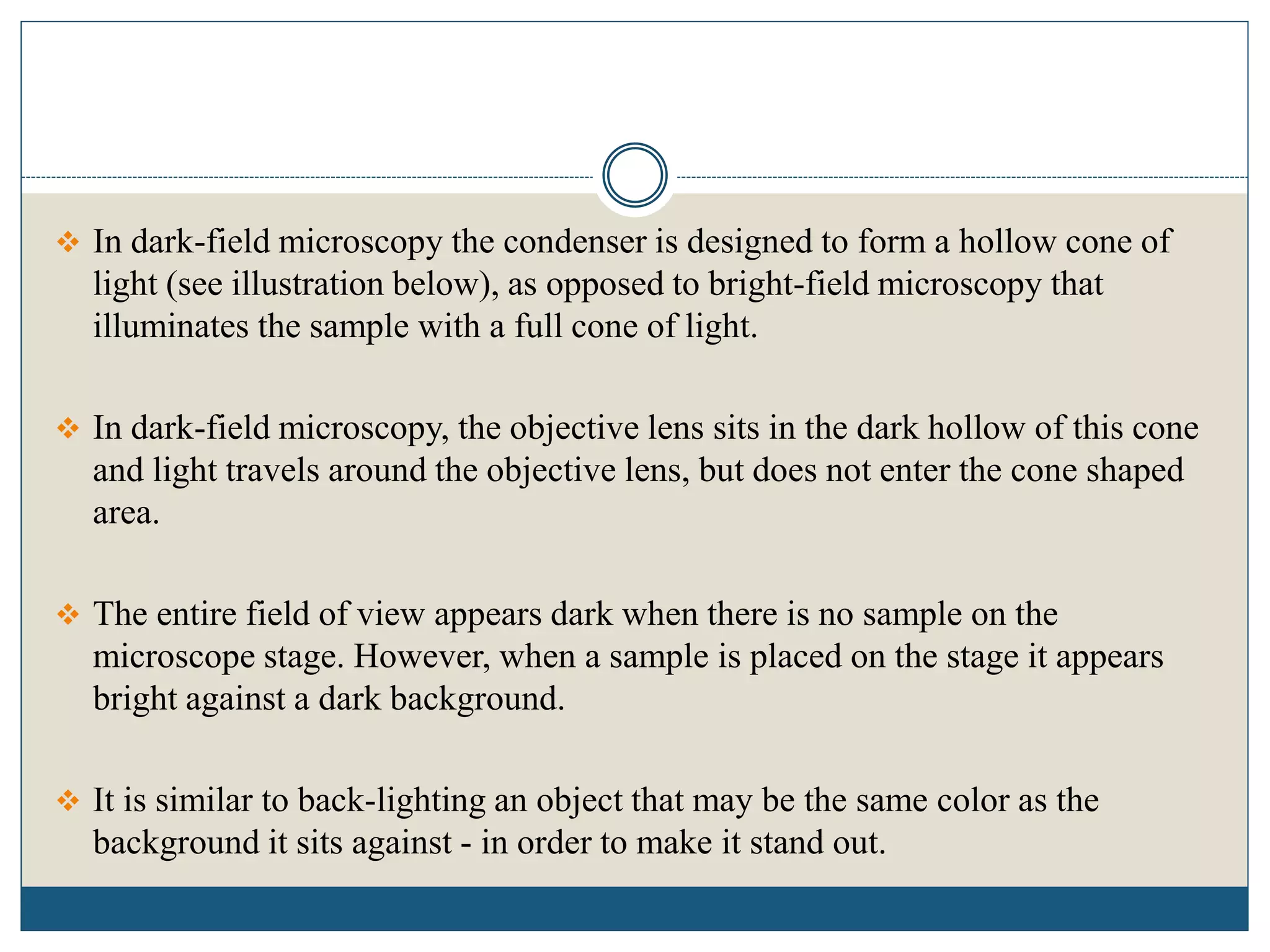
DARK FIELD MICROSCOPY by SIVASANGARI SHANMUGAM
Dark-field microscopy is ideally used to illuminate unstained samples causing them to appear brightly lit against a dark background.
This type of microscope contains a special condenser that scatters light and causes it to reflect off the specimen at an angle
BRIGHT FIELD MICROSCOPY by SIVASANGARI SHANMUGAM
bRIGHT FIELD MICROSCOPY is also called a compound microscope. The name bright - field is derived from the fact that the specimen is dark and contrasted by the surrounding bright viewing field.
Bright field microscopy, Principle and applicationsKAUSHAL SAHU
Introduction
History
Basic Component of Microscope
Light Microscopy
Types of Light Microscopy
What Are Bright Microscopy
Principle of Bright Microscope
Advantage
Disadvantage
Application
Conclusion
Reference
LIGHT MICROSCOPY by SIVASANGARI SHANMUGAM
The optical microscope, The functions of a light microscope is based on its ability to focus a beam of light through, which is very small and transparent, to produce an image.
5. Microsocope ELECTRON MICROSCOPE (TEM & SEM ) - BasicsNethravathi Siri
Basics only
Electron beam is the source of illumination.
Image is produced by magnetic field.
Contrasting features between light microscope and electron microscope are
construction, working principle, specimen preparation, cost-expenses and designed
room (vacuum chamber).
Phase contrast microscopy by sivasangari shanmugam
Phase-contrast microscopy, first described by Dutch physicist Frits Zernike in 1934.
It can be utilized to produce high-contrast images of transparent specimens, such as living cells (usually in culture), microorganisms, thin tissue slices, fibers, latex dispersions, glass fragments, and subcellular particles (including nuclei and other organelles).
Microscopy is the technical field of using microscopes to view objects and areas of objects that cannot be seen with the naked eye (objects that are not within the resolution range of the normal eye). There are three well-known branches of microscopy: optical, electron, and scanning probe microscopy, along with the emerging field of X-ray microscopy.
Electron microscope, principle and applicationKAUSHAL SAHU
Introduction
History
Resolution &Magnification of
Electron microscope
Types of electron microscope
1) Transmission electron microscope (TEM)
- Structural parts of TEM
- Principle & Working of TEM
- Sample preparation for TEM
- Advantages & disadvantages of TEM
Scanning electron microscope (SEM)
- Structural parts of SEM
- Principle & Working of SEM
- Sample preparation for SEM
- Advantages & disadvantages of SEM
3) Scanning transmission electron microscope (STEM)
Applications of electron microscope
Conclusion
References
Dark-field microscopy is ideally used to illuminate unstained samples causing them to appear brightly lit against a dark background. This type of microscope contains a special condenser that scatters light and causes it to reflect off the specimen at an angle
Types of Light Microscopes used in Histological Studies.pptxssuserab552f
Light microscopes relies on glass lenses and visible light to magnify tissue samples. It was
invented in XVII century, and has been improved over the years, resulting in the powerful
modern light microscopes. As individual cellular structures are too small to be seen by the
human eye, microscopy techniques have played a key role in the development of
histological techniques.
Introduction to microscopy
Different parts of a microscope & their function
Different types of microscopy
Different types of optical microscopy
Different types of electron microscopy
Different terms used in microscopy
Staining- Simple, Differential, Special
Gram Staining
Dark-field microscope is a machine or matter that can be used for thisobservation of living, unstained cells and microorganisms. In this microscopy, the specimen is brightly illuminated while the background is dark. It is one type of light microscopes.
Used to examine live microorganisms that are not visible with ordinary light microscope.
3. Microscope simple, compound & stereo - BasicsNethravathi Siri
Basics only
A compound microscope is an optical instrument used to observe the magnified images of small objects on a glass slide. Compound microscopes are so called because they are designed with a compound lens system.
Confocal microscopy is an optical imaging technique for increasing optical resolution and contrast of a micrograph by means of adding a spatial pinhole placed at the confocal plane of the lens to eliminate out-of-focus light.
Bright-field microscopy is the simplest of all the optical microscopy illumination techniques. Sample illumination is transmitted (i.e., illuminated from below and observed from above) white light and contrast in the sample is caused by absorbance of some of the transmitted light in dense areas of the sample.
Microscopy is the technique of using microscopes to observe and analyze objects that are too small to be seen by the naked eye. Microscopes are instruments that magnify and resolve the details of objects, allowing scientists and researchers to study the structure, composition, and behavior of materials and specimens at a microscopic level
It contain more information about Amino acids and their structure. Then , contain both physical and chemical properties. Next Classification of amino acids based on nutritional requirements, based on metabolic fate, Position of NH2 group, etc.,
Transmission electron microscopy (TEM)- by sivasangari Shanmugam. Transmission electron microscopy (TEM) is a technique used to observe the features of very small specimens.
SEM is a type of electron microscope designed for directly studying the surfaces of solid objects, that utilizes a beam of focused electron of relatively low energy as an electron probe that is scanned in a regular manner over the specimen.
Electron microscopy by SIVASANGARI SHANMUGAM.
Electron microscopy is a technique for obtaining high-resolution images of biological and non-biological specimens.
The increased availability of biomedical data, particularly in the public domain, offers the opportunity to better understand human health and to develop effective therapeutics for a wide range of unmet medical needs. However, data scientists remain stymied by the fact that data remain hard to find and to productively reuse because data and their metadata i) are wholly inaccessible, ii) are in non-standard or incompatible representations, iii) do not conform to community standards, and iv) have unclear or highly restricted terms and conditions that preclude legitimate reuse. These limitations require a rethink on data can be made machine and AI-ready - the key motivation behind the FAIR Guiding Principles. Concurrently, while recent efforts have explored the use of deep learning to fuse disparate data into predictive models for a wide range of biomedical applications, these models often fail even when the correct answer is already known, and fail to explain individual predictions in terms that data scientists can appreciate. These limitations suggest that new methods to produce practical artificial intelligence are still needed.
In this talk, I will discuss our work in (1) building an integrative knowledge infrastructure to prepare FAIR and "AI-ready" data and services along with (2) neurosymbolic AI methods to improve the quality of predictions and to generate plausible explanations. Attention is given to standards, platforms, and methods to wrangle knowledge into simple, but effective semantic and latent representations, and to make these available into standards-compliant and discoverable interfaces that can be used in model building, validation, and explanation. Our work, and those of others in the field, creates a baseline for building trustworthy and easy to deploy AI models in biomedicine.
Bio
Dr. Michel Dumontier is the Distinguished Professor of Data Science at Maastricht University, founder and executive director of the Institute of Data Science, and co-founder of the FAIR (Findable, Accessible, Interoperable and Reusable) data principles. His research explores socio-technological approaches for responsible discovery science, which includes collaborative multi-modal knowledge graphs, privacy-preserving distributed data mining, and AI methods for drug discovery and personalized medicine. His work is supported through the Dutch National Research Agenda, the Netherlands Organisation for Scientific Research, Horizon Europe, the European Open Science Cloud, the US National Institutes of Health, and a Marie-Curie Innovative Training Network. He is the editor-in-chief for the journal Data Science and is internationally recognized for his contributions in bioinformatics, biomedical informatics, and semantic technologies including ontologies and linked data.
Earliest Galaxies in the JADES Origins Field: Luminosity Function and Cosmic ...Sérgio Sacani
We characterize the earliest galaxy population in the JADES Origins Field (JOF), the deepest
imaging field observed with JWST. We make use of the ancillary Hubble optical images (5 filters
spanning 0.4−0.9µm) and novel JWST images with 14 filters spanning 0.8−5µm, including 7 mediumband filters, and reaching total exposure times of up to 46 hours per filter. We combine all our data
at > 2.3µm to construct an ultradeep image, reaching as deep as ≈ 31.4 AB mag in the stack and
30.3-31.0 AB mag (5σ, r = 0.1” circular aperture) in individual filters. We measure photometric
redshifts and use robust selection criteria to identify a sample of eight galaxy candidates at redshifts
z = 11.5 − 15. These objects show compact half-light radii of R1/2 ∼ 50 − 200pc, stellar masses of
M⋆ ∼ 107−108M⊙, and star-formation rates of SFR ∼ 0.1−1 M⊙ yr−1
. Our search finds no candidates
at 15 < z < 20, placing upper limits at these redshifts. We develop a forward modeling approach to
infer the properties of the evolving luminosity function without binning in redshift or luminosity that
marginalizes over the photometric redshift uncertainty of our candidate galaxies and incorporates the
impact of non-detections. We find a z = 12 luminosity function in good agreement with prior results,
and that the luminosity function normalization and UV luminosity density decline by a factor of ∼ 2.5
from z = 12 to z = 14. We discuss the possible implications of our results in the context of theoretical
models for evolution of the dark matter halo mass function.
Richard's aventures in two entangled wonderlandsRichard Gill
Since the loophole-free Bell experiments of 2020 and the Nobel prizes in physics of 2022, critics of Bell's work have retreated to the fortress of super-determinism. Now, super-determinism is a derogatory word - it just means "determinism". Palmer, Hance and Hossenfelder argue that quantum mechanics and determinism are not incompatible, using a sophisticated mathematical construction based on a subtle thinning of allowed states and measurements in quantum mechanics, such that what is left appears to make Bell's argument fail, without altering the empirical predictions of quantum mechanics. I think however that it is a smoke screen, and the slogan "lost in math" comes to my mind. I will discuss some other recent disproofs of Bell's theorem using the language of causality based on causal graphs. Causal thinking is also central to law and justice. I will mention surprising connections to my work on serial killer nurse cases, in particular the Dutch case of Lucia de Berk and the current UK case of Lucy Letby.
Cancer cell metabolism: special Reference to Lactate PathwayAADYARAJPANDEY1
Normal Cell Metabolism:
Cellular respiration describes the series of steps that cells use to break down sugar and other chemicals to get the energy we need to function.
Energy is stored in the bonds of glucose and when glucose is broken down, much of that energy is released.
Cell utilize energy in the form of ATP.
The first step of respiration is called glycolysis. In a series of steps, glycolysis breaks glucose into two smaller molecules - a chemical called pyruvate. A small amount of ATP is formed during this process.
Most healthy cells continue the breakdown in a second process, called the Kreb's cycle. The Kreb's cycle allows cells to “burn” the pyruvates made in glycolysis to get more ATP.
The last step in the breakdown of glucose is called oxidative phosphorylation (Ox-Phos).
It takes place in specialized cell structures called mitochondria. This process produces a large amount of ATP. Importantly, cells need oxygen to complete oxidative phosphorylation.
If a cell completes only glycolysis, only 2 molecules of ATP are made per glucose. However, if the cell completes the entire respiration process (glycolysis - Kreb's - oxidative phosphorylation), about 36 molecules of ATP are created, giving it much more energy to use.
IN CANCER CELL:
Unlike healthy cells that "burn" the entire molecule of sugar to capture a large amount of energy as ATP, cancer cells are wasteful.
Cancer cells only partially break down sugar molecules. They overuse the first step of respiration, glycolysis. They frequently do not complete the second step, oxidative phosphorylation.
This results in only 2 molecules of ATP per each glucose molecule instead of the 36 or so ATPs healthy cells gain. As a result, cancer cells need to use a lot more sugar molecules to get enough energy to survive.
introduction to WARBERG PHENOMENA:
WARBURG EFFECT Usually, cancer cells are highly glycolytic (glucose addiction) and take up more glucose than do normal cells from outside.
Otto Heinrich Warburg (; 8 October 1883 – 1 August 1970) In 1931 was awarded the Nobel Prize in Physiology for his "discovery of the nature and mode of action of the respiratory enzyme.
WARNBURG EFFECT : cancer cells under aerobic (well-oxygenated) conditions to metabolize glucose to lactate (aerobic glycolysis) is known as the Warburg effect. Warburg made the observation that tumor slices consume glucose and secrete lactate at a higher rate than normal tissues.
Seminar of U.V. Spectroscopy by SAMIR PANDASAMIR PANDA
Spectroscopy is a branch of science dealing the study of interaction of electromagnetic radiation with matter.
Ultraviolet-visible spectroscopy refers to absorption spectroscopy or reflect spectroscopy in the UV-VIS spectral region.
Ultraviolet-visible spectroscopy is an analytical method that can measure the amount of light received by the analyte.
Slide 1: Title Slide
Extrachromosomal Inheritance
Slide 2: Introduction to Extrachromosomal Inheritance
Definition: Extrachromosomal inheritance refers to the transmission of genetic material that is not found within the nucleus.
Key Components: Involves genes located in mitochondria, chloroplasts, and plasmids.
Slide 3: Mitochondrial Inheritance
Mitochondria: Organelles responsible for energy production.
Mitochondrial DNA (mtDNA): Circular DNA molecule found in mitochondria.
Inheritance Pattern: Maternally inherited, meaning it is passed from mothers to all their offspring.
Diseases: Examples include Leber’s hereditary optic neuropathy (LHON) and mitochondrial myopathy.
Slide 4: Chloroplast Inheritance
Chloroplasts: Organelles responsible for photosynthesis in plants.
Chloroplast DNA (cpDNA): Circular DNA molecule found in chloroplasts.
Inheritance Pattern: Often maternally inherited in most plants, but can vary in some species.
Examples: Variegation in plants, where leaf color patterns are determined by chloroplast DNA.
Slide 5: Plasmid Inheritance
Plasmids: Small, circular DNA molecules found in bacteria and some eukaryotes.
Features: Can carry antibiotic resistance genes and can be transferred between cells through processes like conjugation.
Significance: Important in biotechnology for gene cloning and genetic engineering.
Slide 6: Mechanisms of Extrachromosomal Inheritance
Non-Mendelian Patterns: Do not follow Mendel’s laws of inheritance.
Cytoplasmic Segregation: During cell division, organelles like mitochondria and chloroplasts are randomly distributed to daughter cells.
Heteroplasmy: Presence of more than one type of organellar genome within a cell, leading to variation in expression.
Slide 7: Examples of Extrachromosomal Inheritance
Four O’clock Plant (Mirabilis jalapa): Shows variegated leaves due to different cpDNA in leaf cells.
Petite Mutants in Yeast: Result from mutations in mitochondrial DNA affecting respiration.
Slide 8: Importance of Extrachromosomal Inheritance
Evolution: Provides insight into the evolution of eukaryotic cells.
Medicine: Understanding mitochondrial inheritance helps in diagnosing and treating mitochondrial diseases.
Agriculture: Chloroplast inheritance can be used in plant breeding and genetic modification.
Slide 9: Recent Research and Advances
Gene Editing: Techniques like CRISPR-Cas9 are being used to edit mitochondrial and chloroplast DNA.
Therapies: Development of mitochondrial replacement therapy (MRT) for preventing mitochondrial diseases.
Slide 10: Conclusion
Summary: Extrachromosomal inheritance involves the transmission of genetic material outside the nucleus and plays a crucial role in genetics, medicine, and biotechnology.
Future Directions: Continued research and technological advancements hold promise for new treatments and applications.
Slide 11: Questions and Discussion
Invite Audience: Open the floor for any questions or further discussion on the topic.
Richard's entangled aventures in wonderlandRichard Gill
Since the loophole-free Bell experiments of 2020 and the Nobel prizes in physics of 2022, critics of Bell's work have retreated to the fortress of super-determinism. Now, super-determinism is a derogatory word - it just means "determinism". Palmer, Hance and Hossenfelder argue that quantum mechanics and determinism are not incompatible, using a sophisticated mathematical construction based on a subtle thinning of allowed states and measurements in quantum mechanics, such that what is left appears to make Bell's argument fail, without altering the empirical predictions of quantum mechanics. I think however that it is a smoke screen, and the slogan "lost in math" comes to my mind. I will discuss some other recent disproofs of Bell's theorem using the language of causality based on causal graphs. Causal thinking is also central to law and justice. I will mention surprising connections to my work on serial killer nurse cases, in particular the Dutch case of Lucia de Berk and the current UK case of Lucy Letby.
Introduction:
RNA interference (RNAi) or Post-Transcriptional Gene Silencing (PTGS) is an important biological process for modulating eukaryotic gene expression.
It is highly conserved process of posttranscriptional gene silencing by which double stranded RNA (dsRNA) causes sequence-specific degradation of mRNA sequences.
dsRNA-induced gene silencing (RNAi) is reported in a wide range of eukaryotes ranging from worms, insects, mammals and plants.
This process mediates resistance to both endogenous parasitic and exogenous pathogenic nucleic acids, and regulates the expression of protein-coding genes.
What are small ncRNAs?
micro RNA (miRNA)
short interfering RNA (siRNA)
Properties of small non-coding RNA:
Involved in silencing mRNA transcripts.
Called “small” because they are usually only about 21-24 nucleotides long.
Synthesized by first cutting up longer precursor sequences (like the 61nt one that Lee discovered).
Silence an mRNA by base pairing with some sequence on the mRNA.
Discovery of siRNA?
The first small RNA:
In 1993 Rosalind Lee (Victor Ambros lab) was studying a non- coding gene in C. elegans, lin-4, that was involved in silencing of another gene, lin-14, at the appropriate time in the
development of the worm C. elegans.
Two small transcripts of lin-4 (22nt and 61nt) were found to be complementary to a sequence in the 3' UTR of lin-14.
Because lin-4 encoded no protein, she deduced that it must be these transcripts that are causing the silencing by RNA-RNA interactions.
Types of RNAi ( non coding RNA)
MiRNA
Length (23-25 nt)
Trans acting
Binds with target MRNA in mismatch
Translation inhibition
Si RNA
Length 21 nt.
Cis acting
Bind with target Mrna in perfect complementary sequence
Piwi-RNA
Length ; 25 to 36 nt.
Expressed in Germ Cells
Regulates trnasposomes activity
MECHANISM OF RNAI:
First the double-stranded RNA teams up with a protein complex named Dicer, which cuts the long RNA into short pieces.
Then another protein complex called RISC (RNA-induced silencing complex) discards one of the two RNA strands.
The RISC-docked, single-stranded RNA then pairs with the homologous mRNA and destroys it.
THE RISC COMPLEX:
RISC is large(>500kD) RNA multi- protein Binding complex which triggers MRNA degradation in response to MRNA
Unwinding of double stranded Si RNA by ATP independent Helicase
Active component of RISC is Ago proteins( ENDONUCLEASE) which cleave target MRNA.
DICER: endonuclease (RNase Family III)
Argonaute: Central Component of the RNA-Induced Silencing Complex (RISC)
One strand of the dsRNA produced by Dicer is retained in the RISC complex in association with Argonaute
ARGONAUTE PROTEIN :
1.PAZ(PIWI/Argonaute/ Zwille)- Recognition of target MRNA
2.PIWI (p-element induced wimpy Testis)- breaks Phosphodiester bond of mRNA.)RNAse H activity.
MiRNA:
The Double-stranded RNAs are naturally produced in eukaryotic cells during development, and they have a key role in regulating gene expression .
3. INTRODUCTION
Dark-field microscopy is ideally used to illuminate unstained samples causing them to
appear brightly lit against a dark background.
This type of microscope contains a special condenser that scatters light and causes it to
reflect off the specimen at an angle.
Rather than illuminating the sample with a filled cone of light, the condenser is
designed to form a hollow cone of light.
This microscope uses reflected light instead of transmitted light used in the ordinary
light microscope.
It prevents light from falling directly on the objective lens.
Light rays falling on the object are reflected or scattered onto the objective lens with
the result that the microorganisms appear brightly stained against a dark background.
4. In dark-field microscopy the condenser is designed to form a hollow cone of
light (see illustration below), as opposed to bright-field microscopy that
illuminates the sample with a full cone of light.
In dark-field microscopy, the objective lens sits in the dark hollow of this cone
and light travels around the objective lens, but does not enter the cone shaped
area.
The entire field of view appears dark when there is no sample on the
microscope stage. However, when a sample is placed on the stage it appears
bright against a dark background.
It is similar to back-lighting an object that may be the same color as the
background it sits against - in order to make it stand out.
5. PRINCIPLES
A dark field microscope is arranged so that the light source
is blocked off, causing light to scatter as it hits the
specimen.
This is ideal for making objects with refractive values
similar to the background appear bright against a dark
background.
When light hits an object, rays are scattered in all
directions. The design of the dark field microscope is such
that it removes the dispersed light, so that only the scattered
beams hit the sample.
The introduction of a condenser and/or stop below the stage
ensures that these light rays will hit the specimen at
different angles, rather than as a direct light source
above/below the object.
The result is a “cone of light” where rays are diffracted,
reflected and/or refracted off the object, ultimately,
allowing the individual to view a specimen in dark field.
6. PARTS OF DARK FIELD MICROSCOPE
1. Eye piece
2. Arm
3. Rotating nosepiece
4. Objective lens
5. Condenser
6. Dark disc
7. Light source
8. Course and Fine adjustment
8. HOW DOES IT WORK?
Light reaches the object mounted on the stage through the condenser.
The condenser lens focuses the light towards the sample.
The patch stop provided in the condenser lens blocks light around the central
region allowing light to pass only the periphery of the lens.
The light that enters the specimen, most is directly transmitted, while some is
scattered from the sample.
The scattered light alone enters the objective lens and produce the image.
The directly transmitted light is not collected and is omitted.
Thus the field of vision is rendered dark and the image created is by the scattered
light and therefore, cell components that reflect light are clear in the image.
9. APPLICATIONS
It is useful for the demonstration of very thin bacteria not visible under
ordinary illumination.
Used for rapid demonstration of Treponema pallidum in specimens.
Useful for the demon clinical of motility of flagellated bacteria and
protozoa.
Used to study marine organisms such as algae, plankton, diatoms, insects,
fibers, hairs, yeast and protozoa as well as some minerals and crystals, thin
polymers and some ceramics.
Used to study mounted cells and tissues.
It is more useful in examining external details, such as outlines, edges, grain
boundaries and surface defects than internal structure.
10. ADVANTAGES
Resolution by dark-field microscopy is better than bright-field microscopy.
Improves image contrast without the use of stain, and thus do not kill cells.
Direct detection of non-culturable bacteria present in patient samples.
No sample preparation is required.
Requires no special set up, even a light microscope can be converted to dark
field.
Dark-field microscopy is a very simple yet effective technique.
11. It is well suited for uses involving live and unstained biological samples,
such as a smear from a tissue culture or individual, water-borne, single-celled
organisms.
Considering the simplicity of the setup, the quality of images obtained from
this technique is impressive.
Dark-field microscopy techniques are almost entirely free of artifacts, due to
the nature of the process.
A researcher can achieve a dark field by making modifications to his/her
microscope.
12. DISADVANTAGES
The low light levels seen in the final image.
The sample must be very strongly illuminated, which can cause damage to the
sample.
Necessity to examine wet, moist specimens containing living organisms very
quickly, because visualization of the moving bacteria is essential to detection.
Besides the sample, dust particles also scatter the light and appear bright.
Sample material needs to be spread thinly, dense preparations can grossly
affect the contrast and accuracy of the dark field’s image.
13. REFERENCE
1. https://microbenotes.com/darkfield-microscopy/
2. https://www.slideshare.net/abhishekindurkar/dark-field-microscopy-81857005
3. https://microbeonline.com/dark-field-microscopy-principles-use-advantages-and-
limitations/
4. https://bio.libretexts.org/Bookshelves/Microbiology/Book%3A_Microbiology_(Boun
dless)/3%3A_Microscopy/3.3%3A_Other_Types_of_Microscopy/3.3A%3A_Dark-
Field_Microscopy
5. https://www.microscopeworld.com/t-darkfield_microscopy.aspx
6. Parija S.C. (2012). Textbook of Microbiology & Immunology.(2 ed.). India: Elsevier
India.
7. Cappuccino, J. and Welsh, C. (2014). Microbiology: A Laboratory Manual, Global
Edition. 1st ed. Pearson Education.