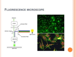
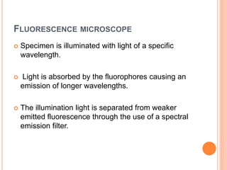
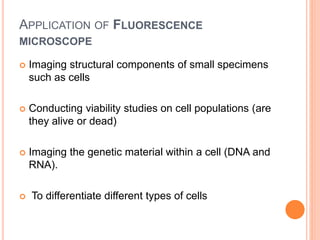
The document discusses different light microscopy techniques including dark field microscopy, phase contrast microscopy, and fluorescence microscopy. Dark field microscopy uses angled illumination to visualize objects that scatter light against a dark background. Phase contrast microscopy converts small phase changes in light passing through a specimen into visible intensity changes, allowing fine specimen details to be seen. Fluorescence microscopy takes advantage of specimens that are inherently fluorescent or can be labeled with fluorescent dyes. These techniques allow better visualization of transparent or unstained specimens compared to traditional bright field microscopy.
The document discusses phase contrast microscopy, which was developed by Fritz Zernike in the 1930s. It allows living or unstained cells and intracellular components to be visible under a microscope. The phase contrast microscope works by converting small phase changes caused by differences in refractive index of cell structures into visible brightness and darkness differences. This makes organelles and other structures visible without using staining. The phase contrast is achieved using an annular diaphragm and phase rings or filters to shift the phase of light passing through or around the specimen.
Microscopy uses lenses to magnify objects too small to see with the naked eye. A microscope contains objective lenses close to the sample that collect light and project an enlarged virtual image, and eyepiece lenses that further magnify the image for viewing. Key components include the condenser, which directs light up through the sample, and various types of objectives and eyepieces optimized for tasks like brightfield, darkfield, phase contrast, or fluorescence microscopy. Objectives are classified by their magnification and degree of correction for chromatic and spherical aberration, from simple achromats to highly-corrected apochromats. Different microscopy techniques employ specific lighting and optics to reveal features of transparent and unstained samples.
The document discusses the simple microscope. It defines a simple microscope as using a single lens for magnification rather than multiple lenses like a compound microscope. A simple microscope works by using a convex lens to produce a virtual, erect and magnified image of an object placed within the lens' focal point. The maximum magnification of a simple microscope is around 10x. Key parts of a simple microscope include a metal stand, stage to hold samples, plano-convex mirror to illuminate samples, and a biconvex lens for magnification. Simple microscopes are used to examine small biological specimens, watch parts, jewelry, book text, soil particles, and skin.
Phase-contrast microscopy is a technique that converts phase shifts in light passing through a transparent specimen to brightness changes in the image, allowing living cells that are otherwise invisible to be seen. It works by separating light rays that pass through a specimen unchanged from those that are diffracted, using an annular diaphragm and phase plate in the light path. Phase-contrast microscopy is widely used in biological research for observing living cells, microorganisms, and other transparent specimens without staining or fixing.
this presentation deals with the introduction of some of the commonly used optical microscopes in forensic labs; compound microscope, stereoscopic microscope, comparison microscope, fluorescence microscope and polarized microscope.
This document discusses phase contrast microscopy. It begins by defining a microscope and microscopy. It then describes the main types of microscopes, including optical, electron, and scanning probe microscopes. It focuses on the light microscope, explaining that it uses visible light and is commonly used in biology to view structures like cells. It defines magnification and resolution. It describes the different modes of light microscopy - bright field, dark field, and phase contrast - and explains that phase contrast microscopy allows observation of living cells by enhancing contrast between structures with small refractive index differences. It notes that phase contrast microscopy was invented by Frits Zernike, for which he won the Nobel Prize in Physics in 1953.
Phase contrast microscopy is a technique that was invented in 1934 by Dutch physicist Frits Zernike, for which he received the Nobel Prize in Physics in 1953. It allows for high-contrast imaging of transparent specimens like living cells without staining. In phase contrast microscopy, variations in the refractive index within a specimen cause some light rays to be retarded, producing an image with areas of different intensities. This converts subtle differences in a sample's density and refractive index into detectable variations in light intensity. Phase contrast microscopy is useful for observing living cells and intracellular structures in their natural state without needing to kill, fix, or stain the specimen. While it provides high resolution living images, it has limitations such as inability to view thick specimens clearly and
The microscope has evolved a lot from the time of Leeuwenhoek. This presentation gives a brief overview about the types of microscope their principle of function and application.
Bright field microscope and its working principleKavimugarajaM
The document discusses the bright field microscope. It notes that the bright field microscope is a simple microscope to use, with few adjustments needed. It can view some specimens without staining and does not alter specimen color. However, it has limitations such as distortion with high contrast and inability to view living bacteria. The document describes how bright field microscopy works by using a light source to illuminate the specimen from below, appearing dark against a bright background, and provides examples of specimens that can be observed with it.
The document discusses the history and development of electron microscopes. It describes how J.J. Thomson discovered electrons in 1897 and how subsequent scientists like de Broglie, Ruska, and Knoll contributed to the development of the first electron microscope in the 1930s. It then explains the basic workings and components of transmission electron microscopes and scanning electron microscopes, how they produce images, and some examples of their applications in biology and materials science.
The document provides information about using a compound light microscope, including:
1) The main parts of a compound light microscope are the ocular lenses, body tube, objective lenses, stage, condenser, iris diaphragm, coarse and fine adjustment knobs, and light source.
2) To examine a specimen, the user selects the appropriate objective lens, focuses using the coarse and fine adjustment knobs, and adjusts the condenser and iris diaphragm to control illumination.
3) For oil immersion objectives, oil is added to the slide and specimen to allow for higher magnification imaging.
Microscope ppt, by jitendra kumar pandey,medical micro,2nd yr, mgm medical co...jitendra Pandey
The document summarizes different types of microscopes used to study microorganisms. It discusses light microscopes like brightfield, darkfield and phase contrast microscopes. It also describes electron microscopes like transmission electron microscopes (TEM) and scanning electron microscopes (SEM) that use electron beams instead of light. TEM images internal structures by transmitting electrons through thin samples while SEM scans sample surfaces using secondary electrons. Sample preparation methods for both light and electron microscopy are also outlined.
This document provides information about different types of microscopes. It discusses simple microscopes, which use a single lens for magnification, and their basic parts and working principle. Compound microscopes are then introduced as using multiple lenses to achieve higher magnification than is possible with a simple lens alone. The key parts of a compound microscope are described, including the eyepiece, objective lenses, body tube, arm, and focus adjustments. It explains that light from the illuminator passes through these parts to produce a magnified image for viewing.
This document provides information on different types of microscopy techniques including bright field, dark field, phase contrast, and polarized light microscopy. It begins with explaining the basics of light and microscopy. It then describes each technique in more detail, including their principles, applications, advantages, and how they are set up optically. Bright field microscopy uses illumination and forms a dark image on a bright background. Dark field uses oblique illumination to see small particles as bright objects on a dark background. Phase contrast converts phase differences into contrast changes to see transparent specimens. Polarized light microscopy uses polarized filters to reveal structural details not otherwise seen.
Dark field microscopy produces bright images of unstained samples against a dark background. It works by using a condenser with an opaque disk to block light entering the objective lens directly, allowing only light reflected off the sample to pass through. This causes specimens to appear bright on a dark background. It is useful for viewing transparent or unstained samples like bacteria, cells, and minerals due to the contrast it provides.
Phase contrast microscopy is a type of light microscopy technique that produces high contrast images of transparent specimens without staining. It works by interfering surrounding light waves and sample-diffracted waves to create an image with the inner regions of the sample appearing darker than the outer regions. Fluorescence microscopy uses fluorescent dyes excited by high-energy light to emit lower-energy light, allowing specific structures within samples to be imaged. It is used to study living cells and view genetic material.
1. MICROSCOPY - introduction + principle (Basics)Nethravathi Siri
Basics only
Microscopy is the technical field that uses microscopes to observe samples which are
not in the resolution range of the normal-unaided eye.
Microscope is a scientific-instrument consisting of magnifying lens that enables an
observer to view the minute features distinctly.
In greek, micro = small
skopein = to view.
Recent advancement in microscopic techniques cryoem and superDanishNigar
Cryo-electron microscopy is an emerging technique that uses electron microscopes to image biomolecules such as proteins that have been rapidly frozen to preserve their structure. It overcomes limitations of other techniques by not requiring crystallization and allowing study of molecules in their natural state. In cryo-EM, a sample is frozen at liquid nitrogen temperatures to vitrify it before being imaged with an electron microscope. This technique has helped determine structures of important proteins and revealed new details about their function. Recent advances in direct electron detectors, image processing, and specimen preparation have led to remarkable improvements in resolution with cryo-EM.
DARK FIELD MICROSCOPY by SIVASANGARI SHANMUGAM
Dark-field microscopy is ideally used to illuminate unstained samples causing them to appear brightly lit against a dark background.
This type of microscope contains a special condenser that scatters light and causes it to reflect off the specimen at an angle
The document discusses different types of microscopes, including compound microscopes and stereomicroscopes. It describes the key parts and principles of operation. Compound microscopes use multiple lenses to magnify specimens and provide a two-dimensional image. Stereomicroscopes use two optical paths to provide a three-dimensional view of surface details. Examples of uses include biology studies, forensics, manufacturing quality control, and more. The document also discusses who may have invented the compound microscope and provides references for further reading.
The document describes the parts and working of a polarizing microscope. It has optical components like polarizers, analyzers and lenses, and mechanical components like the rotating stage. Light from the specimen is polarized and its interaction with the optical components is used to identify properties of minerals and rocks. The polarized microscope allows examination of anisotropic materials and determination of their optical characteristics, which has applications in geology and mineral exploration.
Fluorescence microscopy uses fluorescence to visualize specimens. It works by exciting fluorescent molecules in the sample with high intensity light, causing them to emit light of a longer wavelength. This emitted light is then filtered and used to produce a magnified image of the sample. Modern fluorescence microscopes allow multiple fluorescence filters to be used, and fluorescent markers like dyes, proteins, and antibodies can be introduced to tag specific structures in cells or proteins of interest. This technique is widely used in medical and biological research to study structures and track molecules within living cells.
This document provides an introduction to microscopy. It discusses different types of microscopes including compound microscopes, which have three main systems - the support system, illumination system, and magnification system. Binocular microscopes have two eyepieces. Important microscope terms are also defined, such as magnification, working distance, resolving power, and numerical aperture. Phase-contrast microscopy is described as a technique that converts phase shifts in light to brightness changes, allowing otherwise transparent specimens to be visible.
LIGHT MICROSCOPY by SIVASANGARI SHANMUGAM
The optical microscope, The functions of a light microscope is based on its ability to focus a beam of light through, which is very small and transparent, to produce an image.
Dark field microscopy is a technique that uses a dark field condenser containing an opaque disk to block light entering the objective lens directly. Only light reflected off the specimen enters the lens, causing specimens to appear bright against a dark background. This technique is useful for viewing unstained, transparent specimens like bacteria, algae, and fibers. It has advantages like viewing details on surfaces but disadvantages like image degradation from non-uniform specimens or particles on the optics. Dark field microscopy finds applications in diagnosing syphilis and viewing various microorganisms, minerals, and cells.
- The document discusses the compound microscope, its history, parts, functions, and use.
- A compound microscope has more than one lens and was invented in the 1590s in the Netherlands. It allows higher magnification than simple microscopes.
- The main parts are the mechanical stage, optical system with objectives and eyepieces, illumination system, and adjustment controls. Objectives magnify the specimen while eyepieces provide a final view.
- Focusing involves using coarse and fine adjustments to bring the specimen into view at different magnifications including with immersion oil.
Types of Light Microscopes used in Histological Studies.pptxssuserab552f
Light microscopes relies on glass lenses and visible light to magnify tissue samples. It was
invented in XVII century, and has been improved over the years, resulting in the powerful
modern light microscopes. As individual cellular structures are too small to be seen by the
human eye, microscopy techniques have played a key role in the development of
histological techniques.
Introduction to microscopy
Different parts of a microscope & their function
Different types of microscopy
Different types of optical microscopy
Different types of electron microscopy
Different terms used in microscopy
Staining- Simple, Differential, Special
Gram Staining
3. Microscope simple, compound & stereo - BasicsNethravathi Siri
A simple microscope uses a single lens to magnify objects and forms a virtual image. It provides low magnification and is used to examine things like skin, algae, and soil samples.
A compound microscope has two lens systems that provide higher magnification by compounding the magnification of the objective and eyepiece lenses. It allows detailed examination of stained slides and is commonly used in biology labs and medical diagnostics.
A stereo microscope uses two separate optical paths and lens systems to provide a three-dimensional view of surfaces. It has lower magnification than compound microscopes and is used for examining things like insects and circuit boards.
The document discusses different types of microscopes, their components, principles, and applications. It describes light microscopes like brightfield, darkfield, phase-contrast, and fluorescent microscopes. It also covers electron microscopes. Brightfield microscopes illuminate specimens with light against a bright background. Darkfield microscopes deflect light to view specimens brightly against a dark background. Phase-contrast and fluorescent microscopes use special optics and fluorescent dyes to view unstained or tagged specimens. Electron microscopes use electron beams instead of light for magnifications over 1 million times.
This document provides information about different types of microscopy. It begins with an introduction to microscopy, then discusses the history and key figures in the development of the microscope. It describes different types of microscopes including light/bright field microscopy, dark field microscopy, phase contrast microscopy, fluorescence microscopy, and electron microscopy. For each type, it provides details on the optical principles, components, and applications. The document aims to inform the reader about the basic concepts and techniques of microscopy.
Microscopy - Magnification, Resolving power, Principles, Types and ApplicationsNethravathi Siri
Magnification, Resolving power, Principles and Applications of Simple, Compound, Stereozoom, Phase contrast, Fluorescent and Electron microscopes (TEM & SEM).
Microscopy is the technical field that uses microscopes to observe samples which are not in the resolution range of the normal-unaided eye.
This document discusses various histological tools used to study tissues at the microscopic level. It describes light microscopes, which use visible light and can magnify up to 1000x, and electron microscopes, which use electron beams to achieve much higher magnification up to 1 million times. Key histological techniques mentioned include biopsy, tissue processing, staining, immunohistochemistry, and different types of microscopy like fluorescence and polarizing microscopy. The document provides details on the basic components and functioning of different microscope types used in histology.
This document discusses various histological tools used to study tissues at the microscopic level. It describes light microscopes, which use visible light and magnification to examine thin tissue slices stained with histological dyes. Electron microscopes are also covered, using electron beams instead of light for higher resolution imaging of cell structures. Specific techniques covered include fluorescence microscopy using fluorescent dyes, polarizing microscopy examining birefringence, and transmission electron microscopy producing 2D images of cell organelles. The history and development of microscopy from early simple microscopes to modern compound and electron microscopes is summarized.
Darkfield microscopy uses oblique illumination to make specimens appear bright against a dark background. Light is directed around the specimen so that it is scattered and refracted off of it. This allows thin structures like bacteria to be seen more easily compared to brightfield microscopy. Some applications of darkfield microscopy include viewing unstained live samples, motile organisms, fibers, and external surface details of cells. It has advantages like simplicity, quality images, and lack of artifacts, though light levels are lower.
Light microscopy.pptx bright dark phase contrast microscopeDarshanN65
The document discusses different types of light microscopes, including their basic principles and applications. It describes bright field, dark field, phase contrast, and fluorescent microscopes. Bright field microscopes use light rays to produce a dark image on a bright background and are used to visualize cells. Dark field microscopes illuminate specimens to appear bright on a dark background, allowing observation of unstained living cells. Phase contrast microscopes convert phase shifts in light passing through specimens into brightness changes, while fluorescent microscopes use fluorescent dyes and specific filter sets to detect light emission from excited specimen molecules.
This document discusses various microscopy techniques used in cell biology. It begins by defining microscopy as the use of microscopes to view small objects. Light microscopes use visible light and lenses to examine small specimens. Types of light microscopes include brightfield, darkfield, phase contrast, fluorescence, and confocal microscopes. Electron microscopes like scanning electron microscopes and transmission electron microscopes use electron beams instead of light to provide higher resolution images of cellular structures and components. Microscopy is a powerful tool that is widely used in research, education, and medical diagnostics to study tissues at the cellular level.
This document lists the members of Group One for a microscopy course. It includes 9 students' names and registration numbers. The document then provides an overview of different types of microscopes, including light microscopes, electron microscopes, fluorescence microscopes, and dark field microscopes. It describes the basic setup, working principles, uses and comparisons of these microscopic techniques.
Microscopes and microscopy are introduced. There are two main types of microscopes - light microscopes, which use optical lenses and light, and electron microscopes, which use a beam of electrons. Light microscopes can use different techniques like brightfield, darkfield, fluorescence, and phase contrast. Electron microscopes have higher resolving power and include transmission electron microscopes and scanning electron microscopes. Sample preparation and staining are important for microscopy as they allow small and transparent specimens to be visualized.
Eye microscopy and electron microscopy include differentiation and reflection. Retraction of magnetic fields/electron beams that interact with the image. As well as the scattering of scattered rays or other signals to create the image.
This procedure can be done by inserting a wide-field light sample or by scanning a fine beam over the sample. A microscopy scan probe involves. The interaction of the scanning probe with the surface of the object of interest.
Advances in microscopy transformed living things and exposed the field of histology. And so remain an important strategy for health and natural science.
X-ray microscopy is three-dimensional and unobtrusive. Allowing for repeated photographing of the same sample in situ or 4D subjects. And provides the ability to "see". The sample is readable before devoting it to advanced correction techniques.
The 3D X-ray microscope uses a computed tomography technique, rotating the sample. By 360 degrees and reconstructing images. CT is usually done with a flat panel display. The 3D X-ray microscope uses a series of objectives, e.g., from 4X to 40X, and can include a flat panel.
History of Microscopy
The field of the microscope dates back to at least the 17th century. Early mirrors, single-lens magnifying glasses with limited size. Back to the widespread use of eyeglasses in the 13th century. But the most advanced microscopes first appeared in Europe around 1620 Early.
Microscope doctors included Galileo Galilei, who was discovered in 1610. That he could turn off his telescope to see small objects nearby. And Cornelis Drebbel. Who may have invented the compact microscope in about 1620?
Antonie van Leeuwenhoek developed a simple magnifying microscope. In the 1670s and is often regarded as the first acclaimed microscopist and microbiologist.
Microscope Uses
to view bacteria, parasites, and a variety of human/animal cells
cellular process, cell division
DNA replication
tissue analysis
examining forensic evidence
studying the role of a protein within a cell
studying atomic structures
And in what way are bacteria able to infect human cells, then we use a microscope to study them all. Those studies are done at the micro-level.
We use a microscope to perform the kind of study that we cannot see with the naked eye.
Microscope component
Light
Lence
Optical/Light Microscopy
Bright Field Microscopy
Dark Field Microscopy
Confocal Microscopy
Phase Contrast Microscopy
Fluorescence Microscopy
Electron microscopy
Transmission Electron Microscopy
Scanning Electron Microscopy
Scanning Probe Microscopy
The resolving power of a microscope means
Microscopy is the technical field of using microscopes to view objects that cannot be seen with the naked eye. There are three main types of microscopy - light microscopy, which uses visible light; electron microscopy, which uses electrons; and scanning probe microscopy, which uses a physical probe. Light microscopes like brightfield, darkfield, phase contrast, and fluorescence microscopes are commonly used to view living and stained specimens. Electron microscopes have much higher resolving power than light microscopes and are able to view much smaller structures. Transmission electron microscopes form images using electrons transmitted through thin specimens while scanning electron microscopes form images from electrons emitted from surfaces.
Phase Contrast Microscopy - Microbiology 1st RAHUL PAL
Phase contrast microscopy uses differences in phase shifts of light waves passing through a specimen to visualize unstained living cells. It allows biologists to study living cells and cell division. Dark field microscopy produces a dark background and bright specimen image using oblique illumination. It is used to view unstained or little absorbed objects like bacteria, algae, and diatoms. Electron microscopy uses a beam of accelerated electrons instead of light for higher resolution imaging of nano-scale structures. Types include analytical electron microscopy, scanning transmission electron microscopy, scanning electron microscopy, and transmission electron microscopy.
This document provides an overview of microscopy including:
1. It outlines the historical development of the microscope from the 1500s to present.
2. It describes key microscope components and variables like magnification, resolution, numerical aperture, aberration, and contrast.
3. It explains different microscope types like compound light, darkfield, phase contrast, fluorescence, electron, confocal, and scanning probe microscopes as well as their principles and uses.
4. It provides guidance on microscope care and proper storage, handling of lenses, and care of oil emersion objectives and lamps.
Confocal microscopy was invented by Marvin Minsky in 1957 and aims to improve resolution over traditional microscopy. It uses point illumination and a pinhole to exclude out-of-focus light and produce thin optical sections and high-contrast images. The key components are a laser light source, dichromatic mirror, pinholes, and photodetector. Confocal microscopy finds applications in cell biology and materials science by allowing optical sectioning and 3D reconstruction. It provides advantages like non-invasiveness, live cell imaging, and depth analysis, but has disadvantages such as photobleaching and loss of intensity.
The document defines various types of microscopes and microscopy terms. It describes light microscopes like brightfield, phase contrast, and fluorescence microscopes which use lenses and visible light to magnify small specimens. Electron microscopes like transmission electron microscopes and scanning electron microscopes are also covered, which use electron beams instead of light to achieve higher magnifications. Key microscopy terms defined include magnification, resolution, numerical aperture, refractive index, and aberration. Specific uses and working principles of each microscope type are provided.
Chapter wise All Notes of First year Basic Civil Engineering.pptxDenish Jangid
Chapter wise All Notes of First year Basic Civil Engineering
Syllabus
Chapter-1
Introduction to objective, scope and outcome the subject
Chapter 2
Introduction: Scope and Specialization of Civil Engineering, Role of civil Engineer in Society, Impact of infrastructural development on economy of country.
Chapter 3
Surveying: Object Principles & Types of Surveying; Site Plans, Plans & Maps; Scales & Unit of different Measurements.
Linear Measurements: Instruments used. Linear Measurement by Tape, Ranging out Survey Lines and overcoming Obstructions; Measurements on sloping ground; Tape corrections, conventional symbols. Angular Measurements: Instruments used; Introduction to Compass Surveying, Bearings and Longitude & Latitude of a Line, Introduction to total station.
Levelling: Instrument used Object of levelling, Methods of levelling in brief, and Contour maps.
Chapter 4
Buildings: Selection of site for Buildings, Layout of Building Plan, Types of buildings, Plinth area, carpet area, floor space index, Introduction to building byelaws, concept of sun light & ventilation. Components of Buildings & their functions, Basic concept of R.C.C., Introduction to types of foundation
Chapter 5
Transportation: Introduction to Transportation Engineering; Traffic and Road Safety: Types and Characteristics of Various Modes of Transportation; Various Road Traffic Signs, Causes of Accidents and Road Safety Measures.
Chapter 6
Environmental Engineering: Environmental Pollution, Environmental Acts and Regulations, Functional Concepts of Ecology, Basics of Species, Biodiversity, Ecosystem, Hydrological Cycle; Chemical Cycles: Carbon, Nitrogen & Phosphorus; Energy Flow in Ecosystems.
Water Pollution: Water Quality standards, Introduction to Treatment & Disposal of Waste Water. Reuse and Saving of Water, Rain Water Harvesting. Solid Waste Management: Classification of Solid Waste, Collection, Transportation and Disposal of Solid. Recycling of Solid Waste: Energy Recovery, Sanitary Landfill, On-Site Sanitation. Air & Noise Pollution: Primary and Secondary air pollutants, Harmful effects of Air Pollution, Control of Air Pollution. . Noise Pollution Harmful Effects of noise pollution, control of noise pollution, Global warming & Climate Change, Ozone depletion, Greenhouse effect
Text Books:
1. Palancharmy, Basic Civil Engineering, McGraw Hill publishers.
2. Satheesh Gopi, Basic Civil Engineering, Pearson Publishers.
3. Ketki Rangwala Dalal, Essentials of Civil Engineering, Charotar Publishing House.
4. BCP, Surveying volume 1
How to Build a Module in Odoo 17 Using the Scaffold MethodCeline George
Odoo provides an option for creating a module by using a single line command. By using this command the user can make a whole structure of a module. It is very easy for a beginner to make a module. There is no need to make each file manually. This slide will show how to create a module using the scaffold method.
How to Setup Warehouse & Location in Odoo 17 InventoryCeline George
In this slide, we'll explore how to set up warehouses and locations in Odoo 17 Inventory. This will help us manage our stock effectively, track inventory levels, and streamline warehouse operations.
How to Fix the Import Error in the Odoo 17Celine George
An import error occurs when a program fails to import a module or library, disrupting its execution. In languages like Python, this issue arises when the specified module cannot be found or accessed, hindering the program's functionality. Resolving import errors is crucial for maintaining smooth software operation and uninterrupted development processes.
This document provides an overview of wound healing, its functions, stages, mechanisms, factors affecting it, and complications.
A wound is a break in the integrity of the skin or tissues, which may be associated with disruption of the structure and function.
Healing is the body’s response to injury in an attempt to restore normal structure and functions.
Healing can occur in two ways: Regeneration and Repair
There are 4 phases of wound healing: hemostasis, inflammation, proliferation, and remodeling. This document also describes the mechanism of wound healing. Factors that affect healing include infection, uncontrolled diabetes, poor nutrition, age, anemia, the presence of foreign bodies, etc.
Complications of wound healing like infection, hyperpigmentation of scar, contractures, and keloid formation.
LAND USE LAND COVER AND NDVI OF MIRZAPUR DISTRICT, UPRAHUL
This Dissertation explores the particular circumstances of Mirzapur, a region located in the
core of India. Mirzapur, with its varied terrains and abundant biodiversity, offers an optimal
environment for investigating the changes in vegetation cover dynamics. Our study utilizes
advanced technologies such as GIS (Geographic Information Systems) and Remote sensing to
analyze the transformations that have taken place over the course of a decade.
The complex relationship between human activities and the environment has been the focus
of extensive research and worry. As the global community grapples with swift urbanization,
population expansion, and economic progress, the effects on natural ecosystems are becoming
more evident. A crucial element of this impact is the alteration of vegetation cover, which plays a
significant role in maintaining the ecological equilibrium of our planet.Land serves as the foundation for all human activities and provides the necessary materials for
these activities. As the most crucial natural resource, its utilization by humans results in different
'Land uses,' which are determined by both human activities and the physical characteristics of the
land.
The utilization of land is impacted by human needs and environmental factors. In countries
like India, rapid population growth and the emphasis on extensive resource exploitation can lead
to significant land degradation, adversely affecting the region's land cover.
Therefore, human intervention has significantly influenced land use patterns over many
centuries, evolving its structure over time and space. In the present era, these changes have
accelerated due to factors such as agriculture and urbanization. Information regarding land use and
cover is essential for various planning and management tasks related to the Earth's surface,
providing crucial environmental data for scientific, resource management, policy purposes, and
diverse human activities.
Accurate understanding of land use and cover is imperative for the development planning
of any area. Consequently, a wide range of professionals, including earth system scientists, land
and water managers, and urban planners, are interested in obtaining data on land use and cover
changes, conversion trends, and other related patterns. The spatial dimensions of land use and
cover support policymakers and scientists in making well-informed decisions, as alterations in
these patterns indicate shifts in economic and social conditions. Monitoring such changes with the
help of Advanced technologies like Remote Sensing and Geographic Information Systems is
crucial for coordinated efforts across different administrative levels. Advanced technologies like
Remote Sensing and Geographic Information Systems
9
Changes in vegetation cover refer to variations in the distribution, composition, and overall
structure of plant communities across different temporal and spatial scales. These changes can
occur natural.
Exploiting Artificial Intelligence for Empowering Researchers and Faculty, In...Dr. Vinod Kumar Kanvaria
Exploiting Artificial Intelligence for Empowering Researchers and Faculty,
International FDP on Fundamentals of Research in Social Sciences
at Integral University, Lucknow, 06.06.2024
By Dr. Vinod Kumar Kanvaria
Strategies for Effective Upskilling is a presentation by Chinwendu Peace in a Your Skill Boost Masterclass organisation by the Excellence Foundation for South Sudan on 08th and 09th June 2024 from 1 PM to 3 PM on each day.
Walmart Business+ and Spark Good for Nonprofits.pdfTechSoup
"Learn about all the ways Walmart supports nonprofit organizations.
You will hear from Liz Willett, the Head of Nonprofits, and hear about what Walmart is doing to help nonprofits, including Walmart Business and Spark Good. Walmart Business+ is a new offer for nonprofits that offers discounts and also streamlines nonprofits order and expense tracking, saving time and money.
The webinar may also give some examples on how nonprofits can best leverage Walmart Business+.
The event will cover the following::
Walmart Business + (https://business.walmart.com/plus) is a new shopping experience for nonprofits, schools, and local business customers that connects an exclusive online shopping experience to stores. Benefits include free delivery and shipping, a 'Spend Analytics” feature, special discounts, deals and tax-exempt shopping.
Special TechSoup offer for a free 180 days membership, and up to $150 in discounts on eligible orders.
Spark Good (walmart.com/sparkgood) is a charitable platform that enables nonprofits to receive donations directly from customers and associates.
Answers about how you can do more with Walmart!"
How to Manage Your Lost Opportunities in Odoo 17 CRMCeline George
Odoo 17 CRM allows us to track why we lose sales opportunities with "Lost Reasons." This helps analyze our sales process and identify areas for improvement. Here's how to configure lost reasons in Odoo 17 CRM
A review of the growth of the Israel Genealogy Research Association Database Collection for the last 12 months. Our collection is now passed the 3 million mark and still growing. See which archives have contributed the most. See the different types of records we have, and which years have had records added. You can also see what we have for the future.
2. MICROSCOPY
Microscopy is the technical field of
using microscopes to view objects and areas of
objects that cannot be seen with the naked eye.
“Micro” refers to “tiny” and “Scope” refers to “view or
look at”.
Microscopes are instruments designed to produce
magnified visual or photographic images of objects
too small to be seen with the naked eye and so they
can be studied.
3. CLASSIFICATION OF MICROSCOPE
Depending on number of lenses
i. Single microscope
ii. Compound microscope
Depending on number of eyepiece
i. Monocular microscope
ii. Binocular microscope
Depending on source
i. Light or optical microscope
ii. Electron microscope
4. TYPES OF MICROSCOPE DEPENDING ON SOURCE
Light microscope
1. Bright field Microscope
2. Dark field Microscope
3. Phase contrast Microscope
4. Fluorescence Microscope
Electron microscope
1. Scanning tunneling microscope
2. Transmission electron microscope
5. LIGHT MICROSCOPY
Optical or light microscopy involves passing visible
light transmitted through or reflected from the
sample through a single or multiple lenses to allow
a magnified view of the sample.
The resulting image can be detected directly by the
eye and/or imaged on a photographic plate or
captured digitally.
7. BRIGHT-FIELD
Bright-field microscopy is the simplest microscope
of all the types of optical microscopes.
Sample is illuminated using white light from bottom
side as shown in Figure and the image is observed
from above.
The contrast in the specimen is caused by
absorbance of some of the transmitted light in
dense areas of the sample.
The typical appearance of a bright-field microscopy
image is a dark sample on a bright background
and hence the name.
8. APPLICATION OF BRIGHT-FIELD
Bright field microscopy is very simple to use with
fewer adjustments needed to be made to view
specimens.
Some specimens can be viewed without staining
and the optics used in the bright field technique
don’t alter the color of the specimen.
It is adaptable with new technology and optional
pieces of equipment can be implemented with
bright field illumination to give versatility in the tasks
it can perform.
10. DARK FIELD
Figure shows the light path in a dark-field
microscope. This mode is best for observing the
pale objects.
Only light rays scattered by specimen enter
objective lens and the specimen appears light
against dark background.
The contrast increases, which enables observation
of more details as shown in the Figure , which are
not seen in the bright background.
This mode is used to observe living, unstained
preparations.
11. APPLICATION OF DARK-FIELD
A dark field microscope is ideal for viewing objects
that are unstained, transparent and absorb little or
no light.
You can also use dark field in the research of
live bacterium, as well as
mounted cells and tissues.
It is more useful in examining external details, such
as outlines, edges, grain boundaries and surface
defects than internal structure.
13. PHASE CONTRAST
Phase contrast microscopy is an optical microscopy
technique that converts phase shifts in light passing
through a transparent specimen to brightness
changes in the image.
Phase shifts are invisible, but become visible when
shown as brightness variations.
The basic principle to make phase changes visible
in phase contrast microscopy is to separate the
illuminating background light from the specimen
scattered light (see Figure). This makes up the
foreground details, and to manipulate these
differently.
14. PHASE CONTRAST
Light rays in phase produce brighter images, while
light rays out of phase produce darker images.
Contrast is created because light waves are out of
phase.
Enhances the contrast between intracellular
structures having slight differences in the refractive
index.
Used to examine living organisms or specimens
that would be damaged/altered by attaching them
to slides or staining. Excellent way to observe living
cells.
15. APPLICATION OF PHASE CONTRAST
Small unstained specimens such as a living cell can
be seen.
It makes Highly Transparent objects more visible.
Examining Intracellular components of living cells at
relatively high resolution.
example:- The dynamic motility of Mitochondria, mitotic
chromosomes & vacuoles.
It made it possible for Biologists to study living cells
and how they proliferate through cell division.
17. FLUORESCENCE MICROSCOPE
Specimen is illuminated with light of a specific
wavelength.
Light is absorbed by the fluorophores causing an
emission of longer wavelengths.
The illumination light is separated from weaker
emitted fluorescence through the use of a spectral
emission filter.
18. APPLICATION OF FLUORESCENCE
MICROSCOPE
Imaging structural components of small specimens
such as cells
Conducting viability studies on cell populations (are
they alive or dead)
Imaging the genetic material within a cell (DNA and
RNA).
To differentiate different types of cells