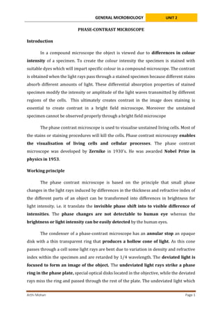
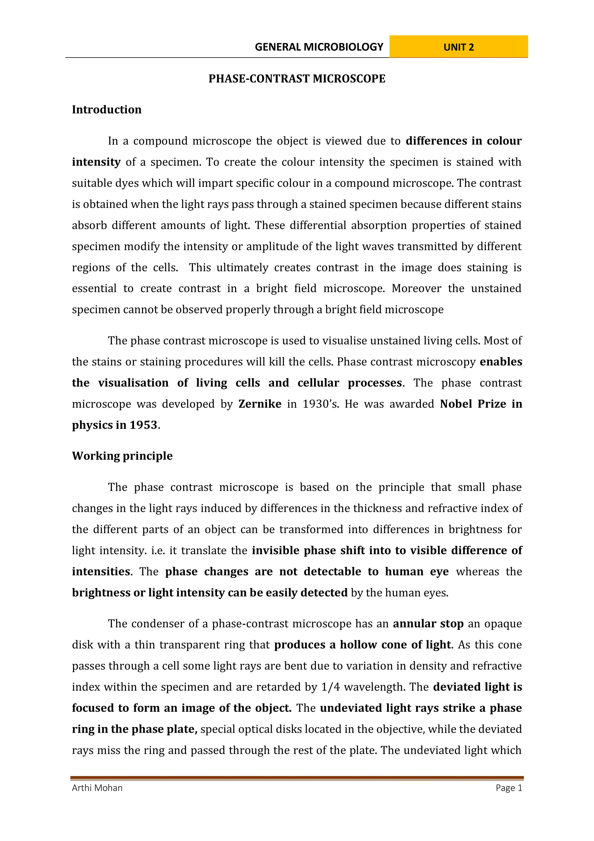
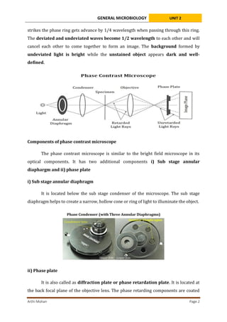
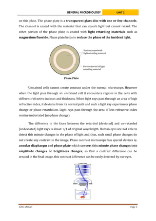
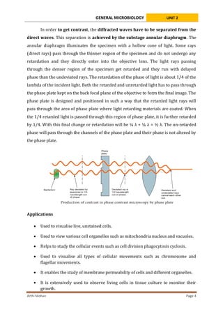
SlideShare a Scribd company logo
1 of 5
GENERAL MICROBIOLOGY UNIT 2
Arthi Mohan Page 1
PHASE-CONTRAST MICROSCOPE
Introduction
In a compound microscope the object is viewed due to differences in colour
intensity of a specimen. To create the colour intensity the specimen is stained with
suitable dyes which will impart specific colour in a compound microscope. The contrast
is obtained when the light rays pass through a stained specimen because different stains
absorb different amounts of light. These differential absorption properties of stained
specimen modify the intensity or amplitude of the light waves transmitted by different
regions of the cells. This ultimately creates contrast in the image does staining is
essential to create contrast in a bright field microscope. Moreover the unstained
specimen cannot be observed properly through a bright field microscope
The phase contrast microscope is used to visualise unstained living cells. Most of
the stains or staining procedures will kill the cells. Phase contrast microscopy enables
the visualisation of living cells and cellular processes. The phase contrast
microscope was developed by Zernike in 1930’s. He was awarded Nobel Prize in
physics in 1953.
Working principle
The phase contrast microscope is based on the principle that small phase
changes in the light rays induced by differences in the thickness and refractive index of
the different parts of an object can be transformed into differences in brightness for
light intensity. i.e. it translate the invisible phase shift into to visible difference of
intensities. The phase changes are not detectable to human eye whereas the
brightness or light intensity can be easily detected by the human eyes.
The condenser of a phase-contrast microscope has an annular stop an opaque
disk with a thin transparent ring that produces a hollow cone of light. As this cone
passes through a cell some light rays are bent due to variation in density and refractive
index within the specimen and are retarded by 1/4 wavelength. The deviated light is
focused to form an image of the object. The undeviated light rays strike a phase
ring in the phase plate, special optical disks located in the objective, while the deviated
rays miss the ring and passed through the rest of the plate. The undeviated light which
GENERAL MICROBIOLOGY UNIT 2
Arthi Mohan Page 2
strikes the phase ring gets advance by 1/4 wavelength when passing through this ring.
The deviated and undeviated waves become 1/2 wavelength to each other and will
cancel each other to come together to form an image. The background formed by
undeviated light is bright while the unstained object appears dark and well-
defined.
Components of phase contrast microscope
The phase contrast microscope is similar to the bright field microscope in its
optical components. It has two additional components i) Sub stage annular
diaphargm and ii) phase plate
i) Sub stage annular diaphragm
It is located below the sub stage condenser of the microscope. The sub stage
diaphragm helps to create a narrow, hollow cone or ring of light to illuminate the object.
ii) Phase plate
It is also called as diffraction plate or phase retardation plate. It is located at
the back focal plane of the objective lens. The phase retarding components are coated
GENERAL MICROBIOLOGY UNIT 2
Arthi Mohan Page 3
on this plate. The phase plate is a transparent glass disc with one or few channels.
The channel is coated with the material that can absorb light but cannot retard. The
other portion of the phase plate is coated with light retarding materials such as
magnesium fluoride. Phase plate helps to reduce the phase of the incident light.
Unstained cells cannot create contrast under the normal microscope. However
when the light pass through an unstained cell it encounters regions in the cells with
different refractive indexes and thickness. When light rays pass through an area of high
refractive index, it deviates from its normal path and such a light ray experiences phase
change or phase retardation. Light rays pass through the area of less refractive index
remine undeviated (no phase change).
The difference in the faces between the retarded (deviated) and un-retarded
(undeviated) light rays is about 1/4 of original wavelength. Human eyes are not able to
detect this minute changes in the phase of light and thus, such small phase changes do
not create any contrast in the image. Phase contrast microscope has special devices iẹ.
annular diaphargm and phase plate which convert this minute phase changes into
amplitude changes or brightness changes, so that a contrast difference can be
created in the final image, this contrast difference can be easily detected by our eyes.
GENERAL MICROBIOLOGY UNIT 2
Arthi Mohan Page 4
In order to get contrast, the diffracted waves have to be separated from the
direct waves. This separation is achieved by the substage annular diaphragm. The
annular diaphragm illuminates the specimen with a hollow cone of light. Some rays
(direct rays) pass through the thinner region of the specimen and do not undergo any
retardation and they directly enter into the objective lens. The light rays passing
through the denser region of the specimen get retarded and they run with delayed
phase than the undeviated rays. The retardation of the phase of light is about 1/4 of the
lambda of the incident light. Both the retarded and unretarded light has to pass through
the phase plate kept on the back focal plane of the objective to form the final image. The
phase plate is designed and positioned in such a way that the retarded light rays will
pass through the area of phase plate where light retarding materials are coated. When
the 1/4 retarded light is passed through this region of phase plate, it is further retarded
by 1/4. With this final change or retardation will be ¼ λ + ¼ λ = ½ λ. The un-retarded
phase will pass through the channels of the phase plate and their phase is not altered by
the phase plate.
Applications
 Used to visualise live, unstained cells.
 Used to view various cell organelles such as mitochondria nucleus and vacuoles.
 Helps to study the cellular events such as cell division phagocytosis cyclosis.
 Used to visualise all types of cellular movements such as chromosome and
flagellar movements.
 It enables the study of membrane permeability of cells and different organelles.
 It is extensively used to observe living cells in tissue culture to monitor their
growth.
GENERAL MICROBIOLOGY UNIT 2
Arthi Mohan Page 5
Advantages
 Provide the clear image of unstained cells.
 Avoid damages off the shelf due to chemical preparation and staining.
 Provide high contrast images highlighting the fine details of the cells.
 The optical construction is simple by adding phase condenser and face objective
 Enables prolonged observation of living cells without losing the viability of the
cells
 Live cell imaging and live process monitoring are possible at affordable cost
Disadvantages
 It produces a bright halo around the images the formation of the halo is due to
the partial or incomplete separation of direct and deviated rays.
 It is only useful for viewing individual cells or thin layer of cells.
References
Madigan Michael T, Bender, Kelly S, Buckley, Daniel H, Sattley, W. Matthew, & Stahl,
David A. (2018). Brock Biology of Microorganisms (15th Edition). Pearson.
Willey, Joanne M, Sherwood, Linda M, & Woolverton, Christopher J. (2016). Prescott’s
Microbiology (10 edition). McGraw-Hill Education.

More Related Content

What's hot (20)

Confocal microscopy by prabhat
Confocal microscopy by prabhatConfocal microscopy by prabhat
Confocal microscopy by prabhat
 
DARK FIELD MICROSCOPY
DARK FIELD MICROSCOPYDARK FIELD MICROSCOPY
DARK FIELD MICROSCOPY
 
1. MICROSCOPY - introduction + principle (Basics)
1. MICROSCOPY -  introduction + principle  (Basics)1. MICROSCOPY -  introduction + principle  (Basics)
1. MICROSCOPY - introduction + principle (Basics)
 
Differential Interference Contrast Microscope.pptx
Differential Interference Contrast Microscope.pptxDifferential Interference Contrast Microscope.pptx
Differential Interference Contrast Microscope.pptx
 
Phase contrast microscope
Phase contrast microscopePhase contrast microscope
Phase contrast microscope
 
Light Microscopy
Light MicroscopyLight Microscopy
Light Microscopy
 
Fluorescence Microscopy
Fluorescence MicroscopyFluorescence Microscopy
Fluorescence Microscopy
 
phase contrast microscope
phase contrast microscopephase contrast microscope
phase contrast microscope
 
Phase contrast microscope
Phase contrast microscopePhase contrast microscope
Phase contrast microscope
 
Phase contrast microscope
Phase contrast microscopePhase contrast microscope
Phase contrast microscope
 
3. Microscope simple, compound & stereo - Basics
3. Microscope   simple, compound & stereo - Basics3. Microscope   simple, compound & stereo - Basics
3. Microscope simple, compound & stereo - Basics
 
PHASE MICROSCOPY
PHASE MICROSCOPYPHASE MICROSCOPY
PHASE MICROSCOPY
 
FLUORESCENCE MICROSCOPY
FLUORESCENCE MICROSCOPYFLUORESCENCE MICROSCOPY
FLUORESCENCE MICROSCOPY
 
Microscopy
MicroscopyMicroscopy
Microscopy
 
Confocal microscope
Confocal microscopeConfocal microscope
Confocal microscope
 
Cell fractionation
Cell fractionationCell fractionation
Cell fractionation
 
Microscopy
MicroscopyMicroscopy
Microscopy
 
Stereo Microscope or Dissecting Miscrscope
Stereo Microscope or Dissecting MiscrscopeStereo Microscope or Dissecting Miscrscope
Stereo Microscope or Dissecting Miscrscope
 
Dark Field microscopy
Dark Field microscopyDark Field microscopy
Dark Field microscopy
 
Light microscope
Light microscopeLight microscope
Light microscope
 

Similar to 4. Phase-contrast microscope.pdf

phasecontrastmicroscope-210805055149 (1).pdf
phasecontrastmicroscope-210805055149 (1).pdfphasecontrastmicroscope-210805055149 (1).pdf
phasecontrastmicroscope-210805055149 (1).pdfrenuka291102
 
PHASE CONTRAST MICRSCOPY characteristics of material .pptx
PHASE CONTRAST MICRSCOPY characteristics of material .pptxPHASE CONTRAST MICRSCOPY characteristics of material .pptx
PHASE CONTRAST MICRSCOPY characteristics of material .pptxkannanchellakannu
 
Microscopy and Micrometry
Microscopy and MicrometryMicroscopy and Micrometry
Microscopy and MicrometrySuprakash Das
 
Phase contrast & Flurosence microscope
Phase contrast & Flurosence microscopePhase contrast & Flurosence microscope
Phase contrast & Flurosence microscopemicrobiology Notes
 
2 Microscopy and staining.pptx
2 Microscopy and staining.pptx2 Microscopy and staining.pptx
2 Microscopy and staining.pptxNeelGamers
 
Phase contrast and Fluorescent microscope
Phase contrast and Fluorescent microscopePhase contrast and Fluorescent microscope
Phase contrast and Fluorescent microscopeNithiya Pappuraj
 
PHASE CONTRAST MICROSCOPE.
PHASE CONTRAST MICROSCOPE.PHASE CONTRAST MICROSCOPE.
PHASE CONTRAST MICROSCOPE.AswathyLavan
 
FLUOROSCENCE & PHASE CONTRAST MICROSCOPY.pptx
FLUOROSCENCE & PHASE CONTRAST MICROSCOPY.pptxFLUOROSCENCE & PHASE CONTRAST MICROSCOPY.pptx
FLUOROSCENCE & PHASE CONTRAST MICROSCOPY.pptxSagarBhakare1
 
Unit – 2 Microscopy
Unit – 2 MicroscopyUnit – 2 Microscopy
Unit – 2 Microscopysanchu yadav
 
Dark ground microscopy
Dark ground microscopyDark ground microscopy
Dark ground microscopy9925752690
 
Types of Light Microscopes used in Histological Studies.pptx
Types of Light Microscopes used in Histological Studies.pptxTypes of Light Microscopes used in Histological Studies.pptx
Types of Light Microscopes used in Histological Studies.pptxssuserab552f
 
Types and applications of Microscopy.ppt
Types and applications of Microscopy.pptTypes and applications of Microscopy.ppt
Types and applications of Microscopy.pptvinothiyagarajan1
 
Phase contrast microscopy
Phase contrast microscopyPhase contrast microscopy
Phase contrast microscopyjakkaas
 

Similar to 4. Phase-contrast microscope.pdf (20)

Phase contrast microscope
Phase contrast microscopePhase contrast microscope
Phase contrast microscope
 
phasecontrastmicroscope-210805055149 (1).pdf
phasecontrastmicroscope-210805055149 (1).pdfphasecontrastmicroscope-210805055149 (1).pdf
phasecontrastmicroscope-210805055149 (1).pdf
 
PHASE CONTRAST MICRSCOPY characteristics of material .pptx
PHASE CONTRAST MICRSCOPY characteristics of material .pptxPHASE CONTRAST MICRSCOPY characteristics of material .pptx
PHASE CONTRAST MICRSCOPY characteristics of material .pptx
 
Microscopy and Micrometry
Microscopy and MicrometryMicroscopy and Micrometry
Microscopy and Micrometry
 
Phase contrast & Flurosence microscope
Phase contrast & Flurosence microscopePhase contrast & Flurosence microscope
Phase contrast & Flurosence microscope
 
Ppt f1
Ppt f1Ppt f1
Ppt f1
 
2 Microscopy and staining.pptx
2 Microscopy and staining.pptx2 Microscopy and staining.pptx
2 Microscopy and staining.pptx
 
Phase contrast and Fluorescent microscope
Phase contrast and Fluorescent microscopePhase contrast and Fluorescent microscope
Phase contrast and Fluorescent microscope
 
PHASE CONTRAST MICROSCOPE.
PHASE CONTRAST MICROSCOPE.PHASE CONTRAST MICROSCOPE.
PHASE CONTRAST MICROSCOPE.
 
FLUOROSCENCE & PHASE CONTRAST MICROSCOPY.pptx
FLUOROSCENCE & PHASE CONTRAST MICROSCOPY.pptxFLUOROSCENCE & PHASE CONTRAST MICROSCOPY.pptx
FLUOROSCENCE & PHASE CONTRAST MICROSCOPY.pptx
 
Unit – 2 Microscopy
Unit – 2 MicroscopyUnit – 2 Microscopy
Unit – 2 Microscopy
 
microscopy
 microscopy microscopy
microscopy
 
Microscopy - Study
Microscopy - StudyMicroscopy - Study
Microscopy - Study
 
Microscopy - Study
Microscopy - StudyMicroscopy - Study
Microscopy - Study
 
Dark ground microscopy
Dark ground microscopyDark ground microscopy
Dark ground microscopy
 
Microscopy
MicroscopyMicroscopy
Microscopy
 
Types of Light Microscopes used in Histological Studies.pptx
Types of Light Microscopes used in Histological Studies.pptxTypes of Light Microscopes used in Histological Studies.pptx
Types of Light Microscopes used in Histological Studies.pptx
 
Microscopy
MicroscopyMicroscopy
Microscopy
 
Types and applications of Microscopy.ppt
Types and applications of Microscopy.pptTypes and applications of Microscopy.ppt
Types and applications of Microscopy.ppt
 
Phase contrast microscopy
Phase contrast microscopyPhase contrast microscopy
Phase contrast microscopy
 

More from MicrobiologyMicro

Structure and types of RNA .pptx
Structure and types of RNA          .pptxStructure and types of RNA          .pptx
Structure and types of RNA .pptxMicrobiologyMicro
 
Transcription - DNA dependent RNA synthesis.pptx
Transcription - DNA dependent RNA synthesis.pptxTranscription - DNA dependent RNA synthesis.pptx
Transcription - DNA dependent RNA synthesis.pptxMicrobiologyMicro
 
. DNA Repair Mechanisms.pptx
.             DNA Repair Mechanisms.pptx.             DNA Repair Mechanisms.pptx
. DNA Repair Mechanisms.pptxMicrobiologyMicro
 
1. Immunology and its history.pptx
1.       Immunology and its history.pptx1.       Immunology and its history.pptx
1. Immunology and its history.pptxMicrobiologyMicro
 
5. ATOMIC FORCE MICROSCOPE (AFM).pdf
5.     ATOMIC FORCE MICROSCOPE (AFM).pdf5.     ATOMIC FORCE MICROSCOPE (AFM).pdf
5. ATOMIC FORCE MICROSCOPE (AFM).pdfMicrobiologyMicro
 
5. Fluorescence Microscope.pdf
5.            Fluorescence Microscope.pdf5.            Fluorescence Microscope.pdf
5. Fluorescence Microscope.pdfMicrobiologyMicro
 
3. Dark-field microscopy.pdf
3.             Dark-field microscopy.pdf3.             Dark-field microscopy.pdf
3. Dark-field microscopy.pdfMicrobiologyMicro
 
2. Types and Bright field microscopy.pdf
2. Types and Bright field microscopy.pdf2. Types and Bright field microscopy.pdf
2. Types and Bright field microscopy.pdfMicrobiologyMicro
 
1. Introduction to Microscopy.pdf
1.        Introduction to Microscopy.pdf1.        Introduction to Microscopy.pdf
1. Introduction to Microscopy.pdfMicrobiologyMicro
 
3. Differences between Prokaryotes and Eukaryotes.pdf
3. Differences between Prokaryotes and Eukaryotes.pdf3. Differences between Prokaryotes and Eukaryotes.pdf
3. Differences between Prokaryotes and Eukaryotes.pdfMicrobiologyMicro
 
History of Microbiology - General microbiology.pdf
History of Microbiology - General microbiology.pdfHistory of Microbiology - General microbiology.pdf
History of Microbiology - General microbiology.pdfMicrobiologyMicro
 
Definition and scope of microbiology.pdf
Definition and scope of microbiology.pdfDefinition and scope of microbiology.pdf
Definition and scope of microbiology.pdfMicrobiologyMicro
 
Transposons, types and transposition.pptx
Transposons, types and transposition.pptxTransposons, types and transposition.pptx
Transposons, types and transposition.pptxMicrobiologyMicro
 
Prokaryotic & Eukaryotic Replication.pptx
Prokaryotic & Eukaryotic Replication.pptxProkaryotic & Eukaryotic Replication.pptx
Prokaryotic & Eukaryotic Replication.pptxMicrobiologyMicro
 
Microbial Assessment of Air Quality.pptx
Microbial Assessment of Air Quality.pptxMicrobial Assessment of Air Quality.pptx
Microbial Assessment of Air Quality.pptxMicrobiologyMicro
 
Composition and Distribution of Air.pptx
Composition and Distribution of Air.pptxComposition and Distribution of Air.pptx
Composition and Distribution of Air.pptxMicrobiologyMicro
 
Interaction of microorganisms with Insects.pptx
Interaction of microorganisms with Insects.pptxInteraction of microorganisms with Insects.pptx
Interaction of microorganisms with Insects.pptxMicrobiologyMicro
 
Interaction of microorganisms with Animals.pptx
Interaction of microorganisms with Animals.pptxInteraction of microorganisms with Animals.pptx
Interaction of microorganisms with Animals.pptxMicrobiologyMicro
 

More from MicrobiologyMicro (20)

Translation .pptx
Translation                         .pptxTranslation                         .pptx
Translation .pptx
 
Structure and types of RNA .pptx
Structure and types of RNA          .pptxStructure and types of RNA          .pptx
Structure and types of RNA .pptx
 
Transcription - DNA dependent RNA synthesis.pptx
Transcription - DNA dependent RNA synthesis.pptxTranscription - DNA dependent RNA synthesis.pptx
Transcription - DNA dependent RNA synthesis.pptx
 
. DNA Repair Mechanisms.pptx
.             DNA Repair Mechanisms.pptx.             DNA Repair Mechanisms.pptx
. DNA Repair Mechanisms.pptx
 
1. Immunology and its history.pptx
1.       Immunology and its history.pptx1.       Immunology and its history.pptx
1. Immunology and its history.pptx
 
5. ATOMIC FORCE MICROSCOPE (AFM).pdf
5.     ATOMIC FORCE MICROSCOPE (AFM).pdf5.     ATOMIC FORCE MICROSCOPE (AFM).pdf
5. ATOMIC FORCE MICROSCOPE (AFM).pdf
 
5. Fluorescence Microscope.pdf
5.            Fluorescence Microscope.pdf5.            Fluorescence Microscope.pdf
5. Fluorescence Microscope.pdf
 
3. Dark-field microscopy.pdf
3.             Dark-field microscopy.pdf3.             Dark-field microscopy.pdf
3. Dark-field microscopy.pdf
 
2. Types and Bright field microscopy.pdf
2. Types and Bright field microscopy.pdf2. Types and Bright field microscopy.pdf
2. Types and Bright field microscopy.pdf
 
1. Introduction to Microscopy.pdf
1.        Introduction to Microscopy.pdf1.        Introduction to Microscopy.pdf
1. Introduction to Microscopy.pdf
 
3. Differences between Prokaryotes and Eukaryotes.pdf
3. Differences between Prokaryotes and Eukaryotes.pdf3. Differences between Prokaryotes and Eukaryotes.pdf
3. Differences between Prokaryotes and Eukaryotes.pdf
 
History of Microbiology - General microbiology.pdf
History of Microbiology - General microbiology.pdfHistory of Microbiology - General microbiology.pdf
History of Microbiology - General microbiology.pdf
 
Definition and scope of microbiology.pdf
Definition and scope of microbiology.pdfDefinition and scope of microbiology.pdf
Definition and scope of microbiology.pdf
 
Transposons, types and transposition.pptx
Transposons, types and transposition.pptxTransposons, types and transposition.pptx
Transposons, types and transposition.pptx
 
Prokaryotic & Eukaryotic Replication.pptx
Prokaryotic & Eukaryotic Replication.pptxProkaryotic & Eukaryotic Replication.pptx
Prokaryotic & Eukaryotic Replication.pptx
 
Microbial Assessment of Air Quality.pptx
Microbial Assessment of Air Quality.pptxMicrobial Assessment of Air Quality.pptx
Microbial Assessment of Air Quality.pptx
 
Air Sanitation.pptx
Air Sanitation.pptxAir Sanitation.pptx
Air Sanitation.pptx
 
Composition and Distribution of Air.pptx
Composition and Distribution of Air.pptxComposition and Distribution of Air.pptx
Composition and Distribution of Air.pptx
 
Interaction of microorganisms with Insects.pptx
Interaction of microorganisms with Insects.pptxInteraction of microorganisms with Insects.pptx
Interaction of microorganisms with Insects.pptx
 
Interaction of microorganisms with Animals.pptx
Interaction of microorganisms with Animals.pptxInteraction of microorganisms with Animals.pptx
Interaction of microorganisms with Animals.pptx
 

Recently uploaded

Formation of low mass protostars and their circumstellar disks
Formation of low mass protostars and their circumstellar disksFormation of low mass protostars and their circumstellar disks
Formation of low mass protostars and their circumstellar disksSérgio Sacani
 
Botany 4th semester series (krishna).pdf
Botany 4th semester series (krishna).pdfBotany 4th semester series (krishna).pdf
Botany 4th semester series (krishna).pdfSumit Kumar yadav
 
Proteomics: types, protein profiling steps etc.
Proteomics: types, protein profiling steps etc.Proteomics: types, protein profiling steps etc.
Proteomics: types, protein profiling steps etc.Silpa
 
Bacterial Identification and Classifications
Bacterial Identification and ClassificationsBacterial Identification and Classifications
Bacterial Identification and ClassificationsAreesha Ahmad
 
Asymmetry in the atmosphere of the ultra-hot Jupiter WASP-76 b
Asymmetry in the atmosphere of the ultra-hot Jupiter WASP-76 bAsymmetry in the atmosphere of the ultra-hot Jupiter WASP-76 b
Asymmetry in the atmosphere of the ultra-hot Jupiter WASP-76 bSérgio Sacani
 
Nanoparticles synthesis and characterization​ ​
Nanoparticles synthesis and characterization​  ​Nanoparticles synthesis and characterization​  ​
Nanoparticles synthesis and characterization​ ​kaibalyasahoo82800
 
SAMASTIPUR CALL GIRL 7857803690 LOW PRICE ESCORT SERVICE
SAMASTIPUR CALL GIRL 7857803690  LOW PRICE  ESCORT SERVICESAMASTIPUR CALL GIRL 7857803690  LOW PRICE  ESCORT SERVICE
SAMASTIPUR CALL GIRL 7857803690 LOW PRICE ESCORT SERVICEayushi9330
 
Factory Acceptance Test( FAT).pptx .
Factory Acceptance Test( FAT).pptx       .Factory Acceptance Test( FAT).pptx       .
Factory Acceptance Test( FAT).pptx .Poonam Aher Patil
 
Labelling Requirements and Label Claims for Dietary Supplements and Recommend...
Labelling Requirements and Label Claims for Dietary Supplements and Recommend...Labelling Requirements and Label Claims for Dietary Supplements and Recommend...
Labelling Requirements and Label Claims for Dietary Supplements and Recommend...Lokesh Kothari
 
Feature-aligned N-BEATS with Sinkhorn divergence (ICLR '24)
Feature-aligned N-BEATS with Sinkhorn divergence (ICLR '24)Feature-aligned N-BEATS with Sinkhorn divergence (ICLR '24)
Feature-aligned N-BEATS with Sinkhorn divergence (ICLR '24)Joonhun Lee
 
American Type Culture Collection (ATCC).pptx
American Type Culture Collection (ATCC).pptxAmerican Type Culture Collection (ATCC).pptx
American Type Culture Collection (ATCC).pptxabhishekdhamu51
 
TEST BANK For Radiologic Science for Technologists, 12th Edition by Stewart C...
TEST BANK For Radiologic Science for Technologists, 12th Edition by Stewart C...TEST BANK For Radiologic Science for Technologists, 12th Edition by Stewart C...
TEST BANK For Radiologic Science for Technologists, 12th Edition by Stewart C...ssifa0344
 
Justdial Call Girls In Indirapuram, Ghaziabad, 8800357707 Escorts Service
Justdial Call Girls In Indirapuram, Ghaziabad, 8800357707 Escorts ServiceJustdial Call Girls In Indirapuram, Ghaziabad, 8800357707 Escorts Service
Justdial Call Girls In Indirapuram, Ghaziabad, 8800357707 Escorts Servicemonikaservice1
 
Connaught Place, Delhi Call girls :8448380779 Model Escorts | 100% verified
Connaught Place, Delhi Call girls :8448380779 Model Escorts | 100% verifiedConnaught Place, Delhi Call girls :8448380779 Model Escorts | 100% verified
Connaught Place, Delhi Call girls :8448380779 Model Escorts | 100% verifiedDelhi Call girls
 
Biogenic Sulfur Gases as Biosignatures on Temperate Sub-Neptune Waterworlds
Biogenic Sulfur Gases as Biosignatures on Temperate Sub-Neptune WaterworldsBiogenic Sulfur Gases as Biosignatures on Temperate Sub-Neptune Waterworlds
Biogenic Sulfur Gases as Biosignatures on Temperate Sub-Neptune WaterworldsSérgio Sacani
 
GBSN - Microbiology (Unit 2)
GBSN - Microbiology (Unit 2)GBSN - Microbiology (Unit 2)
GBSN - Microbiology (Unit 2)Areesha Ahmad
 
Call Girls Alandi Call Me 7737669865 Budget Friendly No Advance Booking
Call Girls Alandi Call Me 7737669865 Budget Friendly No Advance BookingCall Girls Alandi Call Me 7737669865 Budget Friendly No Advance Booking
Call Girls Alandi Call Me 7737669865 Budget Friendly No Advance Bookingroncy bisnoi
 
❤Jammu Kashmir Call Girls 8617697112 Personal Whatsapp Number 💦✅.
❤Jammu Kashmir Call Girls 8617697112 Personal Whatsapp Number 💦✅.❤Jammu Kashmir Call Girls 8617697112 Personal Whatsapp Number 💦✅.
❤Jammu Kashmir Call Girls 8617697112 Personal Whatsapp Number 💦✅.Nitya salvi
 
Pulmonary drug delivery system M.pharm -2nd sem P'ceutics
Pulmonary drug delivery system M.pharm -2nd sem P'ceuticsPulmonary drug delivery system M.pharm -2nd sem P'ceutics
Pulmonary drug delivery system M.pharm -2nd sem P'ceuticssakshisoni2385
 
Seismic Method Estimate velocity from seismic data.pptx
Seismic Method Estimate velocity from seismic  data.pptxSeismic Method Estimate velocity from seismic  data.pptx
Seismic Method Estimate velocity from seismic data.pptxAlMamun560346
 

Recently uploaded (20)

Formation of low mass protostars and their circumstellar disks
Formation of low mass protostars and their circumstellar disksFormation of low mass protostars and their circumstellar disks
Formation of low mass protostars and their circumstellar disks
 
Botany 4th semester series (krishna).pdf
Botany 4th semester series (krishna).pdfBotany 4th semester series (krishna).pdf
Botany 4th semester series (krishna).pdf
 
Proteomics: types, protein profiling steps etc.
Proteomics: types, protein profiling steps etc.Proteomics: types, protein profiling steps etc.
Proteomics: types, protein profiling steps etc.
 
Bacterial Identification and Classifications
Bacterial Identification and ClassificationsBacterial Identification and Classifications
Bacterial Identification and Classifications
 
Asymmetry in the atmosphere of the ultra-hot Jupiter WASP-76 b
Asymmetry in the atmosphere of the ultra-hot Jupiter WASP-76 bAsymmetry in the atmosphere of the ultra-hot Jupiter WASP-76 b
Asymmetry in the atmosphere of the ultra-hot Jupiter WASP-76 b
 
Nanoparticles synthesis and characterization​ ​
Nanoparticles synthesis and characterization​  ​Nanoparticles synthesis and characterization​  ​
Nanoparticles synthesis and characterization​ ​
 
SAMASTIPUR CALL GIRL 7857803690 LOW PRICE ESCORT SERVICE
SAMASTIPUR CALL GIRL 7857803690  LOW PRICE  ESCORT SERVICESAMASTIPUR CALL GIRL 7857803690  LOW PRICE  ESCORT SERVICE
SAMASTIPUR CALL GIRL 7857803690 LOW PRICE ESCORT SERVICE
 
Factory Acceptance Test( FAT).pptx .
Factory Acceptance Test( FAT).pptx       .Factory Acceptance Test( FAT).pptx       .
Factory Acceptance Test( FAT).pptx .
 
Labelling Requirements and Label Claims for Dietary Supplements and Recommend...
Labelling Requirements and Label Claims for Dietary Supplements and Recommend...Labelling Requirements and Label Claims for Dietary Supplements and Recommend...
Labelling Requirements and Label Claims for Dietary Supplements and Recommend...
 
Feature-aligned N-BEATS with Sinkhorn divergence (ICLR '24)
Feature-aligned N-BEATS with Sinkhorn divergence (ICLR '24)Feature-aligned N-BEATS with Sinkhorn divergence (ICLR '24)
Feature-aligned N-BEATS with Sinkhorn divergence (ICLR '24)
 
American Type Culture Collection (ATCC).pptx
American Type Culture Collection (ATCC).pptxAmerican Type Culture Collection (ATCC).pptx
American Type Culture Collection (ATCC).pptx
 
TEST BANK For Radiologic Science for Technologists, 12th Edition by Stewart C...
TEST BANK For Radiologic Science for Technologists, 12th Edition by Stewart C...TEST BANK For Radiologic Science for Technologists, 12th Edition by Stewart C...
TEST BANK For Radiologic Science for Technologists, 12th Edition by Stewart C...
 
Justdial Call Girls In Indirapuram, Ghaziabad, 8800357707 Escorts Service
Justdial Call Girls In Indirapuram, Ghaziabad, 8800357707 Escorts ServiceJustdial Call Girls In Indirapuram, Ghaziabad, 8800357707 Escorts Service
Justdial Call Girls In Indirapuram, Ghaziabad, 8800357707 Escorts Service
 
Connaught Place, Delhi Call girls :8448380779 Model Escorts | 100% verified
Connaught Place, Delhi Call girls :8448380779 Model Escorts | 100% verifiedConnaught Place, Delhi Call girls :8448380779 Model Escorts | 100% verified
Connaught Place, Delhi Call girls :8448380779 Model Escorts | 100% verified
 
Biogenic Sulfur Gases as Biosignatures on Temperate Sub-Neptune Waterworlds
Biogenic Sulfur Gases as Biosignatures on Temperate Sub-Neptune WaterworldsBiogenic Sulfur Gases as Biosignatures on Temperate Sub-Neptune Waterworlds
Biogenic Sulfur Gases as Biosignatures on Temperate Sub-Neptune Waterworlds
 
GBSN - Microbiology (Unit 2)
GBSN - Microbiology (Unit 2)GBSN - Microbiology (Unit 2)
GBSN - Microbiology (Unit 2)
 
Call Girls Alandi Call Me 7737669865 Budget Friendly No Advance Booking
Call Girls Alandi Call Me 7737669865 Budget Friendly No Advance BookingCall Girls Alandi Call Me 7737669865 Budget Friendly No Advance Booking
Call Girls Alandi Call Me 7737669865 Budget Friendly No Advance Booking
 
❤Jammu Kashmir Call Girls 8617697112 Personal Whatsapp Number 💦✅.
❤Jammu Kashmir Call Girls 8617697112 Personal Whatsapp Number 💦✅.❤Jammu Kashmir Call Girls 8617697112 Personal Whatsapp Number 💦✅.
❤Jammu Kashmir Call Girls 8617697112 Personal Whatsapp Number 💦✅.
 
Pulmonary drug delivery system M.pharm -2nd sem P'ceutics
Pulmonary drug delivery system M.pharm -2nd sem P'ceuticsPulmonary drug delivery system M.pharm -2nd sem P'ceutics
Pulmonary drug delivery system M.pharm -2nd sem P'ceutics
 
Seismic Method Estimate velocity from seismic data.pptx
Seismic Method Estimate velocity from seismic  data.pptxSeismic Method Estimate velocity from seismic  data.pptx
Seismic Method Estimate velocity from seismic data.pptx
 

4. Phase-contrast microscope.pdf

  • 1. GENERAL MICROBIOLOGY UNIT 2 Arthi Mohan Page 1 PHASE-CONTRAST MICROSCOPE Introduction In a compound microscope the object is viewed due to differences in colour intensity of a specimen. To create the colour intensity the specimen is stained with suitable dyes which will impart specific colour in a compound microscope. The contrast is obtained when the light rays pass through a stained specimen because different stains absorb different amounts of light. These differential absorption properties of stained specimen modify the intensity or amplitude of the light waves transmitted by different regions of the cells. This ultimately creates contrast in the image does staining is essential to create contrast in a bright field microscope. Moreover the unstained specimen cannot be observed properly through a bright field microscope The phase contrast microscope is used to visualise unstained living cells. Most of the stains or staining procedures will kill the cells. Phase contrast microscopy enables the visualisation of living cells and cellular processes. The phase contrast microscope was developed by Zernike in 1930’s. He was awarded Nobel Prize in physics in 1953. Working principle The phase contrast microscope is based on the principle that small phase changes in the light rays induced by differences in the thickness and refractive index of the different parts of an object can be transformed into differences in brightness for light intensity. i.e. it translate the invisible phase shift into to visible difference of intensities. The phase changes are not detectable to human eye whereas the brightness or light intensity can be easily detected by the human eyes. The condenser of a phase-contrast microscope has an annular stop an opaque disk with a thin transparent ring that produces a hollow cone of light. As this cone passes through a cell some light rays are bent due to variation in density and refractive index within the specimen and are retarded by 1/4 wavelength. The deviated light is focused to form an image of the object. The undeviated light rays strike a phase ring in the phase plate, special optical disks located in the objective, while the deviated rays miss the ring and passed through the rest of the plate. The undeviated light which
  • 2. GENERAL MICROBIOLOGY UNIT 2 Arthi Mohan Page 2 strikes the phase ring gets advance by 1/4 wavelength when passing through this ring. The deviated and undeviated waves become 1/2 wavelength to each other and will cancel each other to come together to form an image. The background formed by undeviated light is bright while the unstained object appears dark and well- defined. Components of phase contrast microscope The phase contrast microscope is similar to the bright field microscope in its optical components. It has two additional components i) Sub stage annular diaphargm and ii) phase plate i) Sub stage annular diaphragm It is located below the sub stage condenser of the microscope. The sub stage diaphragm helps to create a narrow, hollow cone or ring of light to illuminate the object. ii) Phase plate It is also called as diffraction plate or phase retardation plate. It is located at the back focal plane of the objective lens. The phase retarding components are coated
  • 3. GENERAL MICROBIOLOGY UNIT 2 Arthi Mohan Page 3 on this plate. The phase plate is a transparent glass disc with one or few channels. The channel is coated with the material that can absorb light but cannot retard. The other portion of the phase plate is coated with light retarding materials such as magnesium fluoride. Phase plate helps to reduce the phase of the incident light. Unstained cells cannot create contrast under the normal microscope. However when the light pass through an unstained cell it encounters regions in the cells with different refractive indexes and thickness. When light rays pass through an area of high refractive index, it deviates from its normal path and such a light ray experiences phase change or phase retardation. Light rays pass through the area of less refractive index remine undeviated (no phase change). The difference in the faces between the retarded (deviated) and un-retarded (undeviated) light rays is about 1/4 of original wavelength. Human eyes are not able to detect this minute changes in the phase of light and thus, such small phase changes do not create any contrast in the image. Phase contrast microscope has special devices iẹ. annular diaphargm and phase plate which convert this minute phase changes into amplitude changes or brightness changes, so that a contrast difference can be created in the final image, this contrast difference can be easily detected by our eyes.
  • 4. GENERAL MICROBIOLOGY UNIT 2 Arthi Mohan Page 4 In order to get contrast, the diffracted waves have to be separated from the direct waves. This separation is achieved by the substage annular diaphragm. The annular diaphragm illuminates the specimen with a hollow cone of light. Some rays (direct rays) pass through the thinner region of the specimen and do not undergo any retardation and they directly enter into the objective lens. The light rays passing through the denser region of the specimen get retarded and they run with delayed phase than the undeviated rays. The retardation of the phase of light is about 1/4 of the lambda of the incident light. Both the retarded and unretarded light has to pass through the phase plate kept on the back focal plane of the objective to form the final image. The phase plate is designed and positioned in such a way that the retarded light rays will pass through the area of phase plate where light retarding materials are coated. When the 1/4 retarded light is passed through this region of phase plate, it is further retarded by 1/4. With this final change or retardation will be ¼ λ + ¼ λ = ½ λ. The un-retarded phase will pass through the channels of the phase plate and their phase is not altered by the phase plate. Applications  Used to visualise live, unstained cells.  Used to view various cell organelles such as mitochondria nucleus and vacuoles.  Helps to study the cellular events such as cell division phagocytosis cyclosis.  Used to visualise all types of cellular movements such as chromosome and flagellar movements.  It enables the study of membrane permeability of cells and different organelles.  It is extensively used to observe living cells in tissue culture to monitor their growth.
  • 5. GENERAL MICROBIOLOGY UNIT 2 Arthi Mohan Page 5 Advantages  Provide the clear image of unstained cells.  Avoid damages off the shelf due to chemical preparation and staining.  Provide high contrast images highlighting the fine details of the cells.  The optical construction is simple by adding phase condenser and face objective  Enables prolonged observation of living cells without losing the viability of the cells  Live cell imaging and live process monitoring are possible at affordable cost Disadvantages  It produces a bright halo around the images the formation of the halo is due to the partial or incomplete separation of direct and deviated rays.  It is only useful for viewing individual cells or thin layer of cells. References Madigan Michael T, Bender, Kelly S, Buckley, Daniel H, Sattley, W. Matthew, & Stahl, David A. (2018). Brock Biology of Microorganisms (15th Edition). Pearson. Willey, Joanne M, Sherwood, Linda M, & Woolverton, Christopher J. (2016). Prescott’s Microbiology (10 edition). McGraw-Hill Education.