Download to read offline
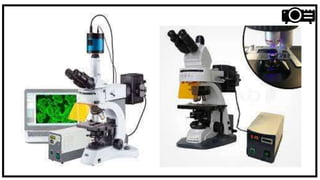
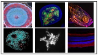


The fluorescence microscope uses fluorescence and phosphorescence instead of, or in addition to, reflection and absorption to study organic or inorganic substances. It works by illuminating a specimen with specific excitation wavelengths that are absorbed by fluorophores, causing them to emit light of longer wavelengths. Key parts include light sources, excitation and emission filters, and a dichroic mirror. Fluorescence microscopy is widely used in biology for applications like imaging cell structures, viability studies, and viewing genetic material. It has advantages like sensitivity and the ability to track multiple molecules simultaneously, but limitations include photobleaching and phototoxicity over long exposures.


