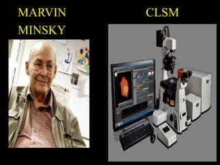
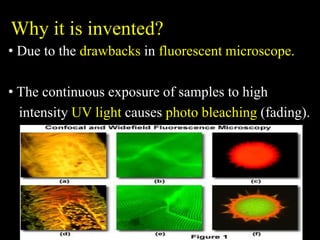
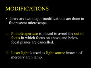
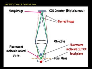
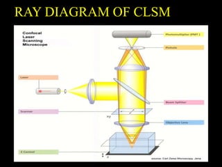
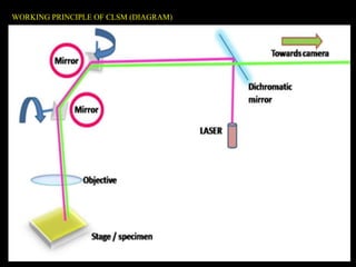
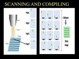
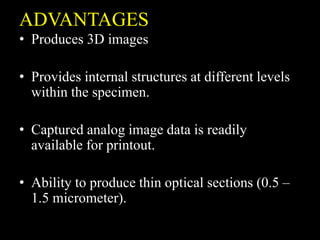
FUNDAMENTALS OF AGRICUTURAL
MICROBIOLOGY (1+1)
LASER SCANNING CONFOCAL
MICROSCOPE
BY TO
BASKAR S Dr.S PARTHASARATHY
BATCH-A 19AGM111
CB.AG.U4AGR19018 ( PLANT PATHOLOGIST)
CONFOCAL MICROSCOPY
• An optical microscopy technique for
increasing optical resolution and contrast of a
microscope.
• The principle of confocal microscopy was
patented by Marvin Minsky in 1957.
• It works on the principle of fluorescence
optics.
• It is a modified version of fluorescent
microscope.
MARVIN CLSM
MINSKY
Why it is invented?
• Due to the drawbacks in fluorescent microscope.
• The continuous exposure of samples to high
intensity UV light causes photo bleaching (fading).
MODIFICATIONS
• There are two major modifications are done in
fluorescent microscope.
i. Pinhole aperture is placed to avoid the out of
focus in which focus on above and below
focal planes are cancelled.
ii. Laser light is used as light source instead of
mercury arch lamp.
MODIFICATIONS & COMPARISON
LASER SCANNING CONFOCAL
MICROSCOPE
• It is similar to the principle of confocal
microscopy.
• Because of using monochromatic laser beam
(He-Ne) as a light source, confocal microscope
is known as laser scanning confocal
microscope.
• In this, detector (PMT) and scanner are placed
to analyse the different focal planes (3D).
COMPONENTS OF CLSM
• Laser beam
• Beam splitter
• Scanner
• Objective lens
• Z-control
• Pinhole
• Photomultiplier tube (PMT)
RAY DIAGRAM OF CLSM
WORKING PRINCIPLE OF CLSM
• Laser beam is focused on the smaller region of
specimen.
• The intensity of laser can be adjusted by beam
splitter (neutral filter).
• Then the laser passes through 2 scanning
mirrors which rotates in an opposite X & Y
direction to focus on another region.
• The laser beam is then brought to the focal
plane of objective lens which focuses onto
sample.
• If the sample is fluorescent, light will pass
back into objective lens (travels on same path
that laser travels).
• Then it passes through the semi transparent
mirror.
• Due to rotating mirrors, laser scans the whole
surface of the specimen.
• Through pinhole, detector (PMT) captures the
images of particular regions of whole
specimen (light signal is converted into
electrical signal).
• Atlast captured individual images are compiled
by software which is connected to detector and
gives real time image.
WORKING PRINCIPLE OF CLSM (DIAGRAM)
SCANNING AND COMPILING
ADVANTAGES
• Produces 3D images
• Provides internal structures at different levels
within the specimen.
• Captured analog image data is readily
available for printout.
• Ability to produce thin optical sections (0.5 –
1.5 micrometer).
DISADVANTAGES
• Monochromatic LASER is harmful to humans.
• It is very costlier (around 1-1.5 crores).
REFERENCES
• Confocal Laser Scanning Microscopy – Colin J.
R. Sheppard
• Microbiology – P.D.Sharma
• https://www.umassmed.edu/globalassets/maps/do
cuments/confocal-explanation.pdf
• https://bitesizebio.com/19958/what-is-confocal-
laser-scanning-microscopy/
THANK YOU

Confocal laser scanning microscopy

  • 1.
    FUNDAMENTALS OF AGRICUTURAL MICROBIOLOGY(1+1) LASER SCANNING CONFOCAL MICROSCOPE BY TO BASKAR S Dr.S PARTHASARATHY BATCH-A 19AGM111 CB.AG.U4AGR19018 ( PLANT PATHOLOGIST)
  • 2.
    CONFOCAL MICROSCOPY • Anoptical microscopy technique for increasing optical resolution and contrast of a microscope. • The principle of confocal microscopy was patented by Marvin Minsky in 1957. • It works on the principle of fluorescence optics. • It is a modified version of fluorescent microscope.
  • 3.
  • 4.
    Why it isinvented? • Due to the drawbacks in fluorescent microscope. • The continuous exposure of samples to high intensity UV light causes photo bleaching (fading).
  • 5.
    MODIFICATIONS • There aretwo major modifications are done in fluorescent microscope. i. Pinhole aperture is placed to avoid the out of focus in which focus on above and below focal planes are cancelled. ii. Laser light is used as light source instead of mercury arch lamp.
  • 6.
  • 7.
    LASER SCANNING CONFOCAL MICROSCOPE •It is similar to the principle of confocal microscopy. • Because of using monochromatic laser beam (He-Ne) as a light source, confocal microscope is known as laser scanning confocal microscope. • In this, detector (PMT) and scanner are placed to analyse the different focal planes (3D).
  • 8.
    COMPONENTS OF CLSM •Laser beam • Beam splitter • Scanner • Objective lens • Z-control • Pinhole • Photomultiplier tube (PMT)
  • 9.
  • 10.
    WORKING PRINCIPLE OFCLSM • Laser beam is focused on the smaller region of specimen. • The intensity of laser can be adjusted by beam splitter (neutral filter). • Then the laser passes through 2 scanning mirrors which rotates in an opposite X & Y direction to focus on another region.
  • 11.
    • The laserbeam is then brought to the focal plane of objective lens which focuses onto sample. • If the sample is fluorescent, light will pass back into objective lens (travels on same path that laser travels). • Then it passes through the semi transparent mirror.
  • 12.
    • Due torotating mirrors, laser scans the whole surface of the specimen. • Through pinhole, detector (PMT) captures the images of particular regions of whole specimen (light signal is converted into electrical signal). • Atlast captured individual images are compiled by software which is connected to detector and gives real time image.
  • 13.
    WORKING PRINCIPLE OFCLSM (DIAGRAM)
  • 14.
  • 15.
    ADVANTAGES • Produces 3Dimages • Provides internal structures at different levels within the specimen. • Captured analog image data is readily available for printout. • Ability to produce thin optical sections (0.5 – 1.5 micrometer).
  • 16.
    DISADVANTAGES • Monochromatic LASERis harmful to humans. • It is very costlier (around 1-1.5 crores).
  • 17.
    REFERENCES • Confocal LaserScanning Microscopy – Colin J. R. Sheppard • Microbiology – P.D.Sharma • https://www.umassmed.edu/globalassets/maps/do cuments/confocal-explanation.pdf • https://bitesizebio.com/19958/what-is-confocal- laser-scanning-microscopy/
  • 18.