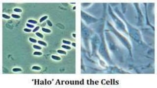
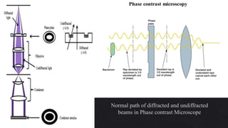
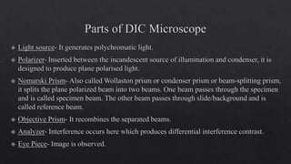
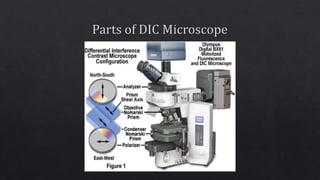
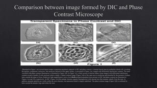
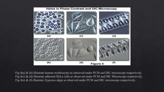
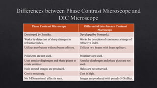
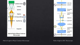
7. Normal path of diffracted and undiffracted
beams in Phase contrast Microscope
8.
9.
10.
11.
12.
13.
14. Illustrated in Figure 1 are several digital images comparing specimens captured in DIC and phase contrast. A human buccal mucosa epithelial (cheek) cell, revealing
the nucleus, cytoplasmic inclusions, and numerous bacteria on the upper surface, is presented in Figure 1(a), imaged with differential interference contrast. The same
viewfield with phase contrast illumination is illustrated in Figure 1(b). In Figure 1(c), a thick section of murine kidney tissue imaged with differential interference
contrast reveals a bundle of cells enclosed within a tubule. A phase contrast image (Figure 1(d)) of the same area is confusing and disturbed by the presence of phase
halos outside the plane of focus. Relatively high magnification views of an Obelia polypoid annulated stem perisarc in DIC and phase contrast are illustrated in
Figures 1(e) and 1(f), respectively. In DIC, (Figure 1(e)) the annular structure appears hemispherical with internal rays that emanate radially from the stem. In
addition, granular particles are visible within the stem structure, but anatomical detail is largely undefined. The phase contrast image (Figure 1(f)) is confused by
halos around the annular rings and within the stem.
15. Fig 4(a) & (b) illustrate human erythrocytes as observed under PCM and DIC microscope respectively.
Fig 4(c) & (d) illustrate adherent HeLa cells as observed under PCM and DIC Microscope respectively.
Fig 4(e) & (f) illustrate Zygnema algae as observed under PCM and DIC microscope respectively.
16. Phase Contrast Microscope Differential Interference Contrast
Microscope
Developed by Zernike. Developed by Nomarski.
Works by detection of sharp changes in
refractive index.
Works by detection of continuous change of
refractive index.
Utilizes two beams without beam splitters. Utilizes two beams with beam splitters.
Polarizers are not used. Polarizers are used.
Uses annular diaphragm and phase plates to
create contrast
Annular diaphragm and phase plate are not
used.
Halo around images are produced. Halos are not observed.
Cost is moderate. Cost is high.
No 3-Dimensional effect is seen. Images are produced with pseudo 3-D effect.
17. Path of Light in Phase Contrast Microscope Path of light in DIC Microscope