Principles and application of light, phase constrast and fluorescence microscope
This document provides an overview of three types of microscopes: light microscopes, phase contrast microscopes, and fluorescence microscopes. It describes the basic principles and components of each microscope type and their applications. Light microscopes use lenses to magnify specimens and are used widely in biology to study cells. Phase contrast microscopes convert phase differences in light passing through specimens into brightness variations, allowing visualization of transparent structures. Fluorescence microscopes use fluorescent dyes and specific wavelengths of light to enhance contrast and study labeled structures within cells.
Overview of microscopes, history by Zacharias Janssen, function of lenses for magnification.
Details on light microscopes including their structure, function, types, and applications in biology and forensics. Applications in biology for disease study, forensic investigation, and mineralogy using thin section.
Introduction to phase contrast microscopes, principle of operation, and their applications in biology.
Overview of fluorescence microscopes, their principles including fluorescence effect, and diverse applications in research.
Principles and application of light, phase constrast and fluorescence microscope
1.
Presented by…
Thakor MaitriM.
M.Sc. (Botany)
Department of life sciences,
H.N.G.U., Patan.
Principles and application of light,
phase contrast, fluorescence
microscope
1
Introduction
3
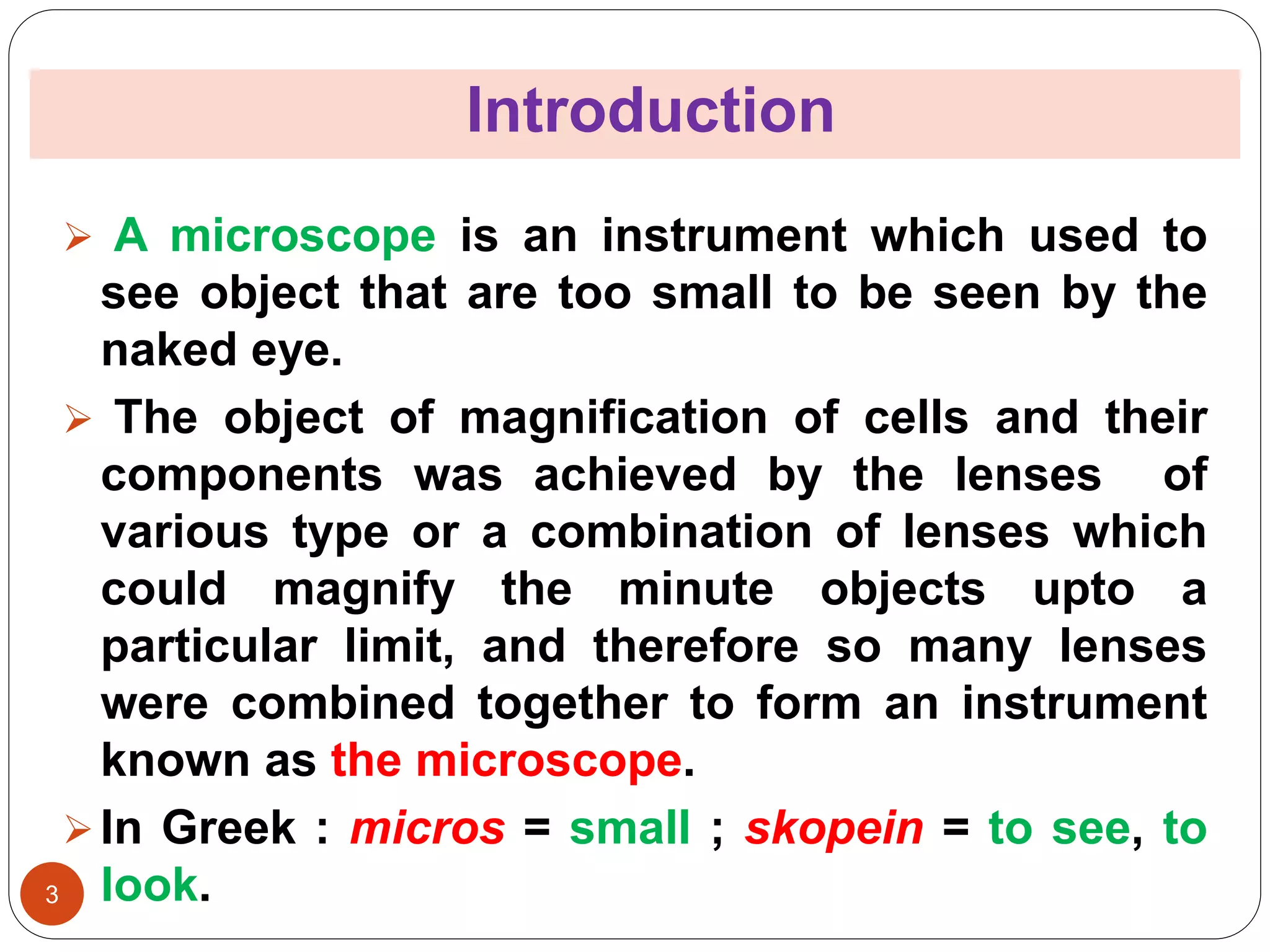
A microscopeis an instrument which used to
see object that are too small to be seen by the
naked eye.
The object of magnification of cells and their
components was achieved by the lenses of
various type or a combination of lenses which
could magnify the minute objects upto a
particular limit, and therefore so many lenses
were combined together to form an instrument
known as the microscope.
In Greek : micros = small ; skopein = to see, to
look.
4.
4
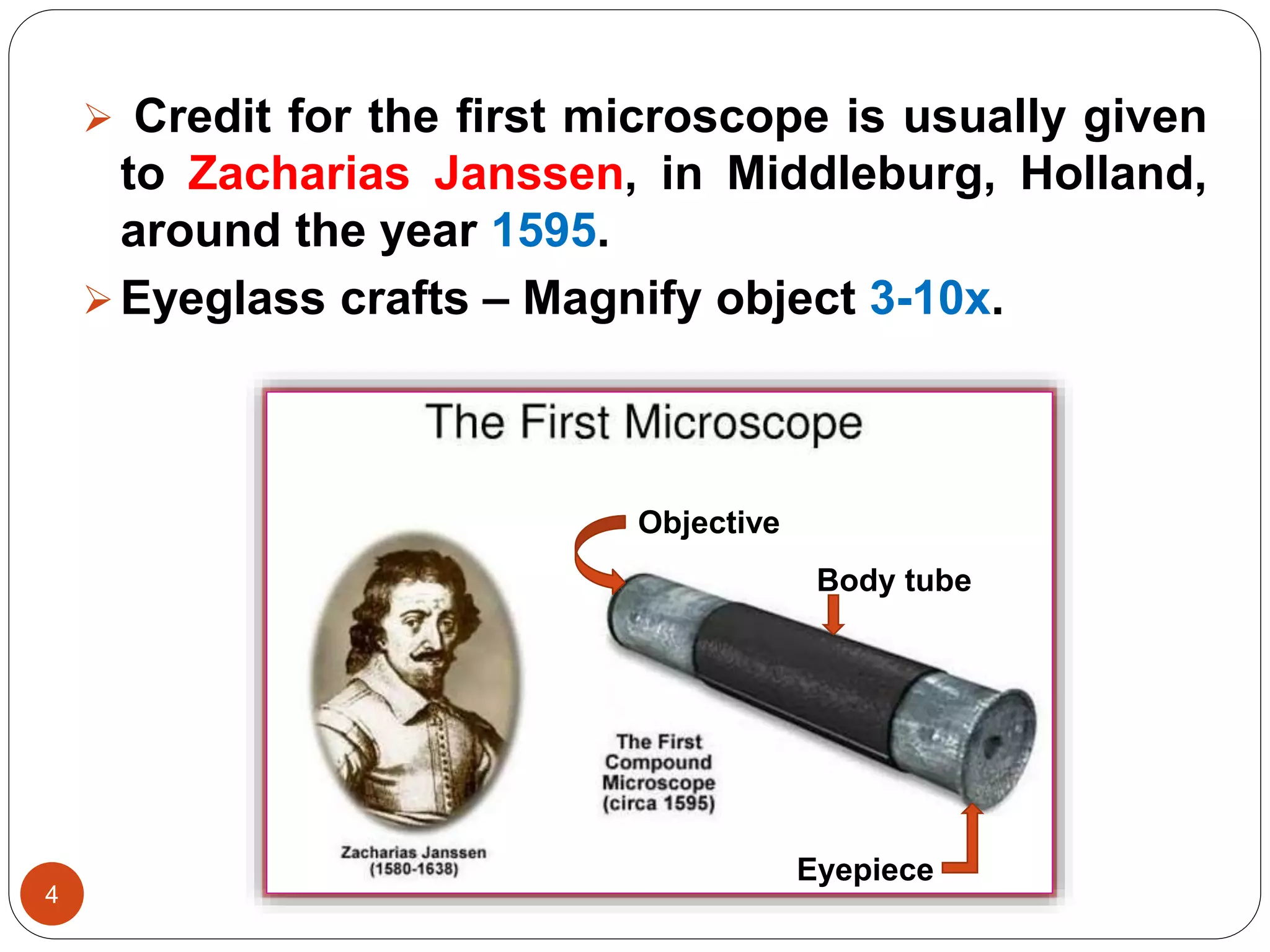
Credit forthe first microscope is usually given
to Zacharias Janssen, in Middleburg, Holland,
around the year 1595.
Eyeglass crafts – Magnify object 3-10x.
Objective
Body tube
Eyepiece
5.
The Light microscope
5
The most important scientific tool for a student of
biology is the light microscope.
The light microscope consists of various
components which gather light and redirect the
light path so that a magnified image viewed object
can be within a short distance.
The student’s microscope or the compound
microscope of twentieth century is the microscope
of much improved and modified type.
6.
6
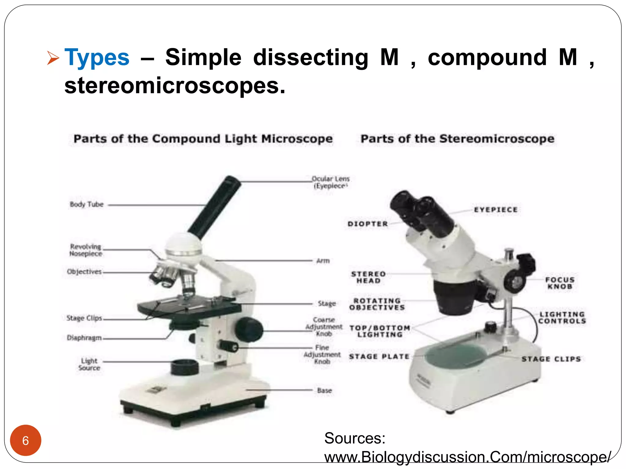
Types – Simpledissecting M , compound M ,
stereomicroscopes.
Sources:
www.Biologydiscussion.Com/microscope/
7.
Principle of lightmicroscope
7
In the light microscope, light is produced from
either an internal or external light sources and
passes through the iris diaphragm, a hole
variable size which controls the amount of
light reaching the specimen.
The main components of the compound light
microscope include a light sources that is
focussed at the specimen by a condenser lens.
8.
8
The slideis held on the stage at 90° to the
path of light which next travels through the
specimen.
The objective lens magnifies the image of the
specimen before the light travels through the
barrel of the microscope.
The light finally passes through the eyepieces
lens and into the viewer’s eye which sends
impulses to the brain which in turn interprets
the image.
9.
How doesit work ?
9
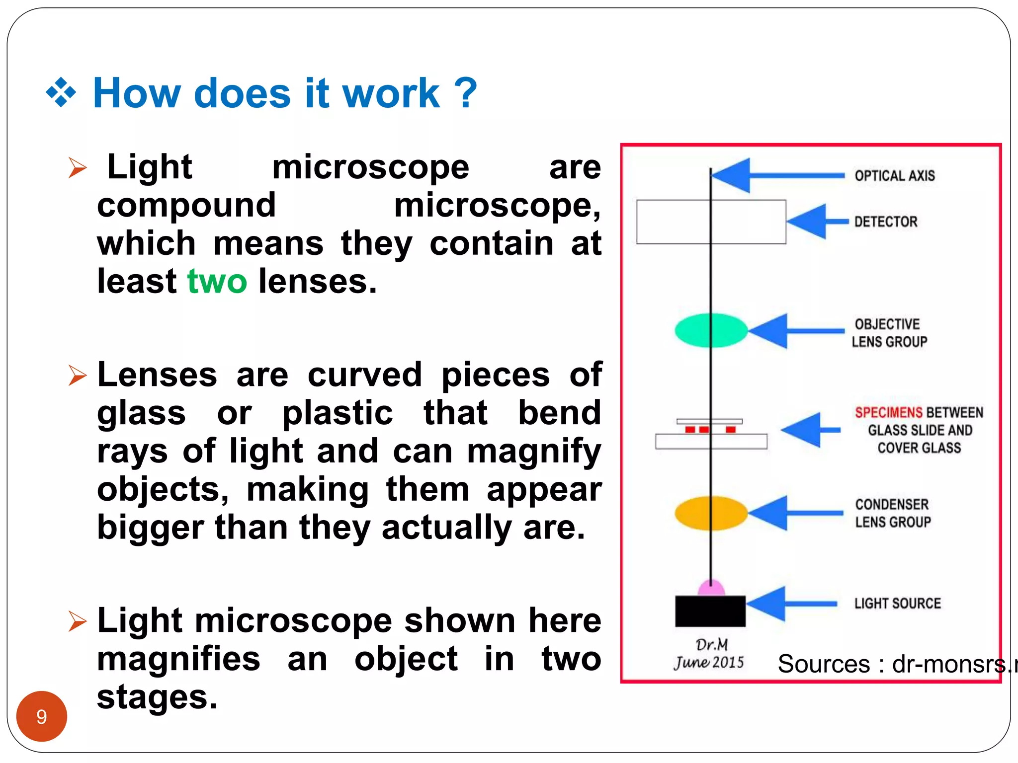
Light microscope are
compound microscope,
which means they contain at
least two lenses.
Lenses are curved pieces of
glass or plastic that bend
rays of light and can magnify
objects, making them appear
bigger than they actually are.
Light microscope shown here
magnifies an object in two
stages.
Sources : dr-monsrs.n
10.
10
Light fromthe mirror is reflected up through
the specimen, or object to be viewed, into the
powerful objective lens, which produce the
one magnification.
The image produced by the objective lens is
than magnified again by the eyepiece lens,
which act as a single magnifying glass.
The magnified image can be seen by looking
into the eyepiece lens.
Important factor in light microscopy include:
Magnification, Resolution, Contrast.
11.
Application of theLight microscope
11
Light microscopes play a large role in today’s
biology. Handy in use.
Biologists use the microscopes to observe
objects and details at a cellular level to learn
more about the building blocks of all
organisms.
Microscopes are also used to observe real
time movement in cells and organisms.
Lastly, microscopes are used in forensics to
help solve many crimes.
12.
12
Microscopes provide thestudents with an
understanding of real cells and their
supporting structures.
Also, microscopes provide students who are
inclined towards the medical field a more
intense look at the career choice and devlop
basic skills.
Lastly, microscopes are used in biology to
study diseases like cancer and AIDS to help
diagnose the disease in patients and to help
find a cure for them.
13.
13
Microscopes areused when studying light and
optics to learn how light refracts through
converging lenses and how a combination of
different lenses with varying focal lengths
affects the properties of the image.
Often times, there will be human evidences left
on the crime scene.
This allows forensic scientists to examine the
evidence under a microscope and match the
results with a database to find the culprit.
14.
14
Minerologist’s alsouse light microscopy, typically
with a special preparation of a sample called thin
section.
As the name imlies, thin section are very thin
slices of a rock. The sample needs to be thin
enough for light to travel through from the light
sources to the user’s eye.
The thin section will allow the shape of different
crystal grains to be seen.
The microscope can be used with different
techniques, like epifluorescences and phase
contrast.
15.
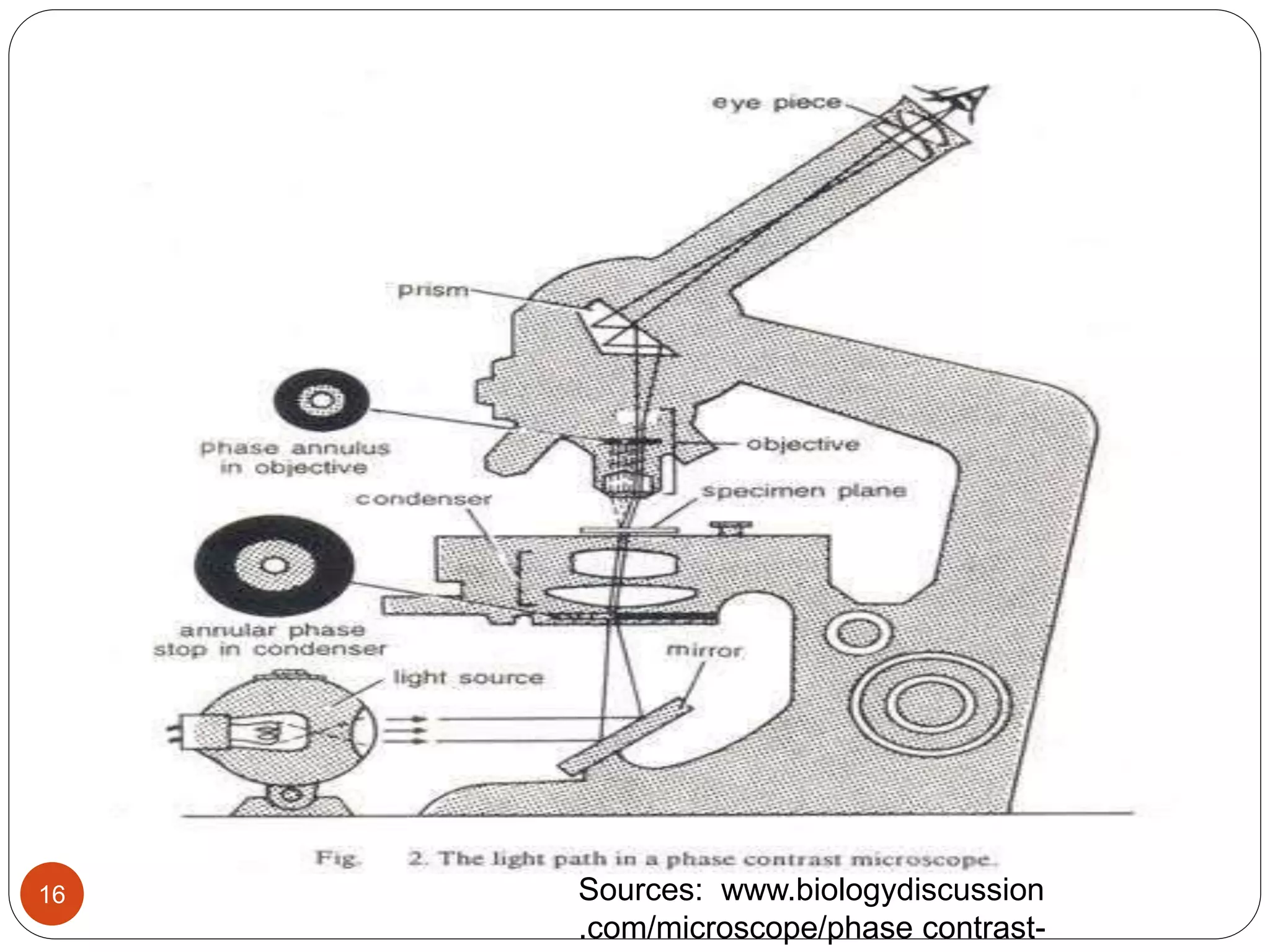
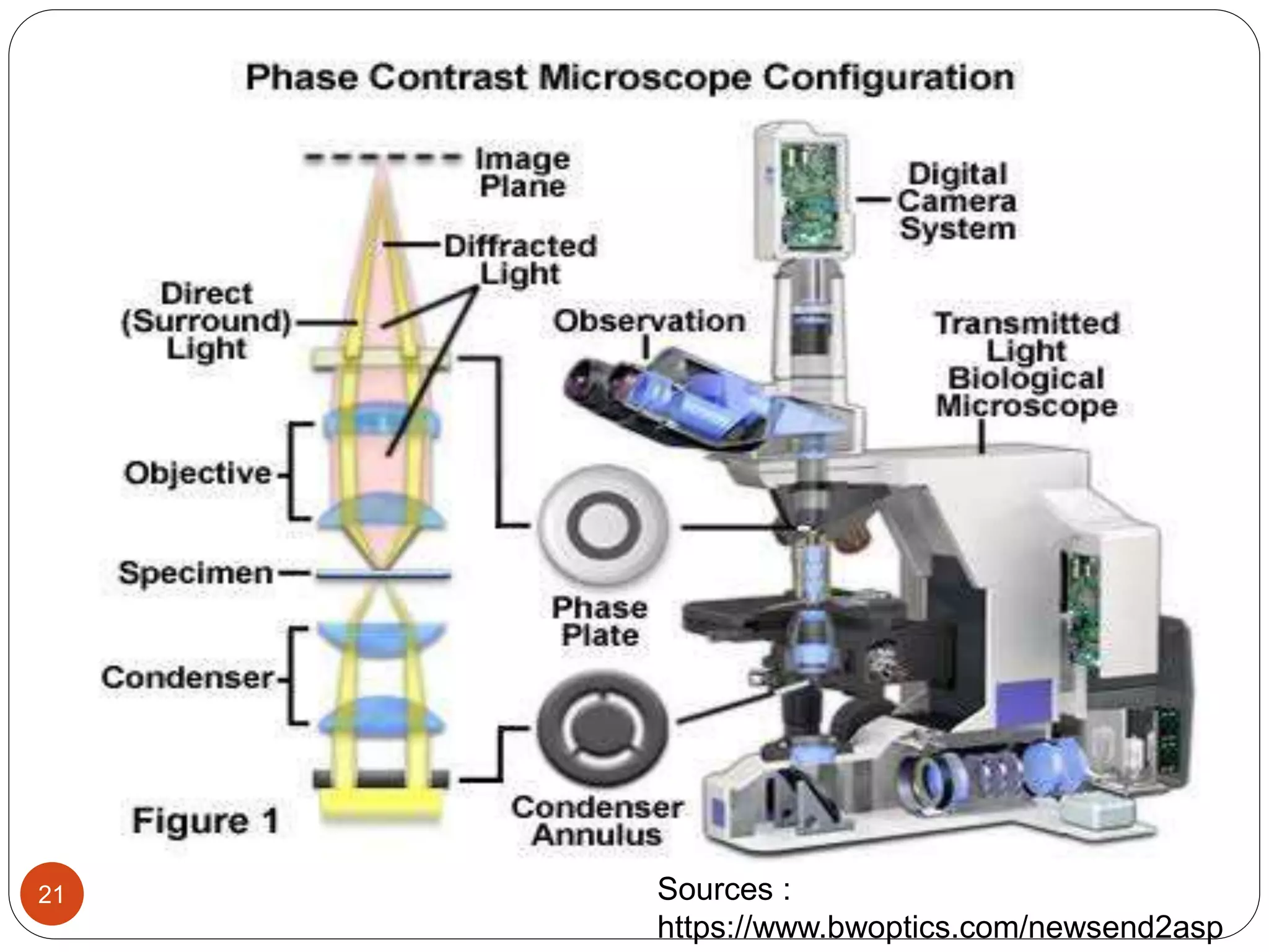
The Phase contrastmicroscope
15
Phase contrast microscopy first described in
1934 by Dutch physicist Fritz zernike, Whom
awarded by Nobel prize in physics in 1953.
A phase contrast microscope makes it possible
by utilizing two characteristics of light,
diffraction and interference, specimens based
on brightness differences.
It requires additional specialized structure
annular diaphragm and phase contrast ring.
Principle of PhaseContrast Microscope
17
It based on the wavelength (nature) of light rays
and the fact that light rays can be in phase or
out of phase.
Different shade of grey are distinguished to our
eyes due to differences in amplitude of light
rays.
PCM converts invisible small phase changes
caused by the cell component in to visible
intensity changes.
18.
18
In a Phasecontrast microscope, one set of
light rays comes directly from the light
sources.
The other set comes from light that is reflected
or diffracted from a particular structure in the
specimen.
The images differences in refractive index of
cellular structure. Light passes through thicker
parts of cell is held up relative to the light that
passes through thinner parts of cytoplasm.
19.
How doesit work ?
19
Light that does not interact with the speciman
is collected by the objective passes through
the phase ring, and is regarded exactly ¼
wavelength.
The Phase shifted is not detectable by the eye
so the resulting image on the image plane in
the microscope appears as a normal bright
background.
20.
20
Light passing throughone material & into
another material of slightly different refractive
index or thickness will undergo a charge in
phase.
This charge in are translated into variations in
brightness of the structures.
Natural light vibrates in many directions but
polarized light only one direction.
Application of PhaseContrast microscope
22
Most commonly used to provide contrast of
transparent specimens such as living cells or
small organisms.
Useful in observing cells cultured in vitro during
mitosis.
Phase contrast enables visualization of internal
cellular components.
It’s used in examination of growth, dynamics, and
behaviour of a wide variety of living cells in cell
culture.
23.
23
It appliedfor equipment from the study of
the living biological specimens, medical
applications, study of live blood cells, and
other biological and science applications.
It’s used in diagnosis of tumour cells.
24.
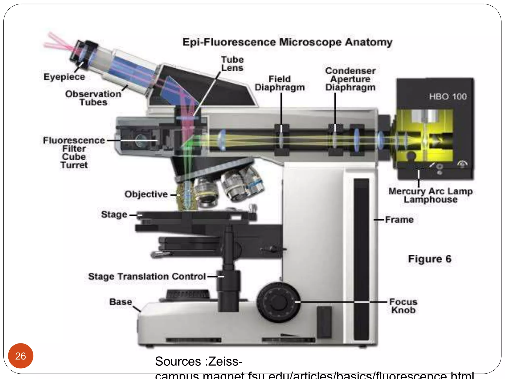
The Fluorescence microscope
24
Thismicroscope additionally requires an
excitation filter, a barrier and a dichromatic
mirror, fluorescent stain.
Fluorescent microscope is much the same as a
conventional light microscope with added
features to enhance its capabilities.
A specific wavelength of light is used to excite
fluorescent molecule in specimen. Light of
higher wavelength is then imaged.
25.
25
It is alsoused to visually enhance 3-D features
at small scales.
This is achieved by using powerful light
sources, such as lasers, that can be focused to
a pinpoint.
This focusing is done repeatedly throughout
one level of a specimen after another.
Most often an image reconstruction program
pieces the multi level image data together into
a 3-4 D reconstruction of the targeted sample.
Principle of theFluorescence microscope
27
When certain compounds are illuminated with high
energy light, they then emit light of a different ,
lower frequency. This effect is known as
Fluorescence.
In most cases the sample of interest is labelled
with a fluorescent substance known as a
fluorophore and then illuminated through the lens
with the higher energy sources.
Often specimens show their own characteristic
auto fluorescence image, based on their chemical
makeup.
28.
28
The keyfeature of fluorescent microscopy is
that it empoys reflected rather than transmitted
light, which means transmitted light techniques
such as phase contrast and DIS can be
combined with fluorescent microscopy.
29.
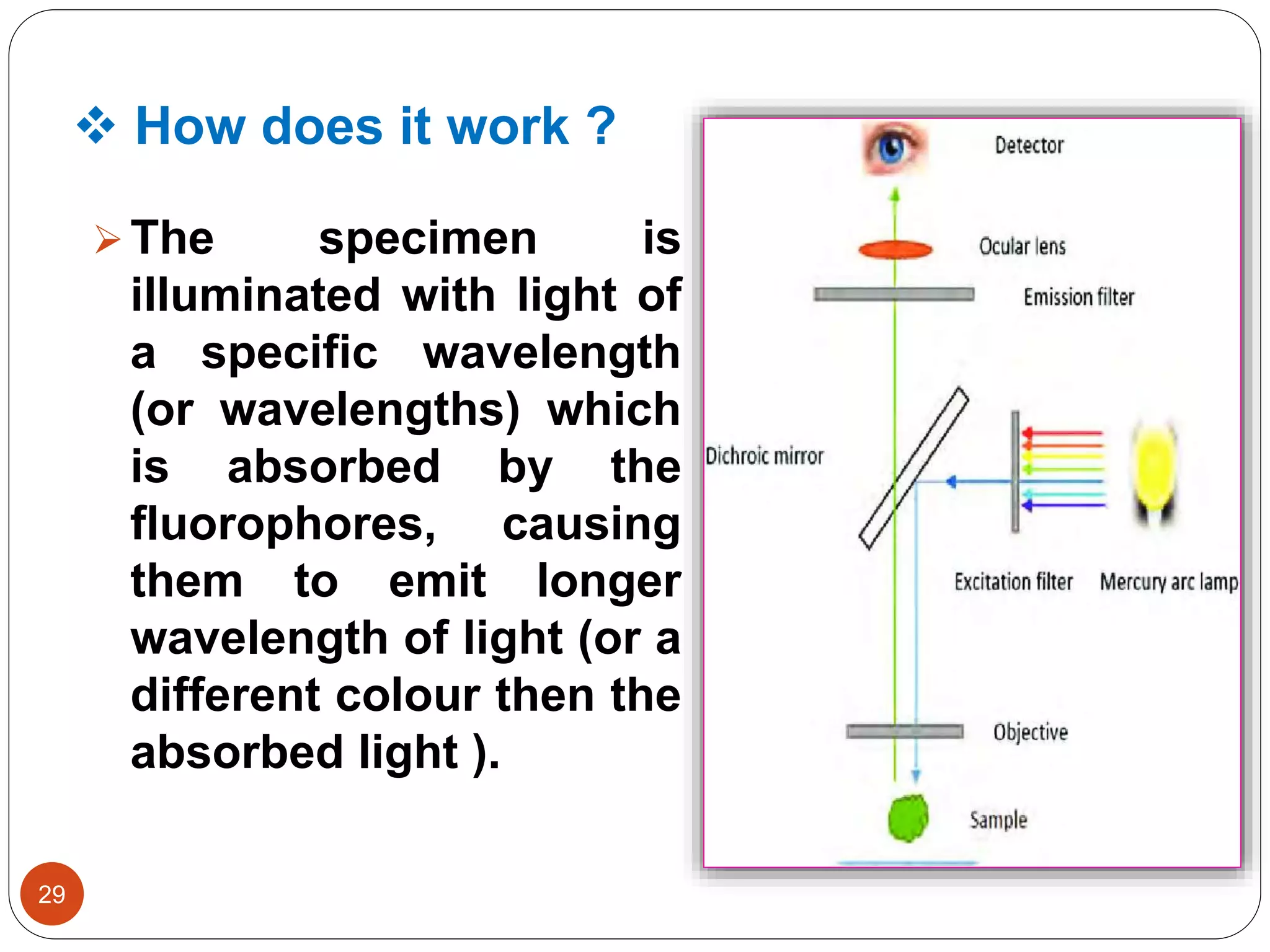
How doesit work ?
29
The specimen is
illuminated with light of
a specific wavelength
(or wavelengths) which
is absorbed by the
fluorophores, causing
them to emit longer
wavelength of light (or a
different colour then the
absorbed light ).
30.
Application of Fluorescencemicroscope
30
Fluorescence microscopy is a critical tool for
academic and pharmaceutical research,
pathology, and clinical medicine.
This method is used for demonstration of
naturally occurring fluorescent material and
other non- fluorescent substances or micro-
organisms after staining with some
fluorescent dyes. e.g.; Mycobacterium
tuberculosis, amyloid, lipids, elastic fibers etc.
31.
31
Imaging structuralcomponents of small
specimens, such as cells.
Conducting viability studies one cell
populations (are they a live or dead ?).
Imaging the genetic material within a cell (
DNA & RNA ).
Viewing specific cells within a larger
populations with techniques such as FISH.
32.
32
1) Biophysics
Author :Vasantha pattabhi ,
N. Gautham.
Edition : 2009 ( Second )
2) Basic Biophysics For Biologist
Author : M. Daniel
Edition : 2003
3) WWW.Slideshare.net