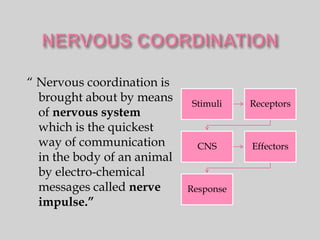
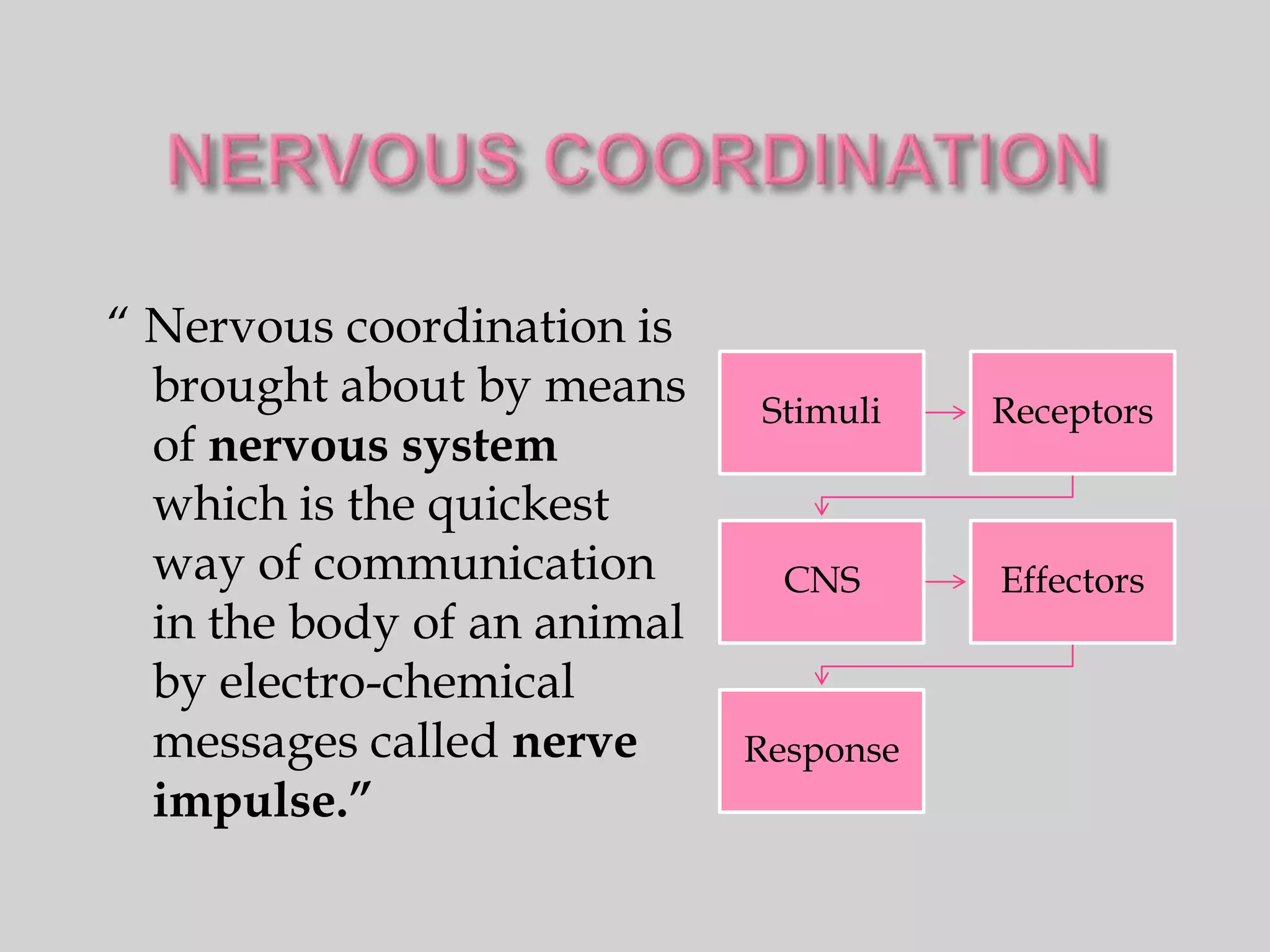
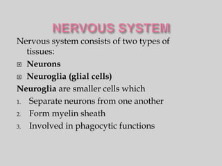
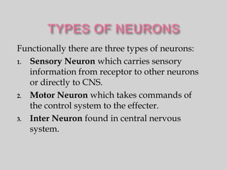
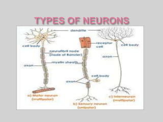
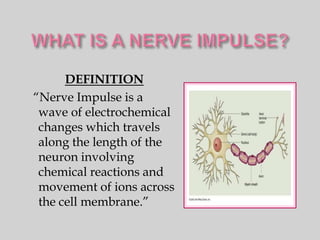
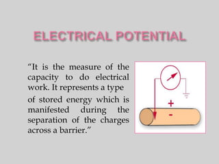
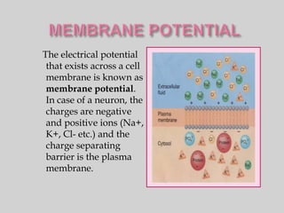
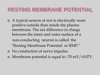
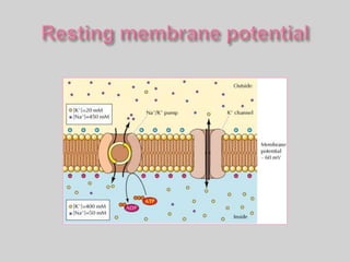
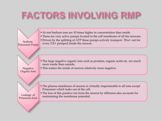
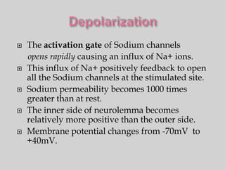
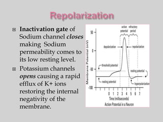
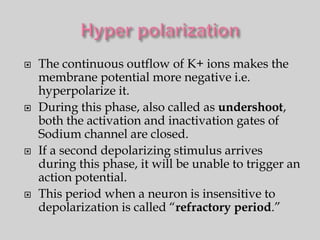
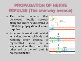
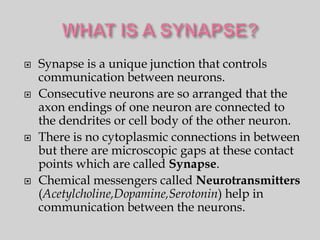
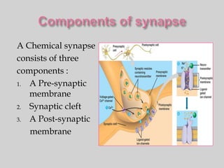
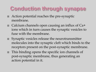
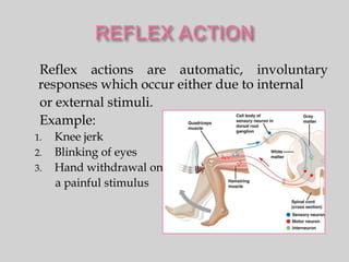
Call Girls in Mehrauli Delhi 💯Call Us 🔝8264348440🔝
Nervous coordination
1. “ Nervous coordination is
brought about by means Stimuli Receptors
of nervous system
which is the quickest
way of communication CNS Effectors
in the body of an animal
by electro-chemical
messages called nerve Response
impulse.”
2. Nervous system consists of two types of
tissues:
Neurons
Neuroglia (glial cells)
Neuroglia are smaller cells which
1. Separate neurons from one another
2. Form myelin sheath
3. Involved in phagocytic functions
3. “A Neuron is a special kind of animal cell which can
generate and conduct electric current.”
Structure:
A Neuron consists of
1. Soma or Cell body
2. Dendrites (Receiving end)
3. Axon (Conducting end)
Function:
Receives and integrate various
stimuli.
Send appropriate instructions
From CNS to effectors.
4. Functionally there are three types of neurons:
1. Sensory Neuron which carries sensory
information from receptor to other neurons
or directly to CNS.
2. Motor Neuron which takes commands of
the control system to the effecter.
3. Inter Neuron found in central nervous
system.
5.
6. DEFINITION
“Nerve Impulse is a
wave of electrochemical
changes which travels
along the length of the
neuron involving
chemical reactions and
movement of ions across
the cell membrane.”
7. “It is the measure of the
capacity to do electrical
work. It represents a type
of stored energy which is
manifested during the
separation of the charges
across a barrier.”
8. The electrical potential
that exists across a cell
membrane is known as
membrane potential.
In case of a neuron, the
charges are negative
and positive ions (Na+,
K+, Cl- etc.) and the
charge separating
barrier is the plasma
membrane.
9. A typical neuron at rest is electrically more
positive outside than inside the plasma
membrane. The net difference in charge
between the inner and outer surface of a
non-conducting neuron is called the
“Resting Membrane Potential or RMP.”
No conduction of nerve impulse.
Membrane potential is equal to -70 mV/-0.07V.
10.
11. •At rest Sodium ions are 10 times higher in concentration than inside.
•These are very active pumps located in the cell membrane of all the neurons.
•Driven by the splitting of ATP these pumps actively transport 3Na+ out for
Sodium-
Potassium Pump every 2 K+ pumped inside the neuron.
•The large negative organic ions such as proteins, organic acids etc. are much
more inside than outside.
Negative •This makes the inside of neuron relatively more negative.
Organic Ions
•The plasma membrane of neuron is virtually impermeable to all ions except
Potassium which leaks out of the cell.
•The loss of this positive ion from the neuron by diffusion also accounts for
Leakage of
maintaining the membrane potential.
Potassium Ions
12.
13. A Nerve Impulse is initiated by an appropriate
stimulus called “threshold stimulus.”
Such a stimulus results in a remarkable localized
change in resting membrane potential which is
replaced by a new potential called “Active
Membrane Potential (AMP) or Action Potential.”
This change(depolarization)is for a brief instant(
perhaps for a few milliseconds) due to the reversal
of charges at the stimulated site of neurolemma.
Conduction of nerve impulse.
Membrane potential becomes +40mV/+0.04 V.
14. Voltage- • Activation
Gated Gate
Sodium • Inactivation
Channels Gate
Voltage- • A Single
Gated Voltage
Potassium Sensitive
Channels Gate
15. The action potential or AMP is actually the
Nerve Impulse.
Once an action potential is triggered, the
membrane potential goes through a
stereotypical sequence of changes which
involves the following steps:
Depolarization
Repolarization
Hyper polarization
16. The activation gate of Sodium channels
opens rapidly causing an influx of Na+ ions.
This influx of Na+ positively feedback to open
all the Sodium channels at the stimulated site.
Sodium permeability becomes 1000 times
greater than at rest.
The inner side of neurolemma becomes
relatively more positive than the outer side.
Membrane potential changes from -70mV to
+40mV.
17. Inactivation gate of
Sodium channel closes
making Sodium
permeability comes to
its low resting level.
Potassium channels
opens causing a rapid
efflux of K+ ions
restoring the internal
negativity of the
membrane.
18. The continuous outflow of K+ ions makes the
membrane potential more negative i.e.
hyperpolarize it.
During this phase, also called as undershoot,
both the activation and inactivation gates of
Sodium channel are closed.
If a second depolarizing stimulus arrives
during this phase, it will be unable to trigger an
action potential.
This period when a neuron is insensitive to
depolarization is called “refractory period.”
19. The action potential that
developed locally spreads
along the entire neurolemma is
called the propagation of nerve
impulse.
A neuron is usually stimulated
at its dendrites or cell body and
resulting action potential is
regenerated anew in a
sequence along the axon to the
other end of the cell until it
reaches Synapse.
20. Synapse is a unique junction that controls
communication between neurons.
Consecutive neurons are so arranged that the
axon endings of one neuron are connected to
the dendrites or cell body of the other neuron.
There is no cytoplasmic connections in between
but there are microscopic gaps at these contact
points which are called Synapse.
Chemical messengers called Neurotransmitters
(Acetylcholine,Dopamine,Serotonin) help in
communication between the neurons.
21. A Chemical synapse
consists of three
components :
1. A Pre-synaptic
membrane
2. Synaptic cleft
3. A Post-synaptic
membrane
22. Action potential reaches the pre-synaptic
membrane.
Calcium channels open causing an influx of Ca+
ions which in turn causes the synaptic vesicles to
fuse with the membrane.
Synaptic vesicles release the neurotransmitter
molecules into the synaptic cleft which binds to the
receptors present on the post-synaptic membrane.
This binding opens the specific ion channels of
post-synaptic membrane, thus generating an
action potential in it.
23. Reflex actions are automatic, involuntary
responses which occur either due to internal
or external stimuli.
Example:
1. Knee jerk
2. Blinking of eyes
3. Hand withdrawal on
a painful stimulus