The presentation contains the description about various parts of morphology of the honey bee viz: head, type of mouthpart, abdomen including the legs and wings, and the abdomen.
the presentation will help you learn more about how the insect eyes really work in field conditions and more over for the better understanding you can take help from from book: THE INSECTS:STRUCTURE AND FUNCTION byR.F.CHAPMAN.....as the contents of my presentation are from that book only.....
The integumentary system comprises the skin and its appendages. Skin + derivatives= Integument.
It aims to protect the body from various kinds of damage, such as loss of water or damages from outside.
The integumentary system in chordates includes hair, scales, feathers, hooves, and nails.
It may serve to water proof, and protect the deeper tissues.
Excrete wastes, and regulate body temperature.
It is the attachment site for sensory receptors to detect pain, sensation, pressure, and temperature.
Insect are animals , but unlike many animals , they have no backbone .
They have an outer support system called an exoskeleton rather than the inner support system ( endoskeleton ) characteristic of most large animals .
Metamorphosis is a complex and tightly regulated process that concludes larval growth and results in the transformation to the adult stage. Since growth only occurs in immature stages, the adult body size is determined by the size the larva attained when it stopped feeding and initiated metamorphosis. In this presentation, I am discussing points about hormonal control of insect metamorphosis.
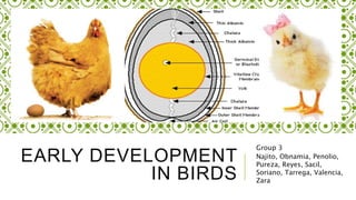
This slide show takes you through the detailed process of development of chick and the various crucial stages of development. It can be a useful resource for science graduation students
DEVELOPMENT OF FACE/ Development of face, palate and jawDishikaBhagwani27
• Introduction, General embryology○ Fertilization ○ Formation of germ layers ○ Development of face – •Pharyngeal arches, pouch & clefts ○ Development of nose. development of maxilla & mandible, development of eyes,development of lips & checks Development of head • Development of skull • Development of face.....
Neural crest cells in 2 parts / dental implant courses by Indian dental academy Indian dental academy
The Indian Dental Academy is the Leader in continuing dental education , training dentists in all aspects of dentistry and
offering a wide range of dental certified courses in different formats.for more details please visit
www.indiandentalacademy.com
Neural crest cells / dental implant courses by Indian dental academy Indian dental academy
The Indian Dental Academy is the Leader in continuing dental education , training dentists in all aspects of dentistry and
offering a wide range of dental certified courses in different formats.for more details please visit
www.indiandentalacademy.com
2137ad - Characters that live in Merindol and are at the center of main storiesluforfor
Kurgan is a russian expatriate that is secretly in love with Sonia Contado. Henry is a british soldier that took refuge in Merindol Colony in 2137ad. He is the lover of Sonia Contado.
2137ad Merindol Colony Interiors where refugee try to build a seemengly norm...luforfor
This are the interiors of the Merindol Colony in 2137ad after the Climate Change Collapse and the Apocalipse Wars. Merindol is a small Colony in the Italian Alps where there are around 4000 humans. The Colony values mainly around meritocracy and selection by effort.
Hadj Ounis's most notable work is his sculpture titled "Metamorphosis." This piece showcases Ounis's mastery of form and texture, as he seamlessly combines metal and wood to create a dynamic and visually striking composition. The juxtaposition of the two materials creates a sense of tension and harmony, inviting viewers to contemplate the relationship between nature and industry.
Explore the multifaceted world of Muntadher Saleh, an Iraqi polymath renowned for his expertise in visual art, writing, design, and pharmacy. This SlideShare delves into his innovative contributions across various disciplines, showcasing his unique ability to blend traditional themes with modern aesthetics. Learn about his impactful artworks, thought-provoking literary pieces, and his vision as a Neo-Pop artist dedicated to raising awareness about Iraq's cultural heritage. Discover why Muntadher Saleh is celebrated as "The Last Polymath" and how his multidisciplinary talents continue to inspire and influence.
3. CLEAVAGE IN BIRD EGGS
Accessible all year
Easily raised
At any particular temperature, developmental stage
can be accurately predicted.
Large numbers of embryos can be obtained at the
same stage.
Chick embryo can be surgically manipulated
Often served as a surrogate for human embryos.
4. CLEAVAGE IN BIRD EGGS
Fertilization occur in the
oviduct before the albumen
and the shell are secreted
upon it.
The egg is telocithal (like
that of a fish)
Eggs undergo discoidal
meroblastic cleavage.
CLEAVAGE IN BIRD EGGS
5. First cleavage furrow appear centrally in the
blastodisc
Equatorial and vertical cleavages divide the
blastoderm into 5-6 cell tissue thick
Subgerminal cavity – space between
blastoderm and yolk.
It is created when blastoderm cell absorb
fluid from the albumin and secrete it between
themselves and the yolk.
At this stage, deep cells in the center of the
blastoderm shed and die, leaving the one cell
thick area pellucida
DISCOIDAL MEROBLASTIC
CLEAVAGE
6. DISCOIDAL MEROBLASTIC
CLEAVAGE
Area pellucida forms most of the
actual embryo.
Area opaca – the peripheral ring of
blastoderm cell that have not shed
their deep cells
Marginal zone – thin layer cell
between area pellucida and area oraca
Some of the marginal zone cells
become very important in determining
cell fate during early chick
development.
DISCOIDAL MEROBLASTIC
CLEAVAGE
9. The time a hen lay an egg, the blastoderm contains about 20, 000 cells.
Most area pellucida cell remain at the surface, froming the epiblast
Other pellucida cells delaminated and migrated individually into the subgerminal
cavity to form the polyinvagination island (primary hypoblast)
It is an archipelago of disconnected clusters containing 5-20 cells each.
A sheet of cells from the posterior margin of the blastoderm (with Koller’s
sickle) migrates anteriorly to join the polyinvagination island, later forming the
secondary hypoblast.
GASTRULATION – THE
HYPOBLAST
10. Figure 3. The primary epiblast
GASTRULATION – THE
HYPOBLAST
Figure 4. Forming of secondary
epiblast
11. GASTRULATION – THE
HYPOBLAST
Two layered blastoderm (epiblast and hypoblast) is jooned together
at the margin of the orea opaca, and the space between then forms a
blastocoel.
The embryo entirely come from the epiblast
Hypoblast cells form portion of external membranes (esp. the yolk
sac and stalk)
Yolk stalk link the yolk mass to the endodermal digestive tube.
All 3 layers are formed from the epiblastic cells.
GASTRULATION – THE
HYPOBLAST
13. GASTRULATION – THE
PRIMITIVE STREAK
Primitive streak –
the major structural
characteristic of
avian, reptilian and
mammalian
gastrulation.
GASTRULATION – THE
HYPOBLAST
Figure 6. Cell migration during gastrulation
14. The streak elongates
toward the future head
region.
At the same time, the
secondary hypoblast cells
continue to migrate
anterior to the posterior
margin of the blastoderm.
The streak extends 60-
75% of the length of area
pellucida.
GASTRULATION – THE PRIMITIVE
STREAK
Figure 7. Anterior and posterior view during
gastrulation
Figure 8. Formation of the
15. The streak defines the
axes of the embryo
(extend from posterior to
anterior, migrate cell from
dorsal side to ventral side)
Those close to the streak
will be the medial
structure, and farther will
be the distal structures
GASTRULATION – THE PRIMITIVE
STREAK
Figure 9. Formation of the foregut and other
structures
16. GASTRULATION – THE
PRIMITIVE STREAK
As cell converge from the streak, a depression forms within the streak (called
primitive groove)
It serves as the opening to the migrating cell into the blastocoel (analogous to
amphibian blastopore)
Primitve knot or Hensen’s node – regional thickening of cells at the anterior
end of the primitive streak.
It is the functional equivalent of the dorsal lip of the amphibian blastopore and
the fish embryonic shield.
Primitve pit – a funnel shape depression at the center of Hensen’s node.
GASTRULATION – THE PRIMITIVE
STREAK
17. GASTRULATION – THE
PRIMITIVE STREAK
As the streak form, epiblast cell begin to migrate through it and into the
blastocoel.
In the blastocoel, they migrate anteriorly, forming the foregut, head
mesoderm, and notochord.
Cell passing laterally of the streak forms the majority of endodermal and
mesodermal tissues.
Scatter factor – a 190-kDA protein thought to decompose the basal
lamina and release cells into the embryo as cell enter the streak. Can
convert epithelial sheets into mesenchymal cells in several ways. Involve
in downregulation of E-cadherin expression and prevention of E-cadherin
to function.
GASTRULATION – THE PRIMITIVE
STREAK
19. First to migrate through Hensen’s node are destined to
become the pharyngeal endoderm of the foregut.
Inside the blastocoel, endodermal cell migrate anteriorly
and displace hypoblast cell to the confined region of area
pellicda anterior portion
Germinal crescent – contain precursors of the germ cells
which later migrate through the blood vessel to the gonads
GASTRULATION – ENDODERM AND
MESODERM FORMATION
20. Next to enter through Hensen’s node move anteriorly but don’t
move far ventrally as the destined foregut endodermal cells,
rather they remain between the endoderm and epiblast to form the
head mesenchyme and prechordal plate mesoderm.
These cells all move anteriorly, pushing the epiblast to form the
head process.
The head of the avian embryo forms anterior (rostral) to Hensen’s
node.
Next cell to migrate through Hensen’s node become
chordamesoderm (notochord) cells
GASTRULATION – ENDODERM AND
MESODERM FORMATION
21. Cells migrating inwardly through the lateral portion of the
primitive streak.
In the blastocoel, these cell separate into 2 layers.
Layer 1 – deep layer join hypoblast along its midline and displace
hypoblast cell to the sides. They give rise to all endodermal organs
and most of the extraembryonic membranes (hypoblast forms the
rest)
Layer 2 – cell spread between the endoderm and epiblast, forming a
loose layer. This generate the mesodermal portion of the embryo
and extraembryonic membranes.
GASTRULATION – ENDODERM AND
MESODERM FORMATION
23. The primitive streak start to regress (Hensen’s node move near the center
of area pellucida)
As the node moves posteriorly the notochord is laid down, starting at the
level of the future midbrain.
The posterior notochord (after somite 17 in the chick) forms from the
condensation of mesodermal tissue that has ingressed through the streak
(not through Hensen’s node).
This portion of the notochord extends posteriorly to form the tail of the
embryo.
GASTRULATION – REGRESSION OF
THE PRIMITIVE STREAK
24. Hensen’s node regresses to its most posterior position, forming the anal
region
At this time, all presumptive endodermal and mesodermal cells have
entered the embryo, and the epiblast is composed entirely of presumptive
ectodermal cells.
Avian (and mammalian) embryos exhibit a distinct anterior-to-posterior
gradient development maturity.
While cell of posterior portion of the embryo undergoes gastrulation, cell at
anterior end starts to form organs.
GASTRULATION – REGRESSION OF
THE PRIMITIVE STREAK
25. GASTRULATION – EPIBOLY OF THE
ECTODERM
Figure 12. Epiboly of ectoderm Figure 13. Notochord length vs. time
26. Ectodermal precursors proliferate while the presumptive mesodermal
and endodermal cells are moving inwardly.
Ectodermal cell migrate to surround the yolk by epiboly. (took 4 days
to complete)
It involves the continuous production of new cellular material and the
migration of the presumptive ectodermal cells along the underside of
the vitelline envelope
Only the cells of the outer edge of the orea opaca attach firmly to the
vitelline envelope.
GASTRULATION – EPIBOLY OF THE
ECTODERM
27. These cells are inherently different from the other blastoderm cells, as they can extend
enormous (500 μm) cytoplasmic processes onto the vitelline envelope.
These elongated filopodia are believed to be the locomotor apparatus of these marginal
cells, by which they pull the other ectodermal cells around the yolk
The filopodia appear to bind to fibronectin, a laminar protein that is a component of the chick
vitelline envelope.
If the contact between the marginal cells and the fibronectin is experimentally broken (by adding
a soluble polypeptide similar to fibronectin), the filopodia retract, and epidermal migration ceases
the ectoderm has surrounded the yolk, the endoderm has replaced the hypoblast, and the
mesoderm has positioned itself between these two regions.
GASTRULATION – EPIBOLY OF THE
ECTODERM
29. Axes are specified early in the cleavage stage.
Formation of these axes are later formed during gastrulation.
AXIS FORMATION IN CHICKS
30. DV axis is established when the
dividing cells of blastoderm form a
barrier between the basic albumin (pH
9.5) above the blastodisc and acidic
subgeminal space below it (6.5).
H2O and Na+ ions are transported
from the albumin to the subgeminal
space and causes a membrane
potential difference of 25 mV.
THE ROLE OF PH IN FORMING THE
DORSAL-VENTRAL AXIS (DV)
The difference in membrane potentials
distinguishes two sides of the epiblast:
1. The side facing the negative and
basic albumin becomes the dorsal
side.
2. The side facing the positive and
acidic subgeminal space fluid
becomes the ventral side.
31. The bilateral symmetry of the chick blastoderm is determined by gravity.
The ovum spins at rate of 10-20 revolutions per hour for about 20 hours through
the hen’s reproductive tract.
The shifting of yolk makes the lighter components to aggregate beneath one
side of the blastoderm.
Lighter components tips up the end of the blastoderm and this end becomes the
posterior portion of the embryo-the part where the primitive streak formation
begins.
THE ROLE OF GRAVITY IN FORMING THE
ANTERIOR-POSTERIOR AXIS (AP)
32. There is still no known interactions that explain how the posterior margin forms
and why it is the site of gastrulation.
The ability to form primitive streak can be seen throughout the marginal zone
and if the blastoderm is separated into parts, each part will form their own
primitive streak.
However, once a posterior marginal zone (PMZ) has formed, it controls the
marginal regions and prevent the other regions to form their own primitive streaks.
Also PMZ cells initiate gastrulation and is regarded as the equivalent of the
amphibian Nieuwkoop center.
PRIMITIVE STREAK FORMATION
33. The PMZ region like the Nieuwkoop center is thought to be the place where the
localization of β-catenin in the nucleus and TGF- β family signal coincide.
Only the PMZ regions secrete VG1 and if the PMZ tissues are grafted to the anterior
marginal zone, that region will able to form primitive streak.
Koller’s sickle
Anterior portion
Forms the Hensen’s node from the epiblast and middle layer cells.
Posterior portion
Contributes to the posterior portion of the primitive streak.
Transplantation of Koller’s sickle can cause formation of new axes and middle layer
cells in the Koller’s sickle express Goosecoid.
PRIMITIVE STREAK FORMATION
34. Regarded as the avian equivalent of the amphibian
dorsal blastopore lip since
it is the site of gastrulation
its cells become the chordamesoderm
it act like the amphibian organizer which can organize a
second embryonic axis when its cells are transplated into
other locations
THE HENSEN’S NODE
35. THE HENSEN’S NODE
Cells of the Hensen’s node secrete chordin, noggin and nodal
proteins which antagonize the BMPs and dorsalize the ectoderm and
mesoderm.
The antagonism of BMPs does not appear to be sufficient for neural
induction.
In chick embryos, fibroblast growth factors (FGFs) generate
neuronal phenotype in epiblast cells rather than BMPs or ectopic
expression of chordin.
FGFs from the Hensen’s node and primitive streaks and beads
induce trunk and hindbrain neuronal expression in the epiblast cells.
THE HENSEN’S NODE
36. This formation is regulated by paracrine factor, Nodal and transcription factor, Pitx2. However,
there is a different mechanism of regulation in chick embryos.
As primitive streak reaches maximum length, the:
transcription of sonic hedgehog genes ceases on the right side of the embryo due to the
expression of activin
activin activated expression of FGF8 and FGF8 prevents the transcription of caronte gene.
The absence of caronte genes ables the bone morphogentic proteins (BMPs) to block the
expression of nodal and lefty 2 which also activates the snail gene (cSNR) that is a characteristic
of the right side of avian embryonic organs.
THE LEFT-RIGHT AXIS FORMATION
(LR)
37. On the left side of the body:
Lefty-1 blocks the FGF8 expression
Hedgehog activates caronte
Caronte genes prevents BMPs to repress nodal and lefty-2 and to inhibit the blocking of
lefty-1 expression on ventral midline structures
On the left side of the body:
Nodal and Lefty-2 activate pitx2 and repress snail (cSNR).
Lefty-1 in the ventral midline prevents the caronte signals from passing to the right side
of the embryo.
THE LEFT-RIGHT AXIS FORMATION
(LR)
Telocithal -With small disc of cytoplasm sitting atop a large yolk.
discoidal meroblastic cleavage- occur in blastodisc, a small disc of cytoplasm 2-3mm in diameter at the animal pole
Continue to create a single layered blastoderm.
Note: cleavage don’t extend to the yolky cytoplasm, early cleavage continues with each other and yolk as their base.
2. These cells become linked together by tight junctions.
Area pellucid
1.Although the shape and formation of the avian blastodisc differ from those of the amphibian, fish and echinoderm blastula, the overall spatial relationship are retained
By 22 hours of incubation , most of the presumptive endodermal cells are in the anterior of the embryo, although presumptive mesodermal cell continue to migrate inward for a longer time.
These cells are inherently different from the other blastoderm cells, as they can extend enormous (500 μm) cytoplasmic processes onto the vitelline envelope. These elongated filopodia are believed to be the locomotor apparatus of these marginal cells, by which they pull the other ectodermal cells around the yolk