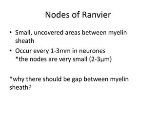
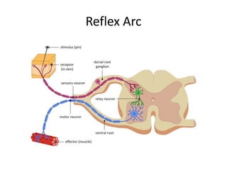
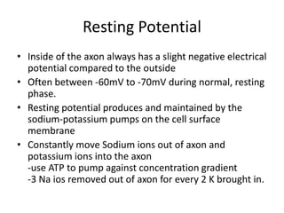
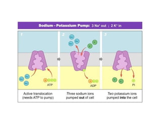
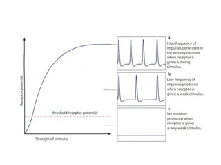
Control and Coordination
Chapter 15
Endocrine Vs. Nervous System
Nervous System Communication
• Consist of Central Nervous System and
Peripheral Nervous System
• Transferred in nerve impulses, along neurons
(nerve cells) directly sent to the target cells
• Coordinates the activitiy of sensory receptors,
decision-making in CNS and effectors (e.g
muscles, glands)
Types of Neurons
1. Sensory Neurons
-Transmit impulses from receptors to CNS
2. Intermediate neurons
-transmit impulses from sensory neurons to
motor neurons
3. Motor Neurons
-transmit impulses from CNS to effectors
Motor Neurons
• Cell body often lies within the spinal cord / brain
• Thin cytoplasmic processes extend from the cell body;
dendrites
-large surface area for axon terminals from other
neurons
• Axon branching further from cell body, conduct
impulses along its pathway
-mitochondria resides in the axon
-mitocondria presents in many amounts at the end of
axon, together with vesicles containing chemicals;
transmitter  passing impulses to the effector cell
• Sensory neurons:
-same basic structur as motor neuron, bt cell
body is located near to the source of the
stimuli; ganglion (swelling of spinal nerves)
• Relay neurons:
-found in CNS
Myelin
• Schwann cells; surrounding the axons by
layers of cell surface membranes
• Rich in phospholipid and thus impermeable to
water and ions in tissue fluid  insulating the
axons
• Not all axons are myelinated; about two-third
are unmyelinated (*why..?)
• The myelin sheath affects the conduction
speed of the impulses
Myelinated Vs. Unmyelinated Neurone
Nodes of Ranvier
• Small, uncovered areas between myelin
sheath
• Occur every 1-3mm in neurones
*the nodes are very small (2-3µm)
*why there should be gap between myelin
sheath?
Reflex Arc
• A pathway along which impulses are transmitted
from a receptor to an effector without involving
‘concious’ region of the brain
• Other reflex arc may not have intermediate
neurones, and the impulses directly passes from
sensory neurone to motor neurone
• There are also reflex arc in the brain; e.g control
of eye focus and allowing light entering the eye
Reflex Arc
• Reflex action; Respond to the stimulus
before there is any voluntary responses
involving the concious region of the brain.
• The impulses passes on the other neurones
that up to the brain.
• At the same time, the impulses travelling
along the motor neurone to the effector.
• Fast, automatic response to the stimulus
• Very useful of responding to danger signals
Impulse Transmission
• Neurones transmit electrical impulses; travel
repidly along the cell surface membrane, and
not a flow of electrons like elctrical power.
• Action Potentials; very brief changes in
distribution of electrical charge across the cell
surface membrane
-caused by the very rapid movement of
sodium and potassium ions into and out of the
axon.
Resting Potential
• Inside of the axon always has a slight negative electrical
potential compared to the outside
• Often between -60mV to -70mV during normal, resting
phase.
• Resting potential produces and maintained by the
sodium-potassium pumps on the cell surface
membrane
• Constantly move Sodium ions out of axon and
potassium ions into the axon
-use ATP to pump against concentration gradient
-3 Na ios removed out of axon for every 2 K brought in.
• The membrane has protein channels for
potassium and sodium that open all the time
• Thus, some K+ ions diffuses back out again much
faster than Na+ ions diffuses back in.
• There are also large, negatively charged moleculs
inside the cell that attract the K+ ions; reducing
the chances that they will diffuse out
• Results; overall excess of negative ions inside the
membrane compared to the outside.
• The membrane is relatively impermeable to
the Na+ ions
• 2 conditions affecting the inward movement
of Na+:
-steep concentration gradient
-negatively charged inside of the membrane
elctrochemical gradient
Action Potentials
• Potential difference across the surface
membrane suddenly switches from -70mV to
+30mV
• It swiftly returns to normal after a brief
‘overshoot’; takes in 3ms
• Caused by the changes of permeability of the
cell surface membrane to Na+ and K+ ions
• As well as channels open at all the time, there
are also other channels that allow Na+ and K+
to pass through.
• They open and close depending on the
electrical potential across the membrane;
voltage-gated channel.
• These channels are closed during resting
potential.
1. Depolarisation
• Electric current used to stimulate the aon causes the opening
of voltage-gated channel  allow Na+ to pass through
• Na+ ions enter through open channel ( due to higher
concentration outside the axon compared to the inside.
• Inward movement causes changes of potential difference
across the membrane  becoming less negative on the inside
depolarisation
• It triggers to open some more channels.
-If the potential difference reacher -50mV, many more
channels will open and th inside reaches the potentials of
+30mV compared to the outside.
• Action potentials are generated if the potential difference
reaches -60mV to -50mV; also called as the threshold
potential
2. Repolarisation
• About 1ms, all Na+ voltage-gated channel closed
stop diffusing Na+ ions into the axon.
• At the same time, another set of voltage-gated
channel for K+ ions opens, allowing diffuse of K+
ions out of axon (down the conc. gradient)
• Outward movement of K+ ions remove positive
charge from inside to outside of axon returning
the potential difference to normal; -70mV
• Potential difference during repolarisation are more
negative than the resting potential
3. Back to Resting Potential
• K+ ions channel protein then closed and Na+ ion
channel protein response to depolarisation again.
• Sodium-potassium pump channel continue pumping all
the time; sodium ions out and potassium ions in
• These will maintain the distribution of Na+ and K+ ions
across the membrane  for the action potential can
continue to re-occur
Action potentials and information
transmission across the axon
1. Action potential (at any point) triggers the
production of action potential on either side of
that point.
2. Temprary depolarisation causes current to flow
in both directions
3. Current flow due to the difference in charge
between the inside and outside the of the axon
membrane.
*depolarisation occurs on both side happens in
the experimental situation only, when stimulus
is applied somewhere on the axon.
• In the body, action potentials begin at one end
o the axon  action potentials are generated
ahead (forward)
-due to the region behind is recovering from
the action potential that has just occured (Na+
voltage-gated channel is closed)
• This recovery is called the refractory period,
where the axon is not responsive.
Refractory period
1. Action potential are discrete event; no merge
into on another
2. There minimum time between action
potentials occuring at any one place on the
neurone
3. Length of refractory period determines the
maximum frequency for impulse
transmissions
-generally, 200-300 impulses per second
Information transmission
• Action potentials do not change in size according
to the stimulus.
-maintain +30mV for whole length of axon
• To determine strong or weak stimulus, is based
on the frequency of action potentials.
-strong stimulus produced higher rate of action
potentials per second compared to the weak
stimulus.
-strong stimulus also will stimulate more
neurones than weak stimulus
• The brain interprets the strenght of stimulus
from the frequency of action potentials, and
number of neurones carrying the action
potentials
• ‘Nature’ of the stimulus determine is deduced
from the position of the sensory neurone
bring the information
-e.g info coming from retina, determined as
‘light’
Impulse Conduction Speed
1. Presence of myelin
• Unmyelinated axons conduct the transmission slow
compared to the myelinated axons (0.5ms/second vs.
100ms/second)
• Myelin speeds up the rate of action potential travel; by
insulating the axons.
-Action potential occurs only at the Nodes of Ranvier,
where the channel proteins and pump protein are
concentrated  saltatory conduction
• This saltatory conduction speeds up the transmission
up to 50 times compared to the unmyelinated axon.
2. Diameter of the
axon
• Thick axon trasmit
faster than thin axon
-greater surface area
 increase the rate
of diffusion
How Action Potential Start?
• Receptor cells; respond to the stimulus by
creating action potential
• These cells are transducer; converting the energy
of the stimuli e.g (light, heat, sound, touch) into
electrical impulses in neurones.
• There are different types of receptor cells;
-Detecting specific type of stimulus and influence
the electrical activity of sensory neurone (e.g
light, chemoreceptor
-The end of the sensory neurones themselves
(e.g touch receptor)
Some types of receptor
All-or-none Law
• The receptors must be stimulated above the
threshold, to initiate action potential
• Action potential has the same amplitude
• Increased stimuli will produce action potential
more frequently; more impulses
Synapses
• Two neurones meet but didn’t touch; forming
a gap called synaptic cleft.
-the area of two neurones near to the cleft
makes up the synapse
• Neurotransmitter, molecules of transmitter
substances are released ti stimulate the next
neurone.
-since the impulse cannot ‘jump’ across the
synapse
Synapse diagram
Synaptic events
1. Action potential occurs at the cell surface of
presynaptic neurone.
2. Action potential cause the release of the
neurotransmitters into the cleft.
3. Neurotransmitters diffuse across the cleft and
bind temporarily to receptors of postsynaptic
neurone.
4. Postsynaptic neurone responds by initiate the
depolarisation
-if reaching above the threshold, it will send
impulses
• There are many type of neurotransmitter;
noradrenaline and Acetylcholine (Ach) found
throughout the nervous system; cholinergic
synapses
• *focus on the Ach type synapse
• Motor neurone forms a motoer end plate with
each muscle fibre and the synapse; 
Neuromuscular junction
• Action potential produce in the muscle fibre,
may cause it to contract.
The Roles of Synapses
1. Ensure one-way transmission
-no chemical transmission can occur at opposite
direction
2. Allow interconnection between nerve pathways
-wider range of behaviour generated where the
neurones ‘wired’ each other
i) Individual sensory & relay neurones have axons
branching with many neurones ready for dangerous
situation
ii) Have many dendrites to provide large surface area
for synapses; allowing neurones to integrate the
informations coming from different parts of body 
essential decision making in the brain

Biology- Control and Coordination.pdf

  • 1.
  • 2.
  • 3.
    Nervous System Communication •Consist of Central Nervous System and Peripheral Nervous System • Transferred in nerve impulses, along neurons (nerve cells) directly sent to the target cells • Coordinates the activitiy of sensory receptors, decision-making in CNS and effectors (e.g muscles, glands)
  • 4.
    Types of Neurons 1.Sensory Neurons -Transmit impulses from receptors to CNS 2. Intermediate neurons -transmit impulses from sensory neurons to motor neurons 3. Motor Neurons -transmit impulses from CNS to effectors
  • 7.
    Motor Neurons • Cellbody often lies within the spinal cord / brain • Thin cytoplasmic processes extend from the cell body; dendrites -large surface area for axon terminals from other neurons • Axon branching further from cell body, conduct impulses along its pathway -mitochondria resides in the axon -mitocondria presents in many amounts at the end of axon, together with vesicles containing chemicals; transmitter  passing impulses to the effector cell
  • 8.
    • Sensory neurons: -samebasic structur as motor neuron, bt cell body is located near to the source of the stimuli; ganglion (swelling of spinal nerves) • Relay neurons: -found in CNS
  • 9.
    Myelin • Schwann cells;surrounding the axons by layers of cell surface membranes • Rich in phospholipid and thus impermeable to water and ions in tissue fluid  insulating the axons • Not all axons are myelinated; about two-third are unmyelinated (*why..?) • The myelin sheath affects the conduction speed of the impulses
  • 10.
  • 11.
    Nodes of Ranvier •Small, uncovered areas between myelin sheath • Occur every 1-3mm in neurones *the nodes are very small (2-3µm) *why there should be gap between myelin sheath?
  • 12.
    Reflex Arc • Apathway along which impulses are transmitted from a receptor to an effector without involving ‘concious’ region of the brain • Other reflex arc may not have intermediate neurones, and the impulses directly passes from sensory neurone to motor neurone • There are also reflex arc in the brain; e.g control of eye focus and allowing light entering the eye
  • 13.
  • 14.
    • Reflex action;Respond to the stimulus before there is any voluntary responses involving the concious region of the brain. • The impulses passes on the other neurones that up to the brain. • At the same time, the impulses travelling along the motor neurone to the effector. • Fast, automatic response to the stimulus • Very useful of responding to danger signals
  • 15.
    Impulse Transmission • Neuronestransmit electrical impulses; travel repidly along the cell surface membrane, and not a flow of electrons like elctrical power. • Action Potentials; very brief changes in distribution of electrical charge across the cell surface membrane -caused by the very rapid movement of sodium and potassium ions into and out of the axon.
  • 16.
    Resting Potential • Insideof the axon always has a slight negative electrical potential compared to the outside • Often between -60mV to -70mV during normal, resting phase. • Resting potential produces and maintained by the sodium-potassium pumps on the cell surface membrane • Constantly move Sodium ions out of axon and potassium ions into the axon -use ATP to pump against concentration gradient -3 Na ios removed out of axon for every 2 K brought in.
  • 18.
    • The membranehas protein channels for potassium and sodium that open all the time • Thus, some K+ ions diffuses back out again much faster than Na+ ions diffuses back in. • There are also large, negatively charged moleculs inside the cell that attract the K+ ions; reducing the chances that they will diffuse out • Results; overall excess of negative ions inside the membrane compared to the outside.
  • 19.
    • The membraneis relatively impermeable to the Na+ ions • 2 conditions affecting the inward movement of Na+: -steep concentration gradient -negatively charged inside of the membrane elctrochemical gradient
  • 20.
    Action Potentials • Potentialdifference across the surface membrane suddenly switches from -70mV to +30mV • It swiftly returns to normal after a brief ‘overshoot’; takes in 3ms • Caused by the changes of permeability of the cell surface membrane to Na+ and K+ ions
  • 21.
    • As wellas channels open at all the time, there are also other channels that allow Na+ and K+ to pass through. • They open and close depending on the electrical potential across the membrane; voltage-gated channel. • These channels are closed during resting potential.
  • 22.
    1. Depolarisation • Electriccurrent used to stimulate the aon causes the opening of voltage-gated channel  allow Na+ to pass through • Na+ ions enter through open channel ( due to higher concentration outside the axon compared to the inside. • Inward movement causes changes of potential difference across the membrane  becoming less negative on the inside depolarisation • It triggers to open some more channels. -If the potential difference reacher -50mV, many more channels will open and th inside reaches the potentials of +30mV compared to the outside. • Action potentials are generated if the potential difference reaches -60mV to -50mV; also called as the threshold potential
  • 25.
    2. Repolarisation • About1ms, all Na+ voltage-gated channel closed stop diffusing Na+ ions into the axon. • At the same time, another set of voltage-gated channel for K+ ions opens, allowing diffuse of K+ ions out of axon (down the conc. gradient) • Outward movement of K+ ions remove positive charge from inside to outside of axon returning the potential difference to normal; -70mV
  • 27.
    • Potential differenceduring repolarisation are more negative than the resting potential 3. Back to Resting Potential • K+ ions channel protein then closed and Na+ ion channel protein response to depolarisation again. • Sodium-potassium pump channel continue pumping all the time; sodium ions out and potassium ions in • These will maintain the distribution of Na+ and K+ ions across the membrane  for the action potential can continue to re-occur
  • 28.
    Action potentials andinformation transmission across the axon 1. Action potential (at any point) triggers the production of action potential on either side of that point. 2. Temprary depolarisation causes current to flow in both directions 3. Current flow due to the difference in charge between the inside and outside the of the axon membrane. *depolarisation occurs on both side happens in the experimental situation only, when stimulus is applied somewhere on the axon.
  • 30.
    • In thebody, action potentials begin at one end o the axon  action potentials are generated ahead (forward) -due to the region behind is recovering from the action potential that has just occured (Na+ voltage-gated channel is closed) • This recovery is called the refractory period, where the axon is not responsive.
  • 32.
    Refractory period 1. Actionpotential are discrete event; no merge into on another 2. There minimum time between action potentials occuring at any one place on the neurone 3. Length of refractory period determines the maximum frequency for impulse transmissions -generally, 200-300 impulses per second
  • 34.
    Information transmission • Actionpotentials do not change in size according to the stimulus. -maintain +30mV for whole length of axon • To determine strong or weak stimulus, is based on the frequency of action potentials. -strong stimulus produced higher rate of action potentials per second compared to the weak stimulus. -strong stimulus also will stimulate more neurones than weak stimulus
  • 36.
    • The braininterprets the strenght of stimulus from the frequency of action potentials, and number of neurones carrying the action potentials • ‘Nature’ of the stimulus determine is deduced from the position of the sensory neurone bring the information -e.g info coming from retina, determined as ‘light’
  • 37.
    Impulse Conduction Speed 1.Presence of myelin • Unmyelinated axons conduct the transmission slow compared to the myelinated axons (0.5ms/second vs. 100ms/second) • Myelin speeds up the rate of action potential travel; by insulating the axons. -Action potential occurs only at the Nodes of Ranvier, where the channel proteins and pump protein are concentrated  saltatory conduction • This saltatory conduction speeds up the transmission up to 50 times compared to the unmyelinated axon.
  • 39.
    2. Diameter ofthe axon • Thick axon trasmit faster than thin axon -greater surface area  increase the rate of diffusion
  • 40.
    How Action PotentialStart? • Receptor cells; respond to the stimulus by creating action potential • These cells are transducer; converting the energy of the stimuli e.g (light, heat, sound, touch) into electrical impulses in neurones. • There are different types of receptor cells; -Detecting specific type of stimulus and influence the electrical activity of sensory neurone (e.g light, chemoreceptor -The end of the sensory neurones themselves (e.g touch receptor)
  • 42.
    Some types ofreceptor
  • 43.
    All-or-none Law • Thereceptors must be stimulated above the threshold, to initiate action potential • Action potential has the same amplitude • Increased stimuli will produce action potential more frequently; more impulses
  • 45.
    Synapses • Two neuronesmeet but didn’t touch; forming a gap called synaptic cleft. -the area of two neurones near to the cleft makes up the synapse • Neurotransmitter, molecules of transmitter substances are released ti stimulate the next neurone. -since the impulse cannot ‘jump’ across the synapse
  • 46.
  • 47.
    Synaptic events 1. Actionpotential occurs at the cell surface of presynaptic neurone. 2. Action potential cause the release of the neurotransmitters into the cleft. 3. Neurotransmitters diffuse across the cleft and bind temporarily to receptors of postsynaptic neurone. 4. Postsynaptic neurone responds by initiate the depolarisation -if reaching above the threshold, it will send impulses
  • 50.
    • There aremany type of neurotransmitter; noradrenaline and Acetylcholine (Ach) found throughout the nervous system; cholinergic synapses • *focus on the Ach type synapse
  • 51.
    • Motor neuroneforms a motoer end plate with each muscle fibre and the synapse;  Neuromuscular junction • Action potential produce in the muscle fibre, may cause it to contract.
  • 52.
    The Roles ofSynapses 1. Ensure one-way transmission -no chemical transmission can occur at opposite direction 2. Allow interconnection between nerve pathways -wider range of behaviour generated where the neurones ‘wired’ each other i) Individual sensory & relay neurones have axons branching with many neurones ready for dangerous situation ii) Have many dendrites to provide large surface area for synapses; allowing neurones to integrate the informations coming from different parts of body  essential decision making in the brain