Downloaded 173 times

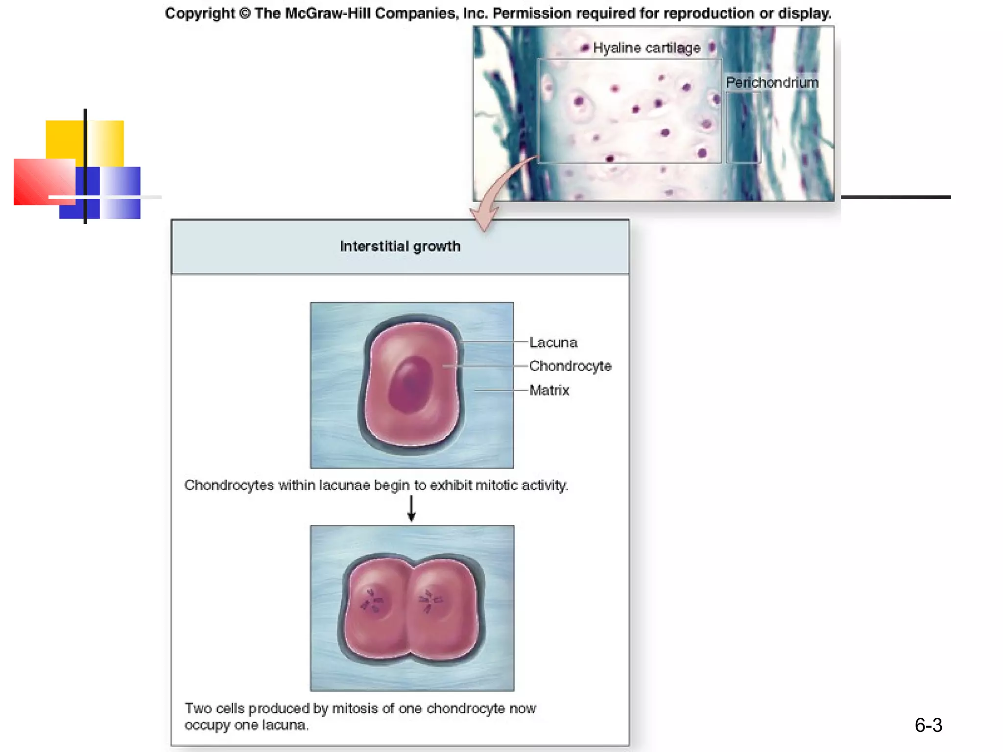
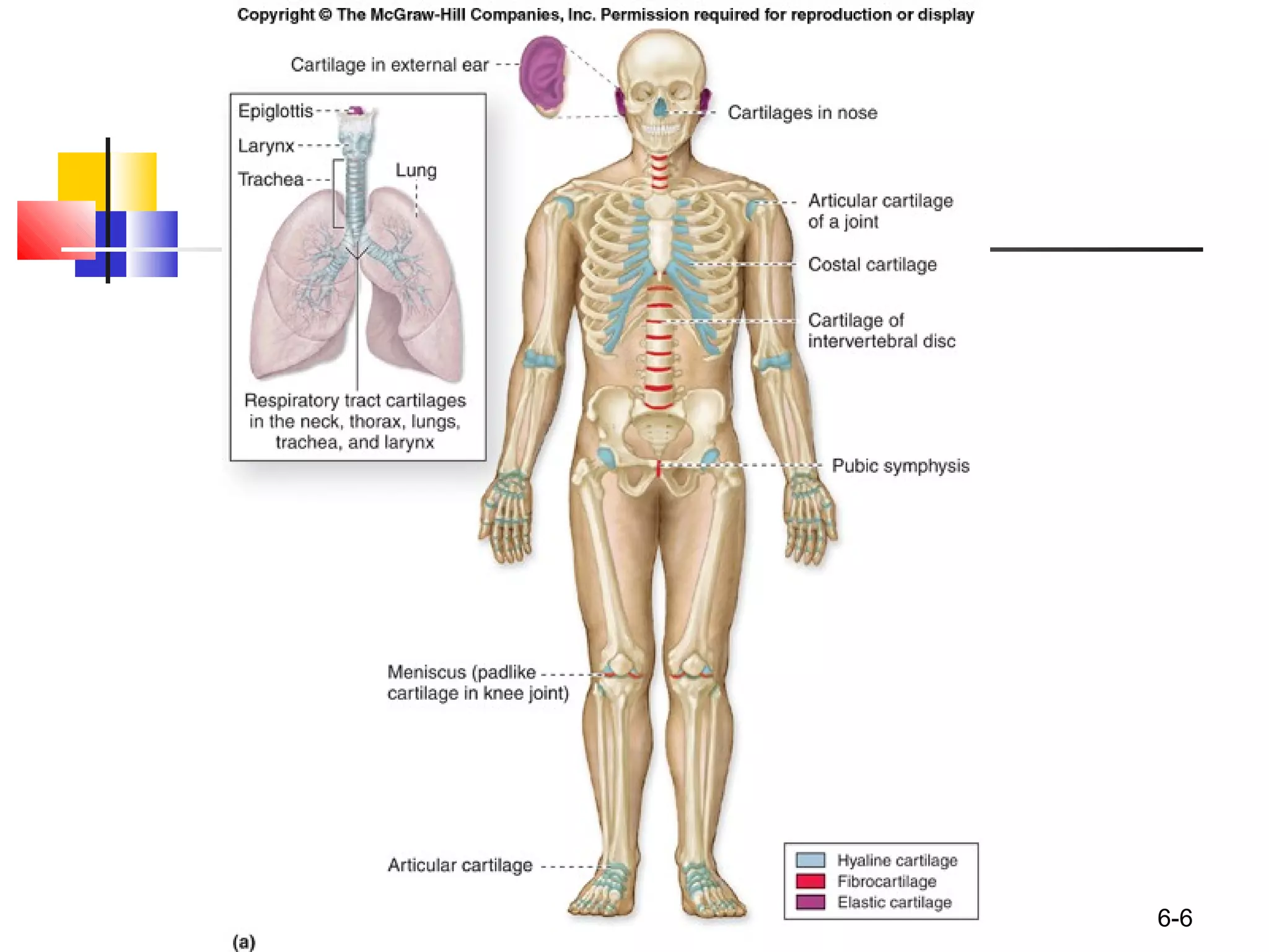
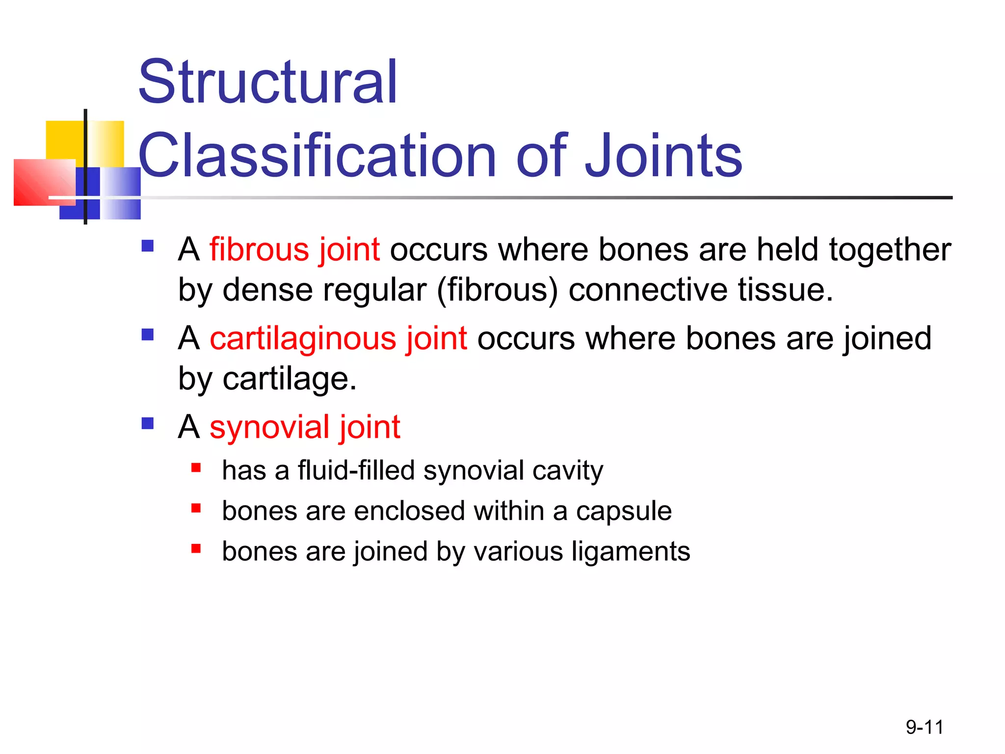
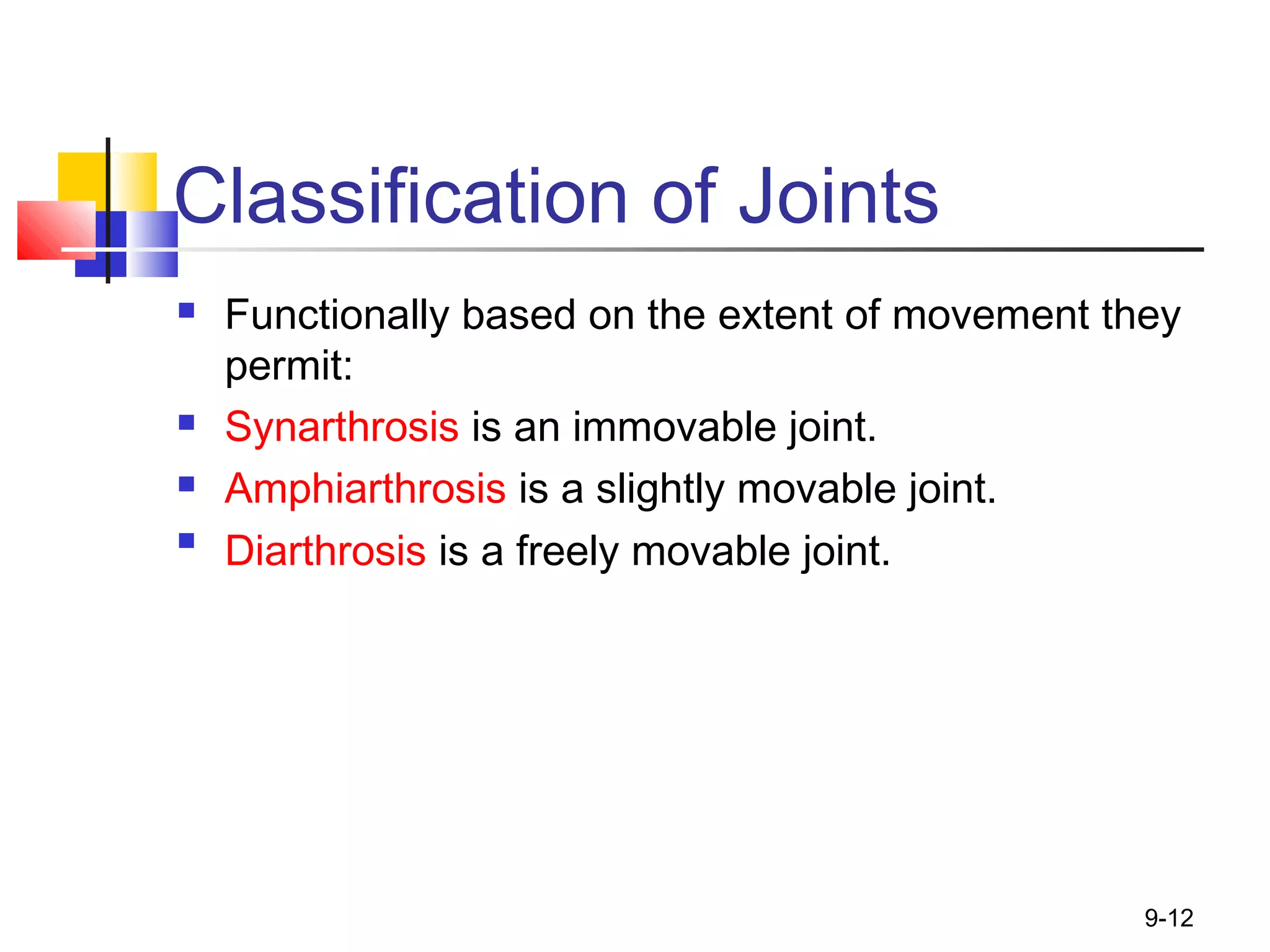
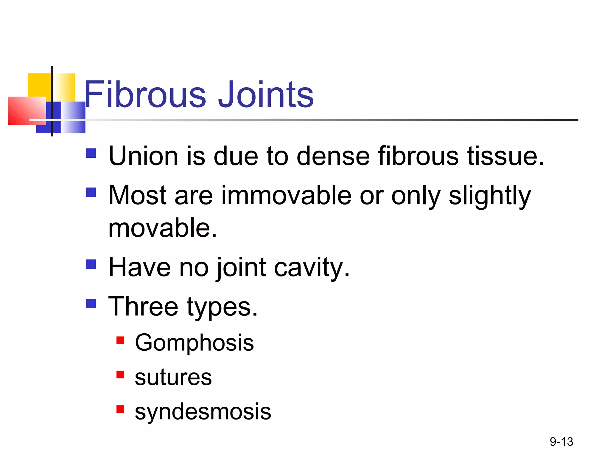

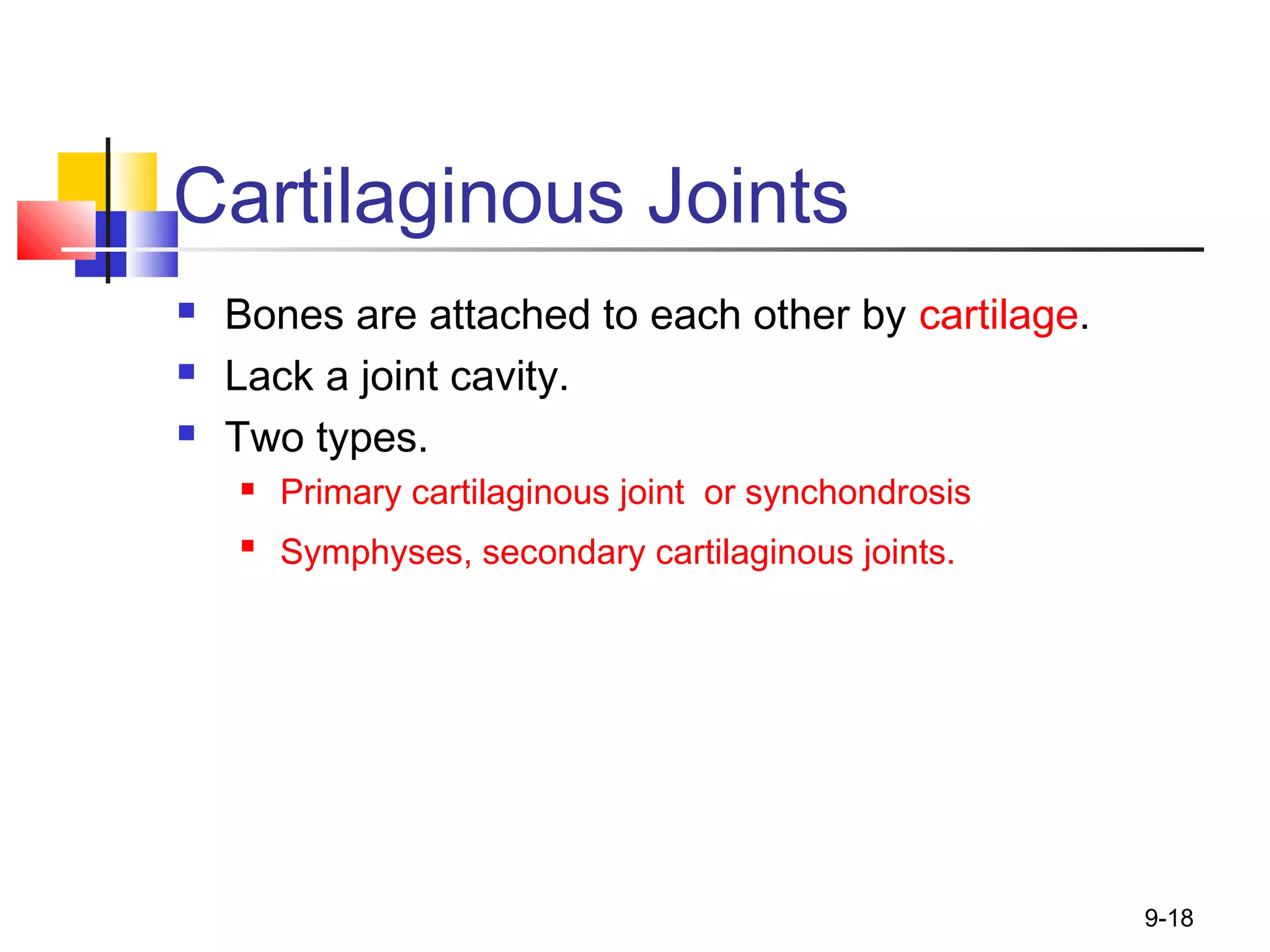


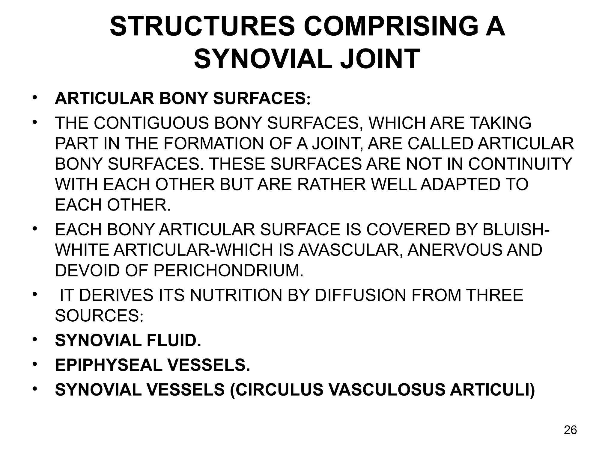
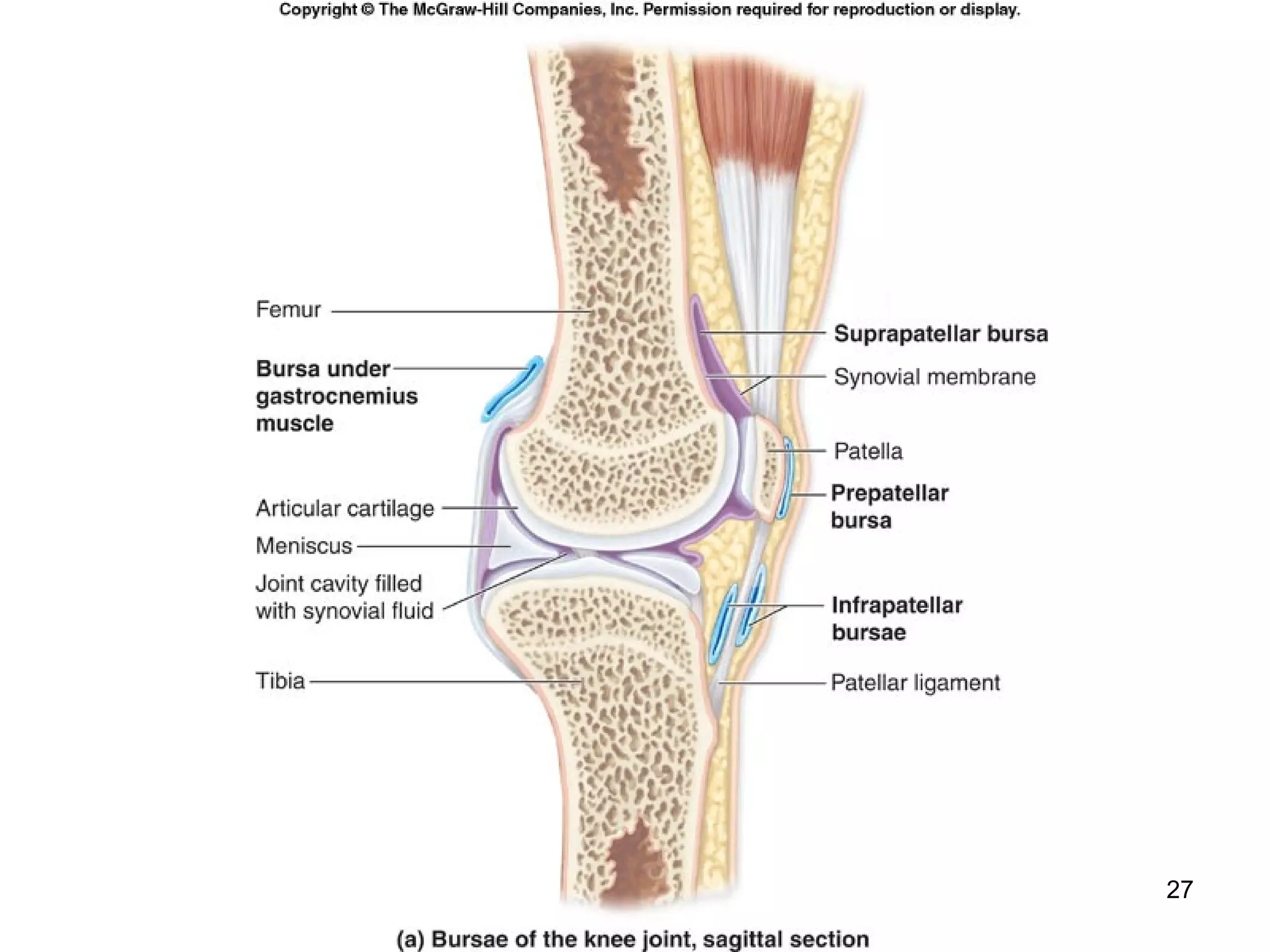


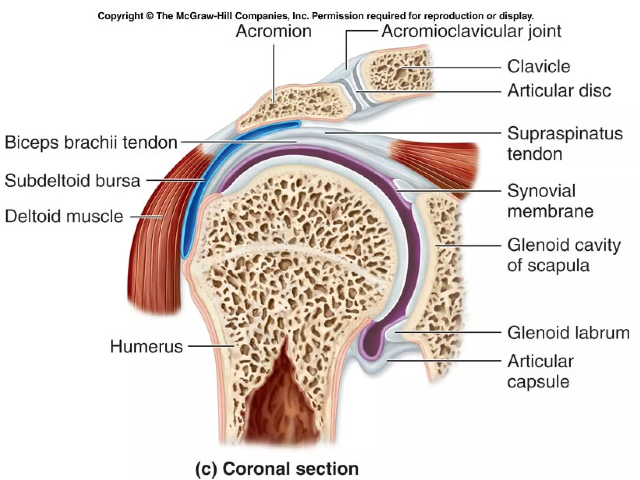
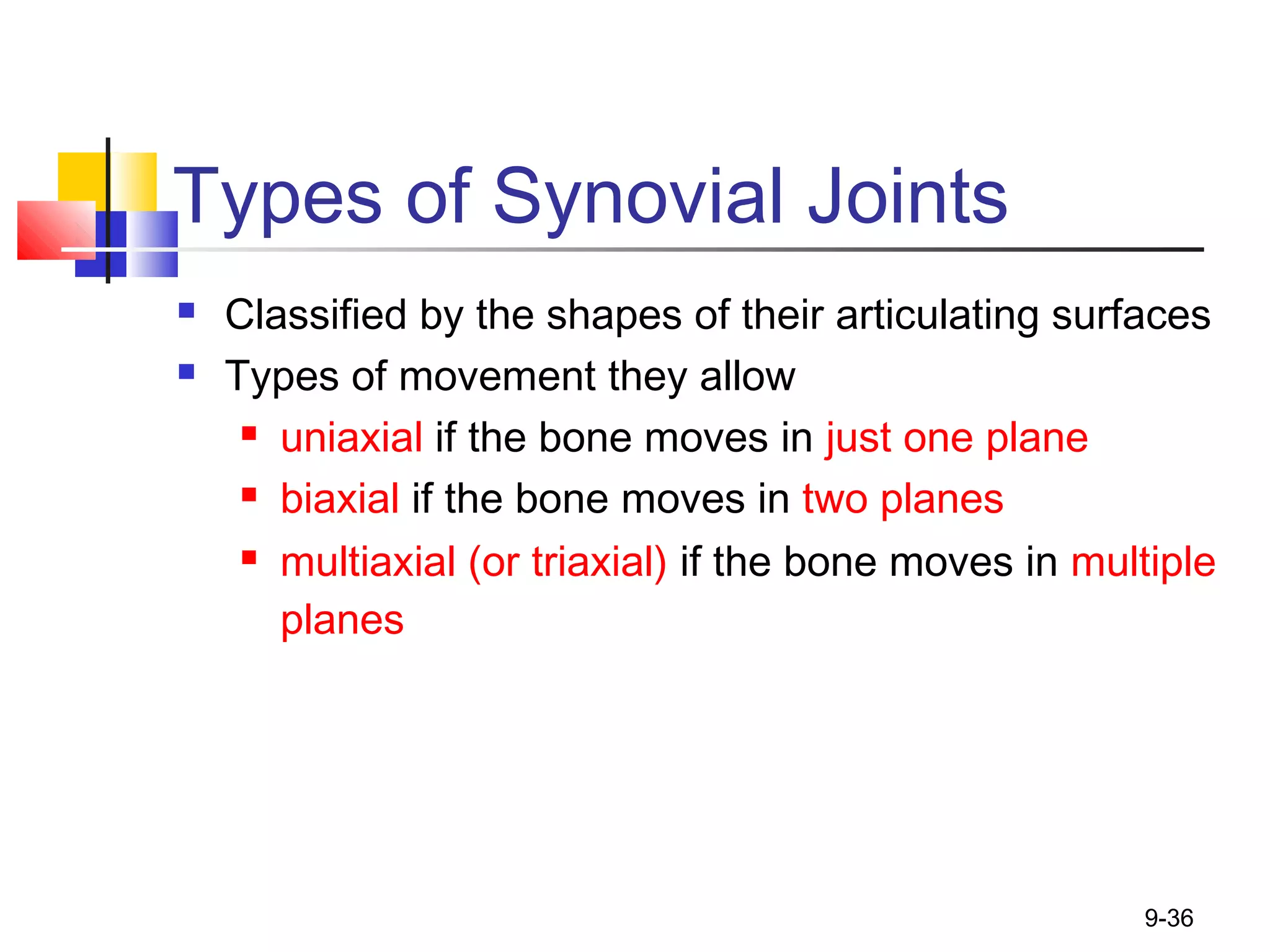
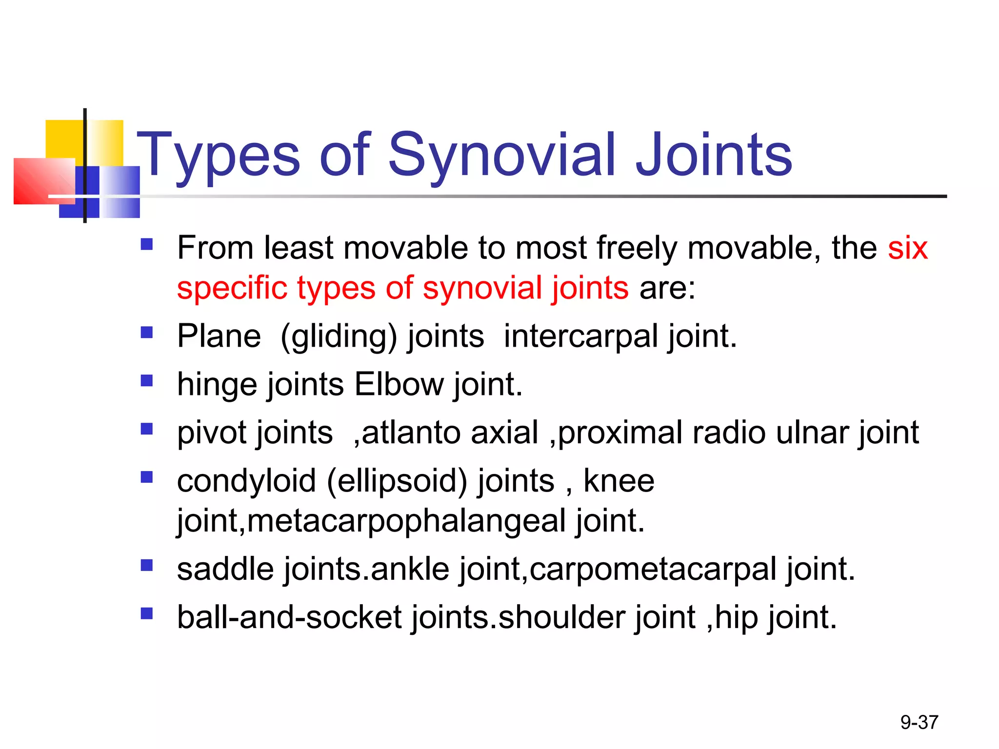
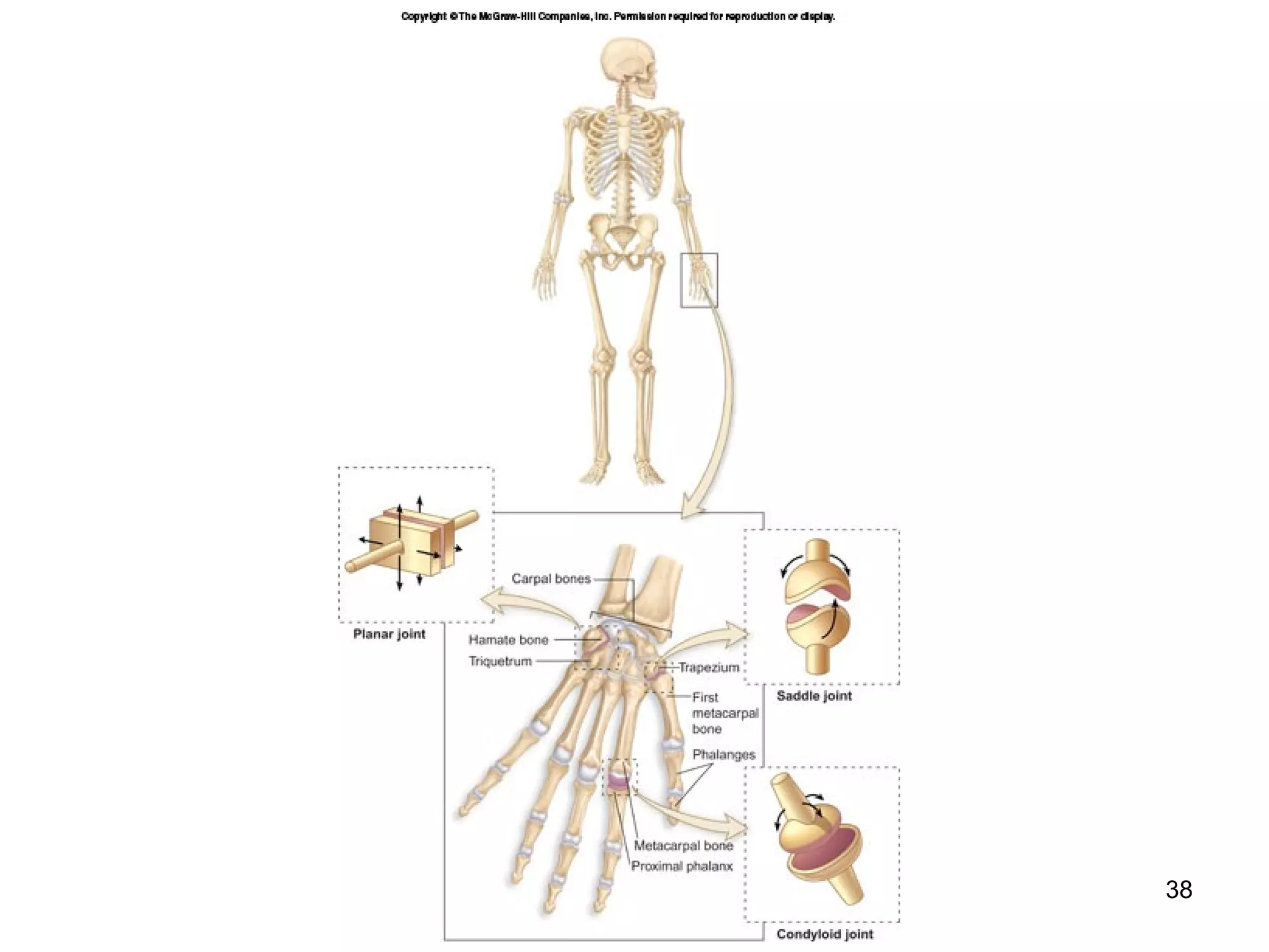
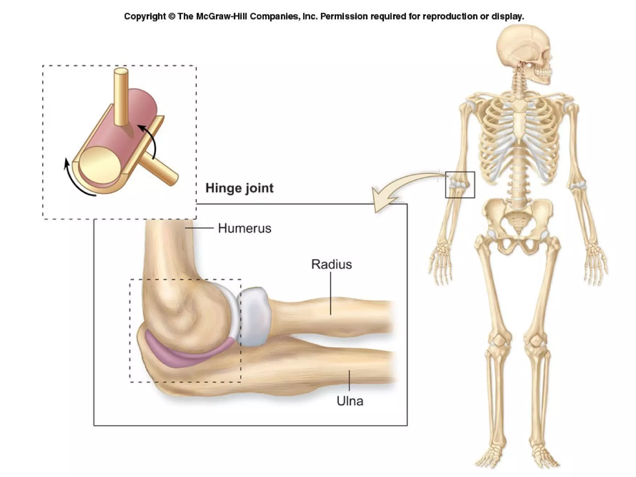
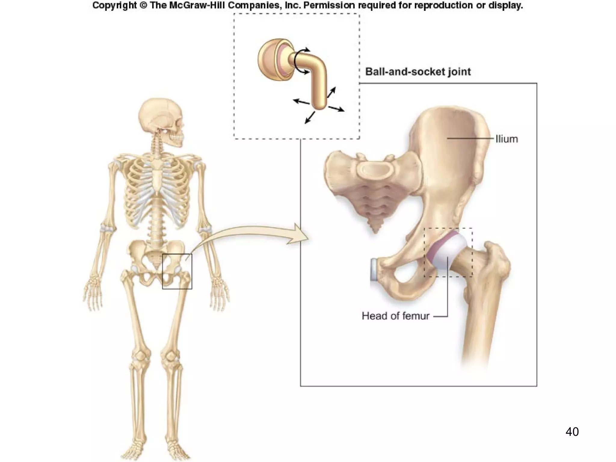
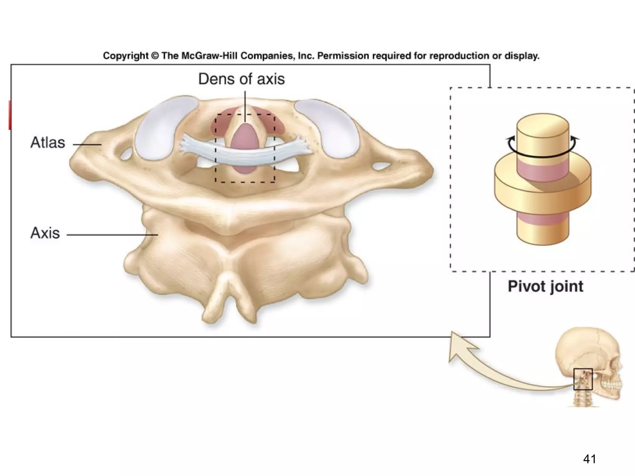


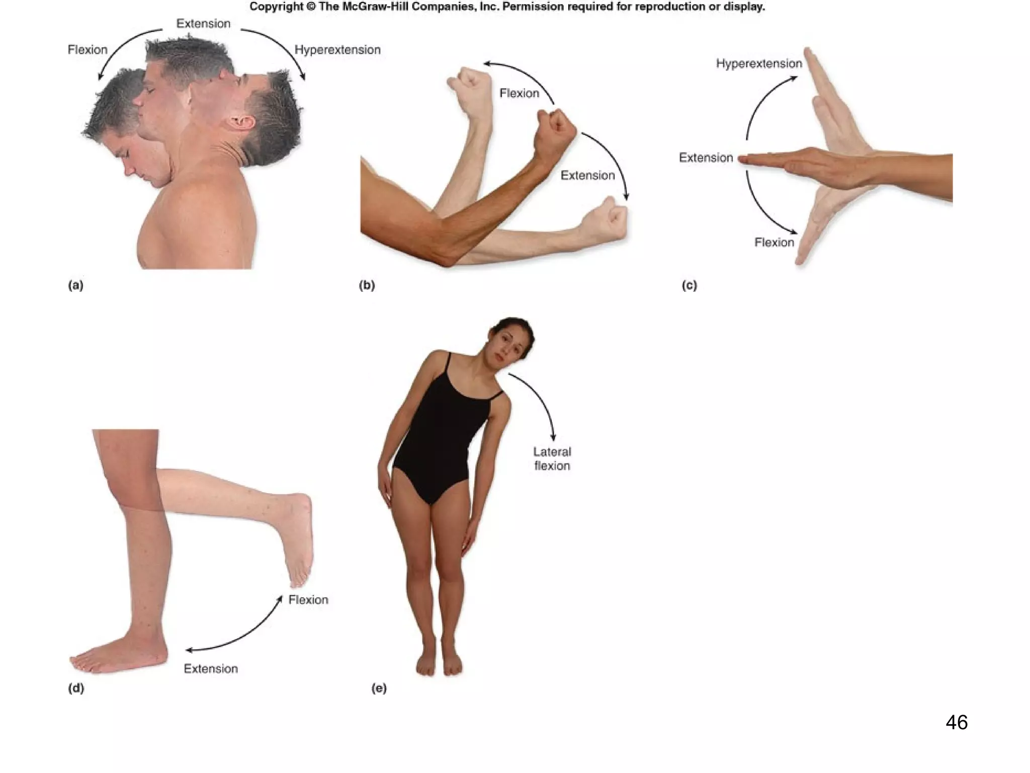
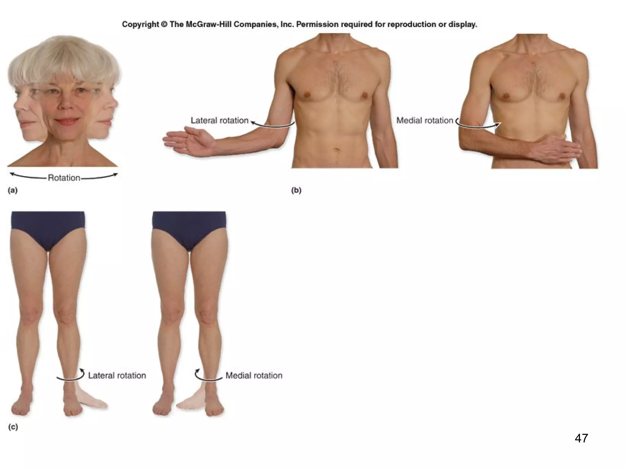
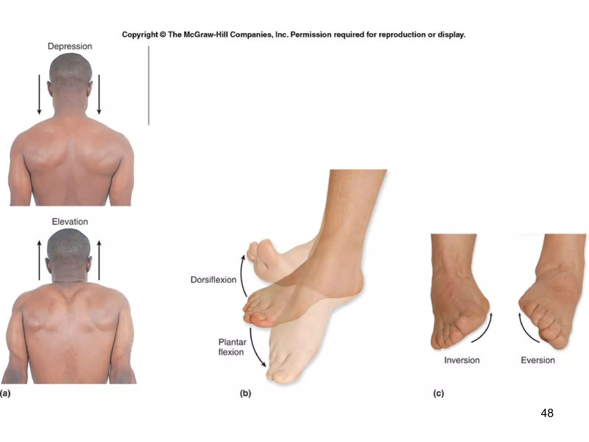
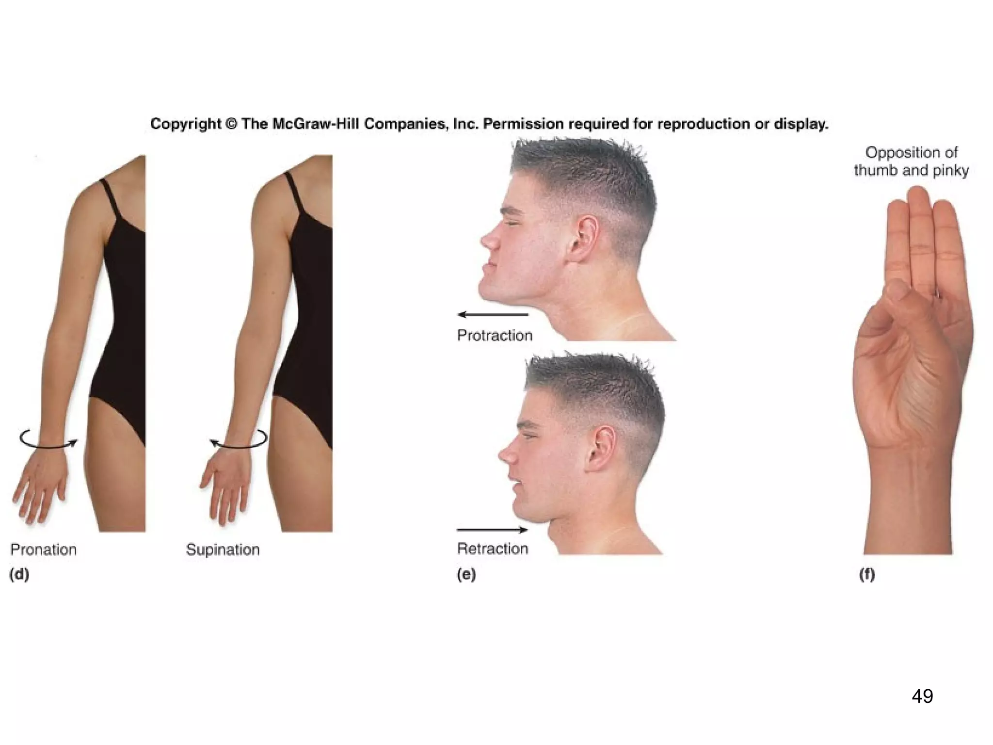

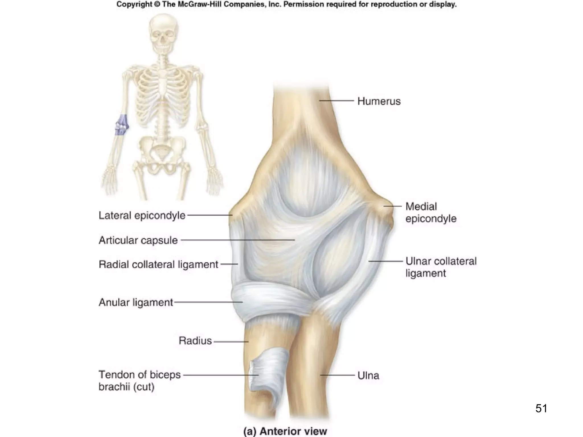
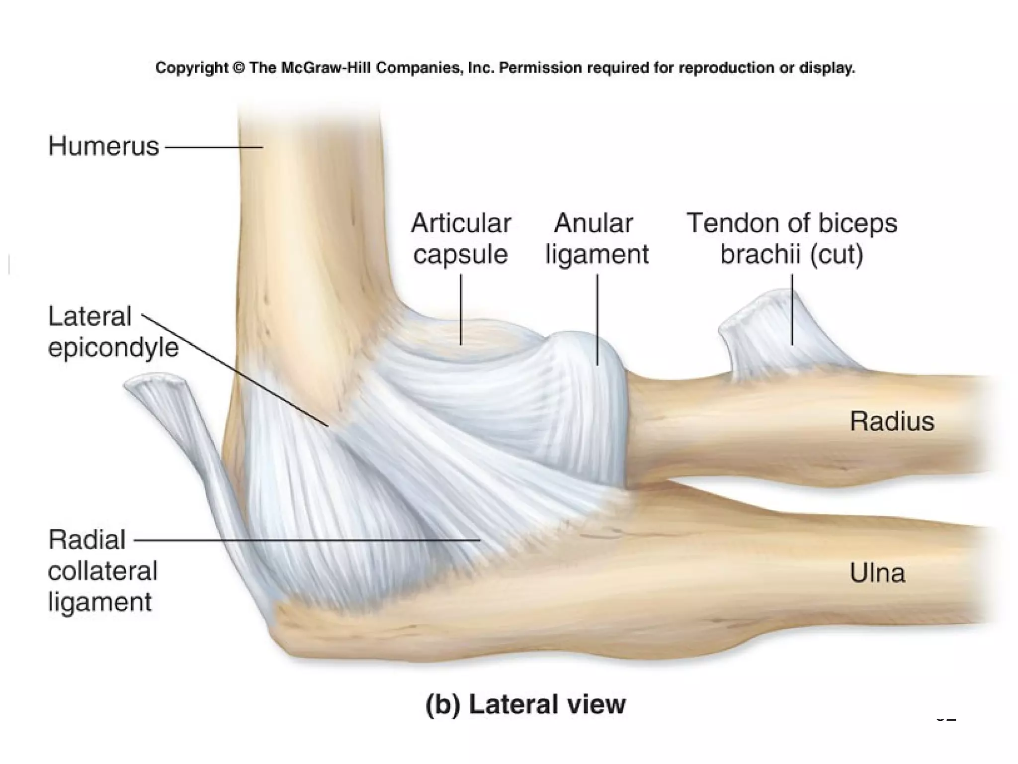
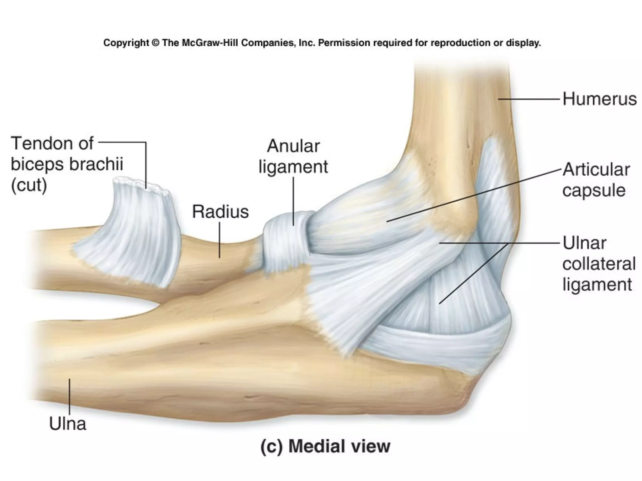


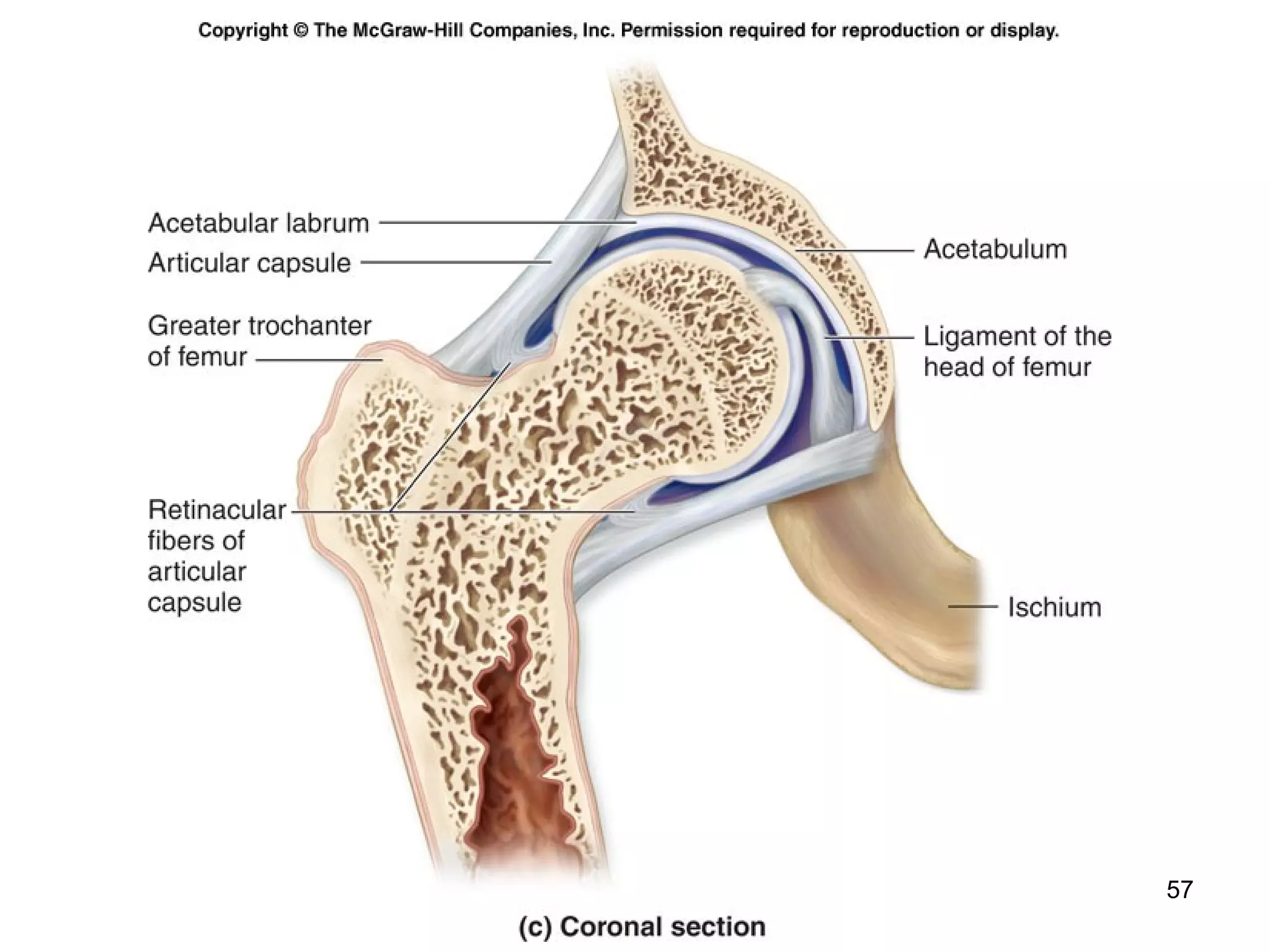


Cartilage is a flexible connective tissue found in joints between bones. There are three main types of cartilage - hyaline, fibrocartilage, and elastic cartilage. Cartilage provides support, acts as a gliding surface in joints, and serves as a template for bone formation. Joints, also called articulations, are places where bones connect. The three major types of joints are fibrous, cartilaginous, and synovial joints. Synovial joints are the most common and include structures like bones, cartilage, synovial fluid, ligaments, and a joint capsule. There are different types of synovial joints based on their shape and movement capabilities, including hinge, ball-and-socket


