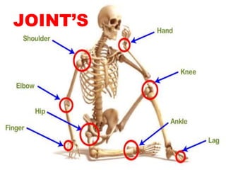
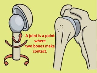
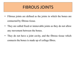
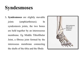
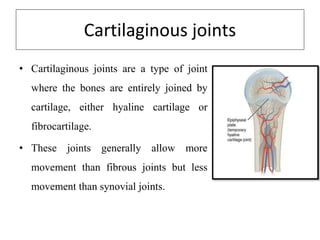
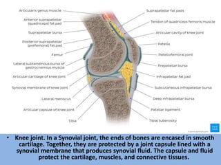
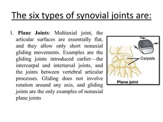
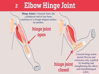
SlideShare a Scribd company logo
JOINT’S
A joint is a point
where
two bones make
contact.
Classification of Joints
Fibrous
Cartilaginous
Synovial
A. Structural Classification
A.
B. Functional Classification
Synarthrosis
Amphiarthrosis
Diarthrosis
IMMOVABLE
SLIGHTLY
IMMOVABLE
FREELY
IMMOVABLE
C. Regional Classification
Skull Type
Vertebral Type
Limb Type
IMMOVABLE
SLIGHTLY
IMMOVABLE
FREELY
IMMOVABLE
FIBROUS JOINTS
• Fibrous joints are defined as the joints in which the bones are
connected by fibrous tissue.
• They are called fixed or immovable joints as they do not allow
any movement between the bones.
• They do not have a joint cavity, and the fibrous tissue which
connects the bones is made up of collage fibres.
These can be grouped in the following
three subtype
1. Sutures are immobile
joints in the cranium. The
plate-like bones of the
skull are slightly mobile at
birth because of the
connective tissue between
them, termed fontanelles.
Conti..
• This initial flexibility allows the infant’s head to get through
the birth canal at delivery and permits the enlargement of the
brain after birth.
• As the skull enlarges, the fontanelles reduce to a narrow layer
of fibrous connective tissue that suture the bony plates
together. Eventually, cranial sutures ossify- the two adjacent
plates fuse to form one bone (termed synostosis).
2. Gomphoses
. Gomphoses are the immobile
joints between the teeth and
their sockets in the mandible
and maxillae. The periodontal
ligament is the fibrous tissue
that connects the tooth to the
socket.
Syndesmoses
3. Syndesmoses are slightly movable
joints (amphiarthroses). In
syndesmosis joints, the two bones
are held together by an interosseous
membrane. Eg Middle Tibiofibular
Joint, a fibrous joint formed by the
interosseus membrane connecting
the shafts of the tibia and the fibula
Cartilaginous joints
• Cartilaginous joints are a type of joint
where the bones are entirely joined by
cartilage, either hyaline cartilage or
fibrocartilage.
• These joints generally allow more
movement than fibrous joints but less
movement than synovial joints.
1.Primary cartilaginous joints
• Primary cartilaginous joints: These cartilaginous joints are
composed entirely of hyaline cartilage and are known as
synchondroses. Most exist between ossification centres of
developing bones and are absent in the mature skeleton, but a
few persist in adults. eg First Sternocostal Joint, between
first rib and manubrium (all other sternocostal joints are plane
synovial joints); Growth plates.
2. secondary cartilaginous joint
• The secondary cartilaginous joint,
also known as symphysis, may
involve either hyaline or
fibrocartilage. These joints are
slightly mobile (amphiarthroses). eg
The pubic symphysis: Intervertebral
discs
Synovial joint
• The primary purpose of the synovial joint is to prevent friction
between the articulating bones of the joint cavity.
• While all synovial joints are diarthroses, the extent of
movement varies among different subtypes and is often limited
by the ligaments that connect the bones.
• Nearly all joints of the limbs and most joints of the body fall
into this class
Synovial joints are the most common
type of joint in the body
• These joints are
termed diarthroses,
meaning they are
freely mobile.
Conti..
• A key structural characteristic for a synovial joint that is not
seen at fibrous or cartilaginous joints is the presence of a joint
cavity.
• The joint cavity contains synovial fluid, secreted by the
synovial membrane (synovium), which lines the articular
capsule.
• This fluid-filled space is the site at which the articulating
surfaces of the bones contact each other.
Conti..
• Hyaline cartilage forms the articular cartilage, covering the
entire articulating surface of each bone.
• The articular cartilage and the synovial membrane are
continuous. A few synovial joints of the body have a
fibrocartilage structure located between the articulating bones.
• This is called an articular disc, which is generally small and
oval-shaped, or a meniscus, which is larger and C-shaped.
• Synovial joints are often further classified by the type of
movements they permit.
• There are six such classifications: hinge (elbow), saddle
(carpometacarpal joint), planar (acromioclavicular joint), pivot
(atlantoaxial joint), condyloid (metacarpophalangeal joint),
and ball and socket (hip joint).
Types of synovial
joint
Features of all Synovial Joints
• Articular capsule with synovial membrane
• Synovial cavity containing synovial fluid
• Hyaline articular cartilage: acts like a Teflon coating over the
bone surface, allowing the articulating bones to move
smoothly against each other without damaging the underlying
bone tissue.
Additional features within some
Synovial Joints
• Fibrocartilage structure located between the articulating bones:
Articular disc, which is generally small and oval-shaped, or a
Meniscus, which is larger and C-shaped. These structures can
serve several functions, depending on the specific joint. Can
serve to smooth the movements between the articulating bones
eg at the temporomandibular joint.
• Intrinsic ligament: fused to or incorporated into the wall of the
articular capsule
• Intracapsular ligament: located inside of the articular capsule.
• Intra-capsular tendons eg. popliteus tendon within the knee
joint
• Intra-articular tendons eg. long head of biceps tendon within
the shoulder joint
• Knee joint. In a Synovial joint, the ends of bones are encased in smooth
cartilage. Together, they are protected by a joint capsule lined with a
synovial membrane that produces synovial fluid. The capsule and fluid
protect the cartilage, muscles, and connective tissues.
The six types of synovial joints are:
1. Plane Joints: Multiaxial joint, the
articular surfaces are essentially flat,
and they allow only short nonaxial
gliding movements. Examples are the
gliding joints introduced earlier—the
intercarpal and intertarsal joints, and
the joints between vertebral articular
processes. Gliding does not involve
rotation around any axis, and gliding
joints are the only examples of nonaxial
plane joints
Hinge Joints: Uniaxial Joint, the
cylindrical end of one bone
conforms to a trough-shaped surface
on another.
Uniaxial hinge joints
permit flexion and
extension only, typified
by bending and
straightening the elbow
and interphalangeal
joints.
2
3. Pivot Joints: Uniaxial Joint, the
rounded end of one bone conforms
to a “sleeve” ring composed of
bone of another. The only
movement allowed is uniaxial
rotation of one bone around its
own long axis. An example is the
joint between the atlas and dens of
the axis, which allows you to
move your head from side to side
to indicate “no.” Another is the
proximal radioulnar joint, where
the head of the radius rotates
within a ringlike ligament secured
to the ulna.
4.
The examples of saddle
joints in the body are the
carpometacarpal joints of
the thumbs.
5.
Movements allowed by
these joints are clearly
demonstrated by
twiddling your thumbs.
These joints are multiaxial and the most
freely moving synovial joints.
6..
Nerve supply of synovial joint
• Sensory and autonomic fibers innervate synovial joints:
• The autonomic nerves are vasomotor in function,
controlling the dilation or constriction of blood vessels.
• The sensory nerves of the articular capsule
and ligaments (articular nerves)
provide proprioceptive feedback from Ruffini endings
and Pacinian corpuscles. Proprioception of the joint
permits reflex control of posture, locomotion, and
movement. Free nerve endings convey pain sensation that
is diffuse and poorly localized. The articular cartilage has
no nerve supply.
Blood Supply
• Synovial joints receive vascular
supply through a rich anastomosis
of arteries extending from either
side of the joint ie the
periarticular plexus. Some vessels
penetrate the fibrous capsule to
form a rich plexus deeper in the
synovial membrane.
Clinical Anatomy
• Arthritis – inflammation that causes stiffness and pain in the
joints eg rheumatoid arthritis or gout, or degeneration
(osteoarthritis)
• Bursitis – inflammation of the bursae (fluid-filled sacs that
cushion and pad bones)
• Tendonitis – inflammation, irritation and swelling of a tendon
that is attached to the joint.
• Injury – including strain or sprain of a ligament or
nearby tendon or muscle, or bone fracture
• Dislocation of joint
• Sprain
• Neuropathic joint

More Related Content

What's hot

Fibrous joints
Fibrous jointsFibrous joints
Fibrous joints
Nabeel Althaf
 
anatomy of joints
anatomy of jointsanatomy of joints
anatomy of joints
dr.supriti verma bhatnagar
 
ANATOMY & PHYSIOLOGY OF JOINT
ANATOMY & PHYSIOLOGY OF JOINT ANATOMY & PHYSIOLOGY OF JOINT
ANATOMY & PHYSIOLOGY OF JOINT
Tun Tan Cheng Lock College Of Nursing
 
Anatomical movement - terms & medical information
Anatomical movement - terms & medical information Anatomical movement - terms & medical information
Anatomical movement - terms & medical information
martinshaji
 
Lower limb bones-joints-muscles-Dr.B.B.Gosai
Lower limb bones-joints-muscles-Dr.B.B.GosaiLower limb bones-joints-muscles-Dr.B.B.Gosai
Lower limb bones-joints-muscles-Dr.B.B.Gosai
Dr.B.B. Gosai
 
Popliteal Fossa.pptx
Popliteal Fossa.pptxPopliteal Fossa.pptx
Popliteal Fossa.pptx
ABHIJIT BHOYAR
 
Joints of upper limb
Joints of upper limb Joints of upper limb
Joints of upper limb
sadhamhussain52
 
Muscles of the back
Muscles of the backMuscles of the back
Muscles of the back
HenanKhan1
 
General anatomy of muscles
General anatomy of muscles General anatomy of muscles
General anatomy of muscles
ritika patel
 
Shoulder girdle
Shoulder girdleShoulder girdle
Shoulder girdle
Mustafa Qamar
 
Radioulnar joints
Radioulnar jointsRadioulnar joints
Radioulnar joints
Idris Siddiqui
 
General Anatomy of Joints
General Anatomy of JointsGeneral Anatomy of Joints
General Anatomy of Joints
MuhammadAteeq31
 
Joints anatomy and physiology
Joints anatomy and physiologyJoints anatomy and physiology
Joints anatomy and physiology
Manthan Kapadiya
 
SKELETAL SYSTEM ANATOMY
SKELETAL SYSTEM ANATOMYSKELETAL SYSTEM ANATOMY
SKELETAL SYSTEM ANATOMY
SUDESHNA BANERJEE
 
Joints and movements
Joints  and movementsJoints  and movements
Introduction,structure,funtions and nutrition of hyaline cartilage
Introduction,structure,funtions and nutrition of hyaline cartilageIntroduction,structure,funtions and nutrition of hyaline cartilage
Introduction,structure,funtions and nutrition of hyaline cartilage
Pramod Yspam
 
Joints
JointsJoints
Skeletal system.pptnursing
Skeletal system.pptnursingSkeletal system.pptnursing
Skeletal system.pptnursing
Jay Patel
 
Layers of muscles of foot
Layers of muscles of footLayers of muscles of foot
Layers of muscles of foot
drasarma1947
 
Joints
JointsJoints

What's hot (20)

Fibrous joints
Fibrous jointsFibrous joints
Fibrous joints
 
anatomy of joints
anatomy of jointsanatomy of joints
anatomy of joints
 
ANATOMY & PHYSIOLOGY OF JOINT
ANATOMY & PHYSIOLOGY OF JOINT ANATOMY & PHYSIOLOGY OF JOINT
ANATOMY & PHYSIOLOGY OF JOINT
 
Anatomical movement - terms & medical information
Anatomical movement - terms & medical information Anatomical movement - terms & medical information
Anatomical movement - terms & medical information
 
Lower limb bones-joints-muscles-Dr.B.B.Gosai
Lower limb bones-joints-muscles-Dr.B.B.GosaiLower limb bones-joints-muscles-Dr.B.B.Gosai
Lower limb bones-joints-muscles-Dr.B.B.Gosai
 
Popliteal Fossa.pptx
Popliteal Fossa.pptxPopliteal Fossa.pptx
Popliteal Fossa.pptx
 
Joints of upper limb
Joints of upper limb Joints of upper limb
Joints of upper limb
 
Muscles of the back
Muscles of the backMuscles of the back
Muscles of the back
 
General anatomy of muscles
General anatomy of muscles General anatomy of muscles
General anatomy of muscles
 
Shoulder girdle
Shoulder girdleShoulder girdle
Shoulder girdle
 
Radioulnar joints
Radioulnar jointsRadioulnar joints
Radioulnar joints
 
General Anatomy of Joints
General Anatomy of JointsGeneral Anatomy of Joints
General Anatomy of Joints
 
Joints anatomy and physiology
Joints anatomy and physiologyJoints anatomy and physiology
Joints anatomy and physiology
 
SKELETAL SYSTEM ANATOMY
SKELETAL SYSTEM ANATOMYSKELETAL SYSTEM ANATOMY
SKELETAL SYSTEM ANATOMY
 
Joints and movements
Joints  and movementsJoints  and movements
Joints and movements
 
Introduction,structure,funtions and nutrition of hyaline cartilage
Introduction,structure,funtions and nutrition of hyaline cartilageIntroduction,structure,funtions and nutrition of hyaline cartilage
Introduction,structure,funtions and nutrition of hyaline cartilage
 
Joints
JointsJoints
Joints
 
Skeletal system.pptnursing
Skeletal system.pptnursingSkeletal system.pptnursing
Skeletal system.pptnursing
 
Layers of muscles of foot
Layers of muscles of footLayers of muscles of foot
Layers of muscles of foot
 
Joints
JointsJoints
Joints
 

Similar to Joints_Skeleton system.pptx

THE JOINTS.pptx
THE JOINTS.pptxTHE JOINTS.pptx
THE JOINTS.pptx
PayalSakhija
 
Share_784_Joints.pdf
Share_784_Joints.pdfShare_784_Joints.pdf
Share_784_Joints.pdf
NITHYA718085
 
Lec-5-Sketetal System- Joints.pdf
Lec-5-Sketetal System- Joints.pdfLec-5-Sketetal System- Joints.pdf
Lec-5-Sketetal System- Joints.pdf
Anand Gowda
 
Joints
JointsJoints
Joints
SUSHIL BHATT
 
GENERAL ANATOMY OF SKELETAL SYSTEM-JOINTS .pptx
GENERAL ANATOMY OF SKELETAL SYSTEM-JOINTS .pptxGENERAL ANATOMY OF SKELETAL SYSTEM-JOINTS .pptx
GENERAL ANATOMY OF SKELETAL SYSTEM-JOINTS .pptx
MuthuRoshni1
 
JOINTS.pptx
JOINTS.pptxJOINTS.pptx
Lecture 1. (introduction to joint)
Lecture 1. (introduction to joint)Lecture 1. (introduction to joint)
Lecture 1. (introduction to joint)
Ayub Abdi
 
JOINTS.pptx
JOINTS.pptxJOINTS.pptx
JOINTS.pptx
PranaliChandurkar2
 
2.2 joint.pptx
2.2  joint.pptx2.2  joint.pptx
2.2 joint.pptx
Keval80
 
Joints
JointsJoints
Joints
Zainab&Sons
 
L.19. JOINTS.pdf
L.19. JOINTS.pdfL.19. JOINTS.pdf
L.19. JOINTS.pdf
JuliusKauki1
 
JOINTS .ppt
JOINTS   .pptJOINTS   .ppt
BSN 4th SEMESTER_Mixed notes_unit 5 joints.ppt
BSN 4th SEMESTER_Mixed notes_unit 5 joints.pptBSN 4th SEMESTER_Mixed notes_unit 5 joints.ppt
BSN 4th SEMESTER_Mixed notes_unit 5 joints.ppt
AbduLLahKhan621805
 
joints and its classification different types of joints .pptx
joints and its classification   different types of joints .pptxjoints and its classification   different types of joints .pptx
joints and its classification different types of joints .pptx
ShraddhaBansal7
 
Cartilage & joints
Cartilage & jointsCartilage & joints
Cartilage & joints
Dr. Noura El Tahawy
 
Basic ap chapter 9 powerpoint 2017
Basic ap chapter 9 powerpoint 2017Basic ap chapter 9 powerpoint 2017
Basic ap chapter 9 powerpoint 2017
Kathy Richards
 
joints of human beings with different types
joints of human beings with different typesjoints of human beings with different types
joints of human beings with different types
g8248418302
 
Skeletal physiology
Skeletal physiologySkeletal physiology
Skeletal physiologytd0297bn
 
joints.pdf
joints.pdfjoints.pdf

Similar to Joints_Skeleton system.pptx (20)

THE JOINTS.pptx
THE JOINTS.pptxTHE JOINTS.pptx
THE JOINTS.pptx
 
Share_784_Joints.pdf
Share_784_Joints.pdfShare_784_Joints.pdf
Share_784_Joints.pdf
 
Lec-5-Sketetal System- Joints.pdf
Lec-5-Sketetal System- Joints.pdfLec-5-Sketetal System- Joints.pdf
Lec-5-Sketetal System- Joints.pdf
 
Joints
JointsJoints
Joints
 
GENERAL ANATOMY OF SKELETAL SYSTEM-JOINTS .pptx
GENERAL ANATOMY OF SKELETAL SYSTEM-JOINTS .pptxGENERAL ANATOMY OF SKELETAL SYSTEM-JOINTS .pptx
GENERAL ANATOMY OF SKELETAL SYSTEM-JOINTS .pptx
 
JOINt.pptx
JOINt.pptxJOINt.pptx
JOINt.pptx
 
JOINTS.pptx
JOINTS.pptxJOINTS.pptx
JOINTS.pptx
 
Lecture 1. (introduction to joint)
Lecture 1. (introduction to joint)Lecture 1. (introduction to joint)
Lecture 1. (introduction to joint)
 
JOINTS.pptx
JOINTS.pptxJOINTS.pptx
JOINTS.pptx
 
2.2 joint.pptx
2.2  joint.pptx2.2  joint.pptx
2.2 joint.pptx
 
Joints
JointsJoints
Joints
 
L.19. JOINTS.pdf
L.19. JOINTS.pdfL.19. JOINTS.pdf
L.19. JOINTS.pdf
 
JOINTS .ppt
JOINTS   .pptJOINTS   .ppt
JOINTS .ppt
 
BSN 4th SEMESTER_Mixed notes_unit 5 joints.ppt
BSN 4th SEMESTER_Mixed notes_unit 5 joints.pptBSN 4th SEMESTER_Mixed notes_unit 5 joints.ppt
BSN 4th SEMESTER_Mixed notes_unit 5 joints.ppt
 
joints and its classification different types of joints .pptx
joints and its classification   different types of joints .pptxjoints and its classification   different types of joints .pptx
joints and its classification different types of joints .pptx
 
Cartilage & joints
Cartilage & jointsCartilage & joints
Cartilage & joints
 
Basic ap chapter 9 powerpoint 2017
Basic ap chapter 9 powerpoint 2017Basic ap chapter 9 powerpoint 2017
Basic ap chapter 9 powerpoint 2017
 
joints of human beings with different types
joints of human beings with different typesjoints of human beings with different types
joints of human beings with different types
 
Skeletal physiology
Skeletal physiologySkeletal physiology
Skeletal physiology
 
joints.pdf
joints.pdfjoints.pdf
joints.pdf
 

More from ABHIJIT BHOYAR

Group Dynamic(presentation for nursing management)
Group Dynamic(presentation for nursing management)Group Dynamic(presentation for nursing management)
Group Dynamic(presentation for nursing management)
ABHIJIT BHOYAR
 
Discipline. pptx (Nursing management) nursing
Discipline. pptx (Nursing management) nursingDiscipline. pptx (Nursing management) nursing
Discipline. pptx (Nursing management) nursing
ABHIJIT BHOYAR
 
Diagnostic importance of enzymes.pptx
Diagnostic importance of enzymes.pptxDiagnostic importance of enzymes.pptx
Diagnostic importance of enzymes.pptx
ABHIJIT BHOYAR
 
Isoenzyme.pptx
Isoenzyme.pptxIsoenzyme.pptx
Isoenzyme.pptx
ABHIJIT BHOYAR
 
Enzymes.pptx
Enzymes.pptxEnzymes.pptx
Enzymes.pptx
ABHIJIT BHOYAR
 
Atherosclerosis. pptx
Atherosclerosis. pptxAtherosclerosis. pptx
Atherosclerosis. pptx
ABHIJIT BHOYAR
 
Lipoproteins & Lipid Profile .pptx
Lipoproteins & Lipid Profile .pptxLipoproteins & Lipid Profile .pptx
Lipoproteins & Lipid Profile .pptx
ABHIJIT BHOYAR
 
Ketone Bodies.pptx
Ketone Bodies.pptxKetone Bodies.pptx
Ketone Bodies.pptx
ABHIJIT BHOYAR
 
Cholesterol.pptx
Cholesterol.pptxCholesterol.pptx
Cholesterol.pptx
ABHIJIT BHOYAR
 
LIPIDS- Metabolism of Lipids.pptx
LIPIDS- Metabolism of Lipids.pptxLIPIDS- Metabolism of Lipids.pptx
LIPIDS- Metabolism of Lipids.pptx
ABHIJIT BHOYAR
 
LIPIDS-Digestion and absorption of Lipids.pptx
LIPIDS-Digestion and absorption of Lipids.pptxLIPIDS-Digestion and absorption of Lipids.pptx
LIPIDS-Digestion and absorption of Lipids.pptx
ABHIJIT BHOYAR
 
Lipids Classification, Essential.pptx
Lipids Classification, Essential.pptxLipids Classification, Essential.pptx
Lipids Classification, Essential.pptx
ABHIJIT BHOYAR
 
2. LIPIDS (Fatty Acids).pptx
2. LIPIDS (Fatty Acids).pptx2. LIPIDS (Fatty Acids).pptx
2. LIPIDS (Fatty Acids).pptx
ABHIJIT BHOYAR
 
lipids, Def. Classification, Function.pptx
lipids, Def. Classification, Function.pptxlipids, Def. Classification, Function.pptx
lipids, Def. Classification, Function.pptx
ABHIJIT BHOYAR
 
UTERUS_Nursing.pptx
UTERUS_Nursing.pptxUTERUS_Nursing.pptx
UTERUS_Nursing.pptx
ABHIJIT BHOYAR
 
Kidney_Nursing.pptx
Kidney_Nursing.pptxKidney_Nursing.pptx
Kidney_Nursing.pptx
ABHIJIT BHOYAR
 
Liver_Nursing.pptx
Liver_Nursing.pptxLiver_Nursing.pptx
Liver_Nursing.pptx
ABHIJIT BHOYAR
 
Pancreas_Nursing.pptx
Pancreas_Nursing.pptxPancreas_Nursing.pptx
Pancreas_Nursing.pptx
ABHIJIT BHOYAR
 
Spleen.pptx
Spleen.pptxSpleen.pptx
Spleen.pptx
ABHIJIT BHOYAR
 
Femoral triangle.
Femoral triangle.Femoral triangle.
Femoral triangle.
ABHIJIT BHOYAR
 

More from ABHIJIT BHOYAR (20)

Group Dynamic(presentation for nursing management)
Group Dynamic(presentation for nursing management)Group Dynamic(presentation for nursing management)
Group Dynamic(presentation for nursing management)
 
Discipline. pptx (Nursing management) nursing
Discipline. pptx (Nursing management) nursingDiscipline. pptx (Nursing management) nursing
Discipline. pptx (Nursing management) nursing
 
Diagnostic importance of enzymes.pptx
Diagnostic importance of enzymes.pptxDiagnostic importance of enzymes.pptx
Diagnostic importance of enzymes.pptx
 
Isoenzyme.pptx
Isoenzyme.pptxIsoenzyme.pptx
Isoenzyme.pptx
 
Enzymes.pptx
Enzymes.pptxEnzymes.pptx
Enzymes.pptx
 
Atherosclerosis. pptx
Atherosclerosis. pptxAtherosclerosis. pptx
Atherosclerosis. pptx
 
Lipoproteins & Lipid Profile .pptx
Lipoproteins & Lipid Profile .pptxLipoproteins & Lipid Profile .pptx
Lipoproteins & Lipid Profile .pptx
 
Ketone Bodies.pptx
Ketone Bodies.pptxKetone Bodies.pptx
Ketone Bodies.pptx
 
Cholesterol.pptx
Cholesterol.pptxCholesterol.pptx
Cholesterol.pptx
 
LIPIDS- Metabolism of Lipids.pptx
LIPIDS- Metabolism of Lipids.pptxLIPIDS- Metabolism of Lipids.pptx
LIPIDS- Metabolism of Lipids.pptx
 
LIPIDS-Digestion and absorption of Lipids.pptx
LIPIDS-Digestion and absorption of Lipids.pptxLIPIDS-Digestion and absorption of Lipids.pptx
LIPIDS-Digestion and absorption of Lipids.pptx
 
Lipids Classification, Essential.pptx
Lipids Classification, Essential.pptxLipids Classification, Essential.pptx
Lipids Classification, Essential.pptx
 
2. LIPIDS (Fatty Acids).pptx
2. LIPIDS (Fatty Acids).pptx2. LIPIDS (Fatty Acids).pptx
2. LIPIDS (Fatty Acids).pptx
 
lipids, Def. Classification, Function.pptx
lipids, Def. Classification, Function.pptxlipids, Def. Classification, Function.pptx
lipids, Def. Classification, Function.pptx
 
UTERUS_Nursing.pptx
UTERUS_Nursing.pptxUTERUS_Nursing.pptx
UTERUS_Nursing.pptx
 
Kidney_Nursing.pptx
Kidney_Nursing.pptxKidney_Nursing.pptx
Kidney_Nursing.pptx
 
Liver_Nursing.pptx
Liver_Nursing.pptxLiver_Nursing.pptx
Liver_Nursing.pptx
 
Pancreas_Nursing.pptx
Pancreas_Nursing.pptxPancreas_Nursing.pptx
Pancreas_Nursing.pptx
 
Spleen.pptx
Spleen.pptxSpleen.pptx
Spleen.pptx
 
Femoral triangle.
Femoral triangle.Femoral triangle.
Femoral triangle.
 

Recently uploaded

678020731-Sumas-y-Restas-Para-Colorear.pdf
678020731-Sumas-y-Restas-Para-Colorear.pdf678020731-Sumas-y-Restas-Para-Colorear.pdf
678020731-Sumas-y-Restas-Para-Colorear.pdf
CarlosHernanMontoyab2
 
Unit 8 - Information and Communication Technology (Paper I).pdf
Unit 8 - Information and Communication Technology (Paper I).pdfUnit 8 - Information and Communication Technology (Paper I).pdf
Unit 8 - Information and Communication Technology (Paper I).pdf
Thiyagu K
 
Home assignment II on Spectroscopy 2024 Answers.pdf
Home assignment II on Spectroscopy 2024 Answers.pdfHome assignment II on Spectroscopy 2024 Answers.pdf
Home assignment II on Spectroscopy 2024 Answers.pdf
Tamralipta Mahavidyalaya
 
2024.06.01 Introducing a competency framework for languag learning materials ...
2024.06.01 Introducing a competency framework for languag learning materials ...2024.06.01 Introducing a competency framework for languag learning materials ...
2024.06.01 Introducing a competency framework for languag learning materials ...
Sandy Millin
 
BÀI TẬP BỔ TRỢ TIẾNG ANH GLOBAL SUCCESS LỚP 3 - CẢ NĂM (CÓ FILE NGHE VÀ ĐÁP Á...
BÀI TẬP BỔ TRỢ TIẾNG ANH GLOBAL SUCCESS LỚP 3 - CẢ NĂM (CÓ FILE NGHE VÀ ĐÁP Á...BÀI TẬP BỔ TRỢ TIẾNG ANH GLOBAL SUCCESS LỚP 3 - CẢ NĂM (CÓ FILE NGHE VÀ ĐÁP Á...
BÀI TẬP BỔ TRỢ TIẾNG ANH GLOBAL SUCCESS LỚP 3 - CẢ NĂM (CÓ FILE NGHE VÀ ĐÁP Á...
Nguyen Thanh Tu Collection
 
1.4 modern child centered education - mahatma gandhi-2.pptx
1.4 modern child centered education - mahatma gandhi-2.pptx1.4 modern child centered education - mahatma gandhi-2.pptx
1.4 modern child centered education - mahatma gandhi-2.pptx
JosvitaDsouza2
 
Supporting (UKRI) OA monographs at Salford.pptx
Supporting (UKRI) OA monographs at Salford.pptxSupporting (UKRI) OA monographs at Salford.pptx
Supporting (UKRI) OA monographs at Salford.pptx
Jisc
 
TESDA TM1 REVIEWER FOR NATIONAL ASSESSMENT WRITTEN AND ORAL QUESTIONS WITH A...
TESDA TM1 REVIEWER  FOR NATIONAL ASSESSMENT WRITTEN AND ORAL QUESTIONS WITH A...TESDA TM1 REVIEWER  FOR NATIONAL ASSESSMENT WRITTEN AND ORAL QUESTIONS WITH A...
TESDA TM1 REVIEWER FOR NATIONAL ASSESSMENT WRITTEN AND ORAL QUESTIONS WITH A...
EugeneSaldivar
 
Additional Benefits for Employee Website.pdf
Additional Benefits for Employee Website.pdfAdditional Benefits for Employee Website.pdf
Additional Benefits for Employee Website.pdf
joachimlavalley1
 
aaaaaaaaaaaaaaaaaaaaaaaaaaaaaaaaaaaaaaaaa
aaaaaaaaaaaaaaaaaaaaaaaaaaaaaaaaaaaaaaaaaaaaaaaaaaaaaaaaaaaaaaaaaaaaaaaaaaaaaaaaaa
aaaaaaaaaaaaaaaaaaaaaaaaaaaaaaaaaaaaaaaaa
siemaillard
 
Introduction to AI for Nonprofits with Tapp Network
Introduction to AI for Nonprofits with Tapp NetworkIntroduction to AI for Nonprofits with Tapp Network
Introduction to AI for Nonprofits with Tapp Network
TechSoup
 
The Challenger.pdf DNHS Official Publication
The Challenger.pdf DNHS Official PublicationThe Challenger.pdf DNHS Official Publication
The Challenger.pdf DNHS Official Publication
Delapenabediema
 
Francesca Gottschalk - How can education support child empowerment.pptx
Francesca Gottschalk - How can education support child empowerment.pptxFrancesca Gottschalk - How can education support child empowerment.pptx
Francesca Gottschalk - How can education support child empowerment.pptx
EduSkills OECD
 
Polish students' mobility in the Czech Republic
Polish students' mobility in the Czech RepublicPolish students' mobility in the Czech Republic
Polish students' mobility in the Czech Republic
Anna Sz.
 
Thesis Statement for students diagnonsed withADHD.ppt
Thesis Statement for students diagnonsed withADHD.pptThesis Statement for students diagnonsed withADHD.ppt
Thesis Statement for students diagnonsed withADHD.ppt
EverAndrsGuerraGuerr
 
Synthetic Fiber Construction in lab .pptx
Synthetic Fiber Construction in lab .pptxSynthetic Fiber Construction in lab .pptx
Synthetic Fiber Construction in lab .pptx
Pavel ( NSTU)
 
Model Attribute Check Company Auto Property
Model Attribute  Check Company Auto PropertyModel Attribute  Check Company Auto Property
Model Attribute Check Company Auto Property
Celine George
 
CLASS 11 CBSE B.St Project AIDS TO TRADE - INSURANCE
CLASS 11 CBSE B.St Project AIDS TO TRADE - INSURANCECLASS 11 CBSE B.St Project AIDS TO TRADE - INSURANCE
CLASS 11 CBSE B.St Project AIDS TO TRADE - INSURANCE
BhavyaRajput3
 
Chapter 3 - Islamic Banking Products and Services.pptx
Chapter 3 - Islamic Banking Products and Services.pptxChapter 3 - Islamic Banking Products and Services.pptx
Chapter 3 - Islamic Banking Products and Services.pptx
Mohd Adib Abd Muin, Senior Lecturer at Universiti Utara Malaysia
 
Embracing GenAI - A Strategic Imperative
Embracing GenAI - A Strategic ImperativeEmbracing GenAI - A Strategic Imperative
Embracing GenAI - A Strategic Imperative
Peter Windle
 

Recently uploaded (20)

678020731-Sumas-y-Restas-Para-Colorear.pdf
678020731-Sumas-y-Restas-Para-Colorear.pdf678020731-Sumas-y-Restas-Para-Colorear.pdf
678020731-Sumas-y-Restas-Para-Colorear.pdf
 
Unit 8 - Information and Communication Technology (Paper I).pdf
Unit 8 - Information and Communication Technology (Paper I).pdfUnit 8 - Information and Communication Technology (Paper I).pdf
Unit 8 - Information and Communication Technology (Paper I).pdf
 
Home assignment II on Spectroscopy 2024 Answers.pdf
Home assignment II on Spectroscopy 2024 Answers.pdfHome assignment II on Spectroscopy 2024 Answers.pdf
Home assignment II on Spectroscopy 2024 Answers.pdf
 
2024.06.01 Introducing a competency framework for languag learning materials ...
2024.06.01 Introducing a competency framework for languag learning materials ...2024.06.01 Introducing a competency framework for languag learning materials ...
2024.06.01 Introducing a competency framework for languag learning materials ...
 
BÀI TẬP BỔ TRỢ TIẾNG ANH GLOBAL SUCCESS LỚP 3 - CẢ NĂM (CÓ FILE NGHE VÀ ĐÁP Á...
BÀI TẬP BỔ TRỢ TIẾNG ANH GLOBAL SUCCESS LỚP 3 - CẢ NĂM (CÓ FILE NGHE VÀ ĐÁP Á...BÀI TẬP BỔ TRỢ TIẾNG ANH GLOBAL SUCCESS LỚP 3 - CẢ NĂM (CÓ FILE NGHE VÀ ĐÁP Á...
BÀI TẬP BỔ TRỢ TIẾNG ANH GLOBAL SUCCESS LỚP 3 - CẢ NĂM (CÓ FILE NGHE VÀ ĐÁP Á...
 
1.4 modern child centered education - mahatma gandhi-2.pptx
1.4 modern child centered education - mahatma gandhi-2.pptx1.4 modern child centered education - mahatma gandhi-2.pptx
1.4 modern child centered education - mahatma gandhi-2.pptx
 
Supporting (UKRI) OA monographs at Salford.pptx
Supporting (UKRI) OA monographs at Salford.pptxSupporting (UKRI) OA monographs at Salford.pptx
Supporting (UKRI) OA monographs at Salford.pptx
 
TESDA TM1 REVIEWER FOR NATIONAL ASSESSMENT WRITTEN AND ORAL QUESTIONS WITH A...
TESDA TM1 REVIEWER  FOR NATIONAL ASSESSMENT WRITTEN AND ORAL QUESTIONS WITH A...TESDA TM1 REVIEWER  FOR NATIONAL ASSESSMENT WRITTEN AND ORAL QUESTIONS WITH A...
TESDA TM1 REVIEWER FOR NATIONAL ASSESSMENT WRITTEN AND ORAL QUESTIONS WITH A...
 
Additional Benefits for Employee Website.pdf
Additional Benefits for Employee Website.pdfAdditional Benefits for Employee Website.pdf
Additional Benefits for Employee Website.pdf
 
aaaaaaaaaaaaaaaaaaaaaaaaaaaaaaaaaaaaaaaaa
aaaaaaaaaaaaaaaaaaaaaaaaaaaaaaaaaaaaaaaaaaaaaaaaaaaaaaaaaaaaaaaaaaaaaaaaaaaaaaaaaa
aaaaaaaaaaaaaaaaaaaaaaaaaaaaaaaaaaaaaaaaa
 
Introduction to AI for Nonprofits with Tapp Network
Introduction to AI for Nonprofits with Tapp NetworkIntroduction to AI for Nonprofits with Tapp Network
Introduction to AI for Nonprofits with Tapp Network
 
The Challenger.pdf DNHS Official Publication
The Challenger.pdf DNHS Official PublicationThe Challenger.pdf DNHS Official Publication
The Challenger.pdf DNHS Official Publication
 
Francesca Gottschalk - How can education support child empowerment.pptx
Francesca Gottschalk - How can education support child empowerment.pptxFrancesca Gottschalk - How can education support child empowerment.pptx
Francesca Gottschalk - How can education support child empowerment.pptx
 
Polish students' mobility in the Czech Republic
Polish students' mobility in the Czech RepublicPolish students' mobility in the Czech Republic
Polish students' mobility in the Czech Republic
 
Thesis Statement for students diagnonsed withADHD.ppt
Thesis Statement for students diagnonsed withADHD.pptThesis Statement for students diagnonsed withADHD.ppt
Thesis Statement for students diagnonsed withADHD.ppt
 
Synthetic Fiber Construction in lab .pptx
Synthetic Fiber Construction in lab .pptxSynthetic Fiber Construction in lab .pptx
Synthetic Fiber Construction in lab .pptx
 
Model Attribute Check Company Auto Property
Model Attribute  Check Company Auto PropertyModel Attribute  Check Company Auto Property
Model Attribute Check Company Auto Property
 
CLASS 11 CBSE B.St Project AIDS TO TRADE - INSURANCE
CLASS 11 CBSE B.St Project AIDS TO TRADE - INSURANCECLASS 11 CBSE B.St Project AIDS TO TRADE - INSURANCE
CLASS 11 CBSE B.St Project AIDS TO TRADE - INSURANCE
 
Chapter 3 - Islamic Banking Products and Services.pptx
Chapter 3 - Islamic Banking Products and Services.pptxChapter 3 - Islamic Banking Products and Services.pptx
Chapter 3 - Islamic Banking Products and Services.pptx
 
Embracing GenAI - A Strategic Imperative
Embracing GenAI - A Strategic ImperativeEmbracing GenAI - A Strategic Imperative
Embracing GenAI - A Strategic Imperative
 

Joints_Skeleton system.pptx

  • 2. A joint is a point where two bones make contact.
  • 5. C. Regional Classification Skull Type Vertebral Type Limb Type IMMOVABLE SLIGHTLY IMMOVABLE FREELY IMMOVABLE
  • 6. FIBROUS JOINTS • Fibrous joints are defined as the joints in which the bones are connected by fibrous tissue. • They are called fixed or immovable joints as they do not allow any movement between the bones. • They do not have a joint cavity, and the fibrous tissue which connects the bones is made up of collage fibres.
  • 7. These can be grouped in the following three subtype 1. Sutures are immobile joints in the cranium. The plate-like bones of the skull are slightly mobile at birth because of the connective tissue between them, termed fontanelles.
  • 8. Conti.. • This initial flexibility allows the infant’s head to get through the birth canal at delivery and permits the enlargement of the brain after birth. • As the skull enlarges, the fontanelles reduce to a narrow layer of fibrous connective tissue that suture the bony plates together. Eventually, cranial sutures ossify- the two adjacent plates fuse to form one bone (termed synostosis).
  • 9. 2. Gomphoses . Gomphoses are the immobile joints between the teeth and their sockets in the mandible and maxillae. The periodontal ligament is the fibrous tissue that connects the tooth to the socket.
  • 10. Syndesmoses 3. Syndesmoses are slightly movable joints (amphiarthroses). In syndesmosis joints, the two bones are held together by an interosseous membrane. Eg Middle Tibiofibular Joint, a fibrous joint formed by the interosseus membrane connecting the shafts of the tibia and the fibula
  • 11. Cartilaginous joints • Cartilaginous joints are a type of joint where the bones are entirely joined by cartilage, either hyaline cartilage or fibrocartilage. • These joints generally allow more movement than fibrous joints but less movement than synovial joints.
  • 12. 1.Primary cartilaginous joints • Primary cartilaginous joints: These cartilaginous joints are composed entirely of hyaline cartilage and are known as synchondroses. Most exist between ossification centres of developing bones and are absent in the mature skeleton, but a few persist in adults. eg First Sternocostal Joint, between first rib and manubrium (all other sternocostal joints are plane synovial joints); Growth plates.
  • 13. 2. secondary cartilaginous joint • The secondary cartilaginous joint, also known as symphysis, may involve either hyaline or fibrocartilage. These joints are slightly mobile (amphiarthroses). eg The pubic symphysis: Intervertebral discs
  • 14. Synovial joint • The primary purpose of the synovial joint is to prevent friction between the articulating bones of the joint cavity. • While all synovial joints are diarthroses, the extent of movement varies among different subtypes and is often limited by the ligaments that connect the bones. • Nearly all joints of the limbs and most joints of the body fall into this class
  • 15. Synovial joints are the most common type of joint in the body • These joints are termed diarthroses, meaning they are freely mobile.
  • 16. Conti.. • A key structural characteristic for a synovial joint that is not seen at fibrous or cartilaginous joints is the presence of a joint cavity. • The joint cavity contains synovial fluid, secreted by the synovial membrane (synovium), which lines the articular capsule. • This fluid-filled space is the site at which the articulating surfaces of the bones contact each other.
  • 17. Conti.. • Hyaline cartilage forms the articular cartilage, covering the entire articulating surface of each bone. • The articular cartilage and the synovial membrane are continuous. A few synovial joints of the body have a fibrocartilage structure located between the articulating bones. • This is called an articular disc, which is generally small and oval-shaped, or a meniscus, which is larger and C-shaped.
  • 18. • Synovial joints are often further classified by the type of movements they permit. • There are six such classifications: hinge (elbow), saddle (carpometacarpal joint), planar (acromioclavicular joint), pivot (atlantoaxial joint), condyloid (metacarpophalangeal joint), and ball and socket (hip joint).
  • 20. Features of all Synovial Joints • Articular capsule with synovial membrane • Synovial cavity containing synovial fluid • Hyaline articular cartilage: acts like a Teflon coating over the bone surface, allowing the articulating bones to move smoothly against each other without damaging the underlying bone tissue.
  • 21. Additional features within some Synovial Joints • Fibrocartilage structure located between the articulating bones: Articular disc, which is generally small and oval-shaped, or a Meniscus, which is larger and C-shaped. These structures can serve several functions, depending on the specific joint. Can serve to smooth the movements between the articulating bones eg at the temporomandibular joint. • Intrinsic ligament: fused to or incorporated into the wall of the articular capsule
  • 22. • Intracapsular ligament: located inside of the articular capsule. • Intra-capsular tendons eg. popliteus tendon within the knee joint • Intra-articular tendons eg. long head of biceps tendon within the shoulder joint
  • 23. • Knee joint. In a Synovial joint, the ends of bones are encased in smooth cartilage. Together, they are protected by a joint capsule lined with a synovial membrane that produces synovial fluid. The capsule and fluid protect the cartilage, muscles, and connective tissues.
  • 24. The six types of synovial joints are: 1. Plane Joints: Multiaxial joint, the articular surfaces are essentially flat, and they allow only short nonaxial gliding movements. Examples are the gliding joints introduced earlier—the intercarpal and intertarsal joints, and the joints between vertebral articular processes. Gliding does not involve rotation around any axis, and gliding joints are the only examples of nonaxial plane joints
  • 25. Hinge Joints: Uniaxial Joint, the cylindrical end of one bone conforms to a trough-shaped surface on another. Uniaxial hinge joints permit flexion and extension only, typified by bending and straightening the elbow and interphalangeal joints. 2
  • 26. 3. Pivot Joints: Uniaxial Joint, the rounded end of one bone conforms to a “sleeve” ring composed of bone of another. The only movement allowed is uniaxial rotation of one bone around its own long axis. An example is the joint between the atlas and dens of the axis, which allows you to move your head from side to side to indicate “no.” Another is the proximal radioulnar joint, where the head of the radius rotates within a ringlike ligament secured to the ulna.
  • 27. 4.
  • 28. The examples of saddle joints in the body are the carpometacarpal joints of the thumbs. 5. Movements allowed by these joints are clearly demonstrated by twiddling your thumbs.
  • 29. These joints are multiaxial and the most freely moving synovial joints. 6..
  • 30. Nerve supply of synovial joint • Sensory and autonomic fibers innervate synovial joints: • The autonomic nerves are vasomotor in function, controlling the dilation or constriction of blood vessels. • The sensory nerves of the articular capsule and ligaments (articular nerves) provide proprioceptive feedback from Ruffini endings and Pacinian corpuscles. Proprioception of the joint permits reflex control of posture, locomotion, and movement. Free nerve endings convey pain sensation that is diffuse and poorly localized. The articular cartilage has no nerve supply.
  • 31. Blood Supply • Synovial joints receive vascular supply through a rich anastomosis of arteries extending from either side of the joint ie the periarticular plexus. Some vessels penetrate the fibrous capsule to form a rich plexus deeper in the synovial membrane.
  • 32. Clinical Anatomy • Arthritis – inflammation that causes stiffness and pain in the joints eg rheumatoid arthritis or gout, or degeneration (osteoarthritis) • Bursitis – inflammation of the bursae (fluid-filled sacs that cushion and pad bones) • Tendonitis – inflammation, irritation and swelling of a tendon that is attached to the joint. • Injury – including strain or sprain of a ligament or nearby tendon or muscle, or bone fracture
  • 33. • Dislocation of joint • Sprain • Neuropathic joint