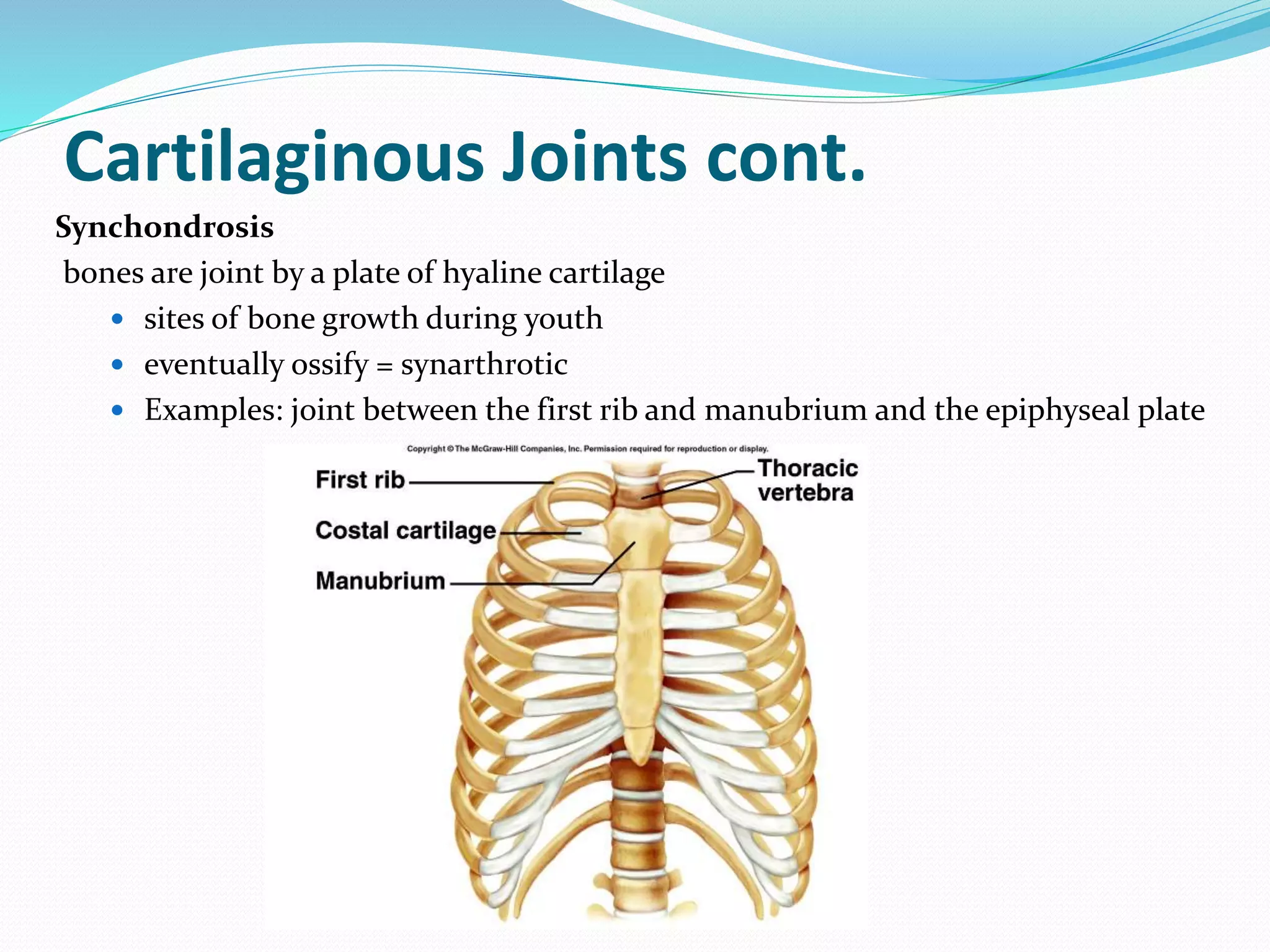
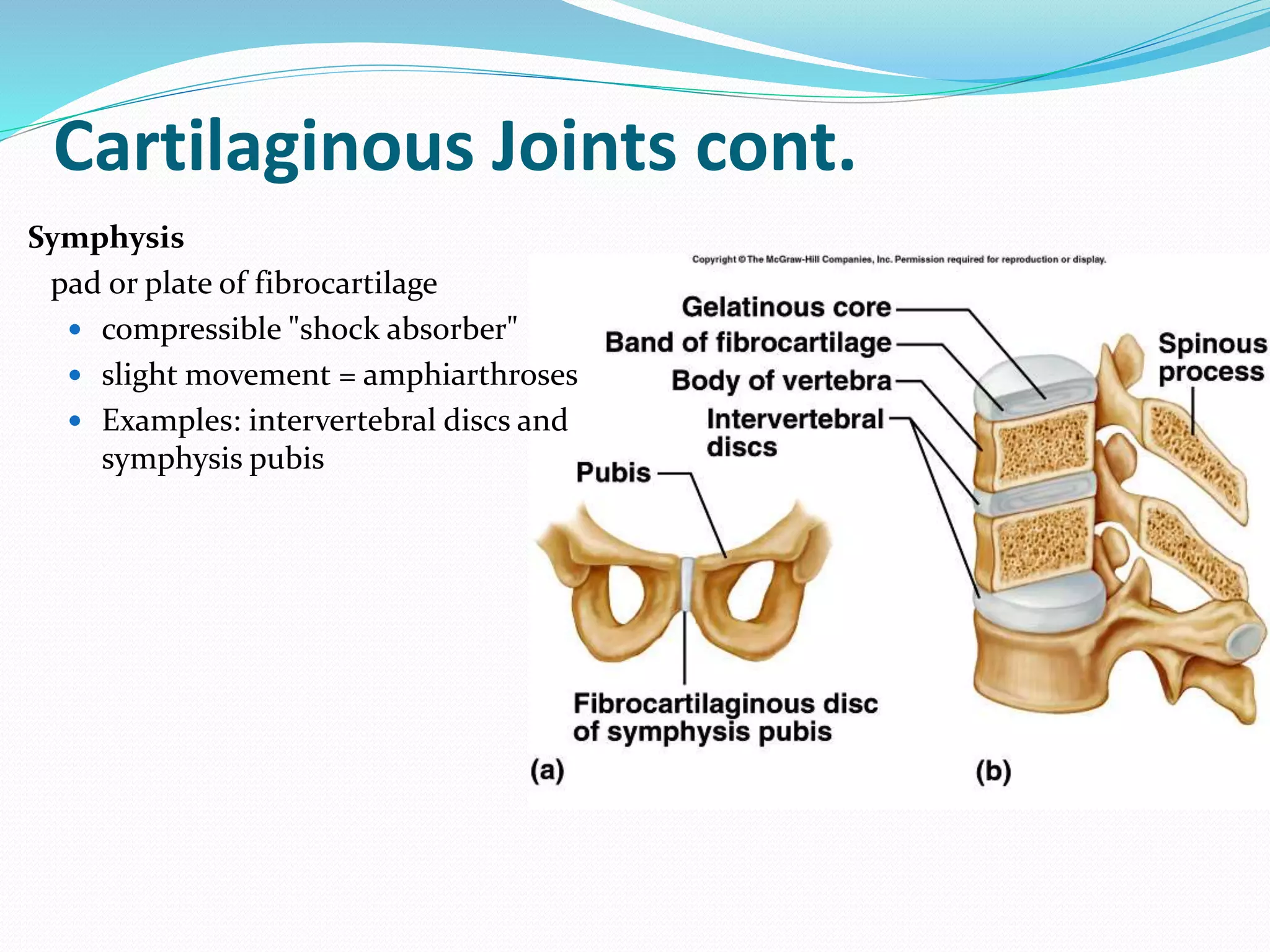
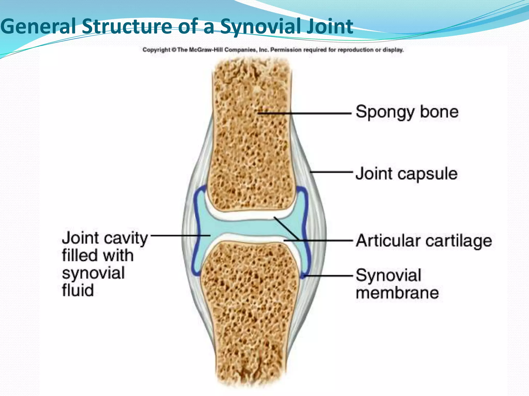
Dr Asma Lashari
University of Health Sciences
Lahore, Pakistan
Joints of the Skeletal System
 A joint or articulation is a connation between two or
more bones.
 Long bones articulate by their ends, flat bones by
margins, whereas in short or irregular bones surfaces
are articular.
 Functional junctions between bones.
 Bind parts of skeletal system together.
 Permit parts of the skeleton to change shape during
childbirth.
 Enable body to move in response to skeletal muscle
contraction.
Functional Classification Of Joints
 Classification=based on the amount of movement allowed
 3 types:
 Synarthroses = immovable joints
 Example = sutures of skull
 Amphiarthroses = slightly movable joints.
 Example = intervertebral discs between vertebrae
 Diarthroses = freely movable joints
 Examples = joints of appendicular skeleton
Structural Classification Of Joints
 Classification= Based on material, which joins bones
 3 types:
 Fibrous
 Fibrous joints are connected by dense connective tissue consisting mainly of collagen.
 These joints are also called fixed or immovable joints because they do not move.
 Fibrous joints have no joint cavity and are connected via fibrous connective tissue.
 The skull bones are connected by fibrous joints called sutures.
 Cartilaginous
 These joints are connected entirely by cartilage (fibrocartilage or hyaline).
 These joints allow more movement between bones than a fibrous joint but less than the
highly mobile synovial joint
 Synovial
 Also known as diarthrosis, joins bones with a fibrous joint capsule that is continuous with
the periosteum of the joined bones,
 Constitutes the outer boundary of a synovial cavity, surrounds the bones' articulating
surfaces.
 The synovial cavity/joint is filled with synovial fluid.
Fibrous Joints
 joints composed of fibrous tissue
 no joint cavity is present
 3 types:
 Syndesmosis
 Suture
 Gomphosis
Fibrous Joints cont.
Syndesmosis
is a fibrous joint between two bones and linked by
ligaments and a strong membrane.
Example = distal tibiofibular joint
Fibrous Joints cont.
Sutures = short fibrous joint only present in skull
 Synarthroses, provides for a strong connection between the adjacent bones
 Only found in skull
Fibrous Joints cont.
Gomphosis
tooth within its bony socket (alveolar fossa)
 short periodontal ligament
Cartilaginous Joints
 joints composed of cartilage
 no joint cavity
 2 types:
 Synchondrosis
 Symphysis
Cartilaginous Joints cont.
Synchondrosis
bones are joint by a plate of hyaline cartilage
 sites of bone growth during youth
 eventually ossify = synarthrotic
 Examples: joint between the first rib and manubrium and the epiphyseal plate
Cartilaginous Joints cont.
Symphysis
pad or plate of fibrocartilage
 compressible "shock absorber"
 slight movement = amphiarthroses
 Examples: intervertebral discs and
symphysis pubis
Synovial Joints
Fluid-filled joint cavity
 free movement = diarthrosis
General Structure Of A Synovial Joint
General Structure Of A Synovial Joint = 5 distinct features:
1. Articular cartilage = hyaline cartilage covers the surface of each bone
2. Joint cavity = a potential space between the two bones, filled with synovial fluid
3. Synovial fluid = viscous lubricating fluid within cavity
 reduces friction between cartilages of 2 bones
 provide lubrication
 nourish cartilage
 contain phagocytes
4. Articular capsule = double layered capsule surrounding cavity:
 External, tough flexible fibrous capsule (continuous with periosteum of the
bones)
 Synovial membrane = loose CT lining of fibrous capsule, that also covers all
internal joint surfaces excluding hyaline cartilage
5. Reinforcing ligaments = ligaments that strengthen joint
 Definition: A ligament joins a bone to another bone across a synovial joint
 usually thickened portions of fibrous capsule (intrinsic or capsular)
General Structure of a Synovial Joint
Synovial Joints cont.
 Other joint features:
 fatty pads (hip & knee)
 menisci or articular discs or that separate cavity into 2 compartments (knee,
jaw, sternoclavicular)
 bursa = flattened fibrous sacs with a synovial membrane and fluid that act as "ball
bearings" to prevent friction on adjacent structures during joint activity
 cushion the movement of one body part over another;
 located between skin and bone (where skin rubs over bone), and between muscle,
tendons, ligaments and bone.
Types Of Synovial Joints
 Ball-and-socket joints = most freely
movable joints; all angular movement
 The head of one bone fits into the
socket of another
 Examples = hip and shoulder
 Condyloid joints = permit all angular
motion, except rotation
 Examples = wrists and knuckles
Types Of Synovial Joints cont.
 Gliding joints = cartilaginous joints
 Example = intervertebral discs
 Hinge joints = permit flexion &
extension only
 Examples = elbow and knee
Types Of Synovial Joints cont.
 Pivot joints = permit rotation
 Example = first intervertebral
joint (atlantoaxial joint)
 Saddle joints = thumb
THANK YOU

Joints in skeletal system

  • 1.
    Dr Asma Lashari Universityof Health Sciences Lahore, Pakistan
  • 2.
    Joints of theSkeletal System  A joint or articulation is a connation between two or more bones.  Long bones articulate by their ends, flat bones by margins, whereas in short or irregular bones surfaces are articular.  Functional junctions between bones.  Bind parts of skeletal system together.  Permit parts of the skeleton to change shape during childbirth.  Enable body to move in response to skeletal muscle contraction.
  • 3.
    Functional Classification OfJoints  Classification=based on the amount of movement allowed  3 types:  Synarthroses = immovable joints  Example = sutures of skull  Amphiarthroses = slightly movable joints.  Example = intervertebral discs between vertebrae  Diarthroses = freely movable joints  Examples = joints of appendicular skeleton
  • 4.
    Structural Classification OfJoints  Classification= Based on material, which joins bones  3 types:  Fibrous  Fibrous joints are connected by dense connective tissue consisting mainly of collagen.  These joints are also called fixed or immovable joints because they do not move.  Fibrous joints have no joint cavity and are connected via fibrous connective tissue.  The skull bones are connected by fibrous joints called sutures.  Cartilaginous  These joints are connected entirely by cartilage (fibrocartilage or hyaline).  These joints allow more movement between bones than a fibrous joint but less than the highly mobile synovial joint  Synovial  Also known as diarthrosis, joins bones with a fibrous joint capsule that is continuous with the periosteum of the joined bones,  Constitutes the outer boundary of a synovial cavity, surrounds the bones' articulating surfaces.  The synovial cavity/joint is filled with synovial fluid.
  • 5.
    Fibrous Joints  jointscomposed of fibrous tissue  no joint cavity is present  3 types:  Syndesmosis  Suture  Gomphosis
  • 6.
    Fibrous Joints cont. Syndesmosis isa fibrous joint between two bones and linked by ligaments and a strong membrane. Example = distal tibiofibular joint
  • 7.
    Fibrous Joints cont. Sutures= short fibrous joint only present in skull  Synarthroses, provides for a strong connection between the adjacent bones  Only found in skull
  • 8.
    Fibrous Joints cont. Gomphosis toothwithin its bony socket (alveolar fossa)  short periodontal ligament
  • 9.
    Cartilaginous Joints  jointscomposed of cartilage  no joint cavity  2 types:  Synchondrosis  Symphysis
  • 10.
    Cartilaginous Joints cont. Synchondrosis bonesare joint by a plate of hyaline cartilage  sites of bone growth during youth  eventually ossify = synarthrotic  Examples: joint between the first rib and manubrium and the epiphyseal plate
  • 11.
    Cartilaginous Joints cont. Symphysis pador plate of fibrocartilage  compressible "shock absorber"  slight movement = amphiarthroses  Examples: intervertebral discs and symphysis pubis
  • 12.
    Synovial Joints Fluid-filled jointcavity  free movement = diarthrosis
  • 13.
    General Structure OfA Synovial Joint General Structure Of A Synovial Joint = 5 distinct features: 1. Articular cartilage = hyaline cartilage covers the surface of each bone 2. Joint cavity = a potential space between the two bones, filled with synovial fluid 3. Synovial fluid = viscous lubricating fluid within cavity  reduces friction between cartilages of 2 bones  provide lubrication  nourish cartilage  contain phagocytes 4. Articular capsule = double layered capsule surrounding cavity:  External, tough flexible fibrous capsule (continuous with periosteum of the bones)  Synovial membrane = loose CT lining of fibrous capsule, that also covers all internal joint surfaces excluding hyaline cartilage 5. Reinforcing ligaments = ligaments that strengthen joint  Definition: A ligament joins a bone to another bone across a synovial joint  usually thickened portions of fibrous capsule (intrinsic or capsular)
  • 14.
    General Structure ofa Synovial Joint
  • 15.
    Synovial Joints cont. Other joint features:  fatty pads (hip & knee)  menisci or articular discs or that separate cavity into 2 compartments (knee, jaw, sternoclavicular)  bursa = flattened fibrous sacs with a synovial membrane and fluid that act as "ball bearings" to prevent friction on adjacent structures during joint activity  cushion the movement of one body part over another;  located between skin and bone (where skin rubs over bone), and between muscle, tendons, ligaments and bone.
  • 16.
    Types Of SynovialJoints  Ball-and-socket joints = most freely movable joints; all angular movement  The head of one bone fits into the socket of another  Examples = hip and shoulder  Condyloid joints = permit all angular motion, except rotation  Examples = wrists and knuckles
  • 17.
    Types Of SynovialJoints cont.  Gliding joints = cartilaginous joints  Example = intervertebral discs  Hinge joints = permit flexion & extension only  Examples = elbow and knee
  • 18.
    Types Of SynovialJoints cont.  Pivot joints = permit rotation  Example = first intervertebral joint (atlantoaxial joint)  Saddle joints = thumb
  • 19.