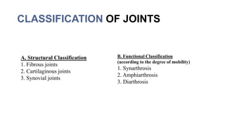
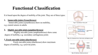
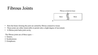
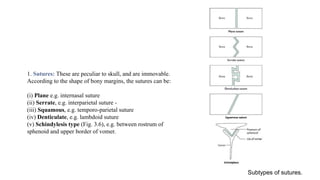
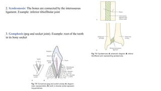
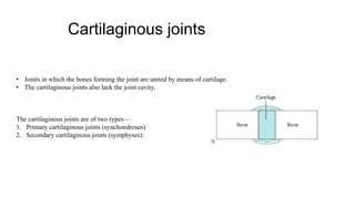
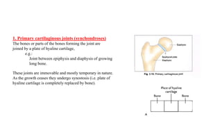
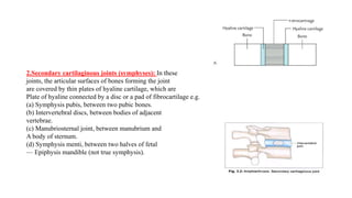
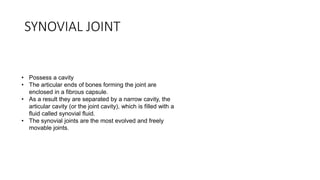
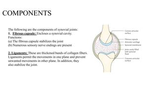
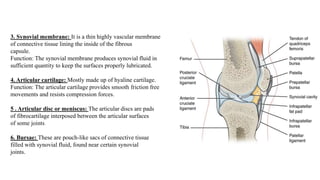
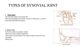
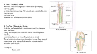
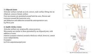
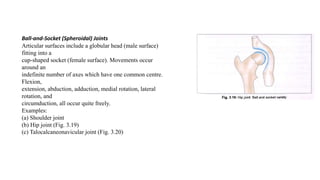
2. INTRODUCTION
• Joint is a junction between two or more bones or cartilages.
• It is a device to permit movements.
There are more joints in a child than in an adult because as growth
proceeds some of the bones fuse together.
e.g. The two halves of the infant frontal bone,
3. CLASSIFICATION OF JOINTS
A. Structural Classification
1. Fibrous joints
2. Cartilaginous joints
3. Synovial joints
B. Functional Classification
(according to the degree of mobility)
1. Synarthrosis
2. Amphiarthrosis
3. Diarthrosis
4. Functional Classification
It is based upon the degree of mobility of the joint. They are of three types:
1. Immovable joints (Synarthroses)
Immovable joints (synarthroses) show no mobility,
e.g. cranial sutures in adults
2. Slightly movable joints (amphiarthroses)
Slightly movable joints (amphiarthroses) show some
degree of mobility, e.g. secondary cartilaginous joints
3. Freely movable joints (diarthroses)
Freely movable joints (diarthroses) show maximum
degree of mobility, e.g. synovial joints.
5. Fibrous Joints
• Here the bones forming the joint are united by fibrous connective tissue.
• These joints are either immovable or permit only a slight degree of movement.
• A fibrous joint lacks joint cavity.
The fibrous joints are of three types—
1. Sutures,
2. Syndesmoses
3. Gomphoses.
6. 1. Sutures: These are peculiar to skull, and are immovable.
According to the shape of bony margins, the sutures can be:
(i) Plane e.g. internasal suture
(ii) Serrate, e.g. interparietal suture -
(iii) Squamous, e.g. temporo-parietal suture
(iv) Denticulate, e.g. lambdoid suture
(v) Schindylesis type (Fig. 3.6), e.g. between rostrum of
sphenoid and upper border of vomer.
Subtypes of sutures.
7. 2. Syndesmosis: The bones are connected by the interosseous
ligament. Example: inferior tibiofibular joint
3. Gomphosis (peg and socket joint). Example: root of the tooth
in its bony socket
8. Cartilaginous joints
• Joints in which the bones forming the joint are united by means of cartilage.
• The cartilaginous joints also lack the joint cavity.
The cartilaginous joints are of two types—
1. Primary cartilaginous joints (synchondroses)
2. Secondary cartilaginous joints (symphyses):
9. 1. Primary cartilaginous joints (synchondroses)
The bones or parts of the bones forming the joint are
joined by a plate of hyaline cartilage,
e.g.:
Joint between epiphysis and diaphysis of growing
long bone.
These joints are immovable and mostly temporary in nature.
As the growth ceases they undergo synostosis (i.e. plate of
hyaline cartilage is completely replaced by bone).
10. 2.Secondary cartilaginous joints (symphyses): In these
joints, the articular surfaces of bones forming the joint
are covered by thin plates of hyaline cartilage, which are
Plate of hyaline connected by a disc or a pad of fibrocartilage e.g.
(a) Symphysis pubis, between two pubic bones.
(b) Intervertebral discs, between bodies of adjacent
vertebrae.
(c) Manubriosternal joint, between manubrium and
A body of sternum.
(d) Symphysis menti, between two halves of fetal
— Epiphysis mandible (not true symphysis).
11. SYNOVIAL JOINT
• Possess a cavity
• The articular ends of bones forming the joint are
enclosed in a fibrous capsule.
• As a result they are separated by a narrow cavity, the
articular cavity (or the joint cavity), which is filled with a
fluid called synovial fluid.
• The synovial joints are the most evolved and freely
movable joints.
12. COMPONENTS
The following are the components of synovial joints:
1. Fibrous capsule: Encloses a synovial cavity.
Functions:
(a) The fibrous capsule stabilizes the joint
(b) Numerous sensory nerve endings are present
2. Ligaments: These are thickened bands of collagen fibers.
Ligaments permit the movements in one plane and prevent
unwanted movements in other plane. In addition, they
also stabilize the joint.
13. 3. Synovial membrane: It is a thin highly vascular membrane
of connective tissue lining the inside of the fibrous
capsule.
Function: The synovial membrane produces synovial fluid in
sufficient quantity to keep the surfaces properly lubricated.
4. Articular cartilage: Mostly made up of hyaline cartilage.
Function: The articular cartilage provides smooth friction free
movements and resists compression forces.
5 . Articular disc or meniscus: The articular discs are pads
of fibrocartilage interposed between the articular surfaces
of some joints
6. Bursae: These are pouch-like sacs of connective tissue
filled with synovial fluid, found near certain synovial
joints.
14. TYPES OF SYNOVIAL JOINT
1. Plane joints
The articular surfaces are nearly flat
They permit gliding movements in various directions, i.e. side to
side, back and forth movements
2. Hinge Joints
Articular surfaces are pulley-shaped.
There are strong collateral ligaments.
Movements are permitted in one plane around a transverse axis.
Examples:
Elbow joint
15. 3. Pivot (Trochoid) Joints
Articular surfaces comprise a central bony pivot (peg)
surrounded by
an osteoligamentous ring. Movements are permitted in one
plane around
a vertical axis.
Superior and inferior radio-ulnar joints
4. Condylar (Bicondylar) Joints
Articular surfaces include two distinct condyles (convex
male surfaces)
fitting into reciprocally concave female surfaces (which
are also,
sometimes, known as condyles, such as in tibia).
These joints permit movements mainly in one plane around
a transverse axis, but partly in another plane (rotation)
around a vertical axis.
Examples:
Knee joint
16. 5. Ellipsoid Joints
Articular surfaces include an oval, convex, male surface fitting into an
elliptical, concave female surface.
Free movements are permitted around both the axes, flexion and
extension around the transverse axis,
and abduction and adduction around the anteroposterior axis.
Eg Wrist joint
6. Saddle (Sellar) Joints
Articular surfaces are reciprocally concavoconvex.
Movements are similar to those permitted by an ellipsoid joint, with
addition of some
rotation (conjunct rotation) around a third axis which, however, cannot
occur independently.
Examples:
(a) First carpometacarpal joint
17. Ball-and-Socket (Spheroidal) Joints
Articular surfaces include a globular head (male surface)
fitting into a
cup-shaped socket (female surface). Movements occur
around an
indefinite number of axes which have one common centre.
Flexion,
extension, abduction, adduction, medial rotation, lateral
rotation, and
circumduction, all occur quite freely.
Examples:
(a) Shoulder joint
(b) Hip joint (Fig. 3.19)
(c) Talocalcaneonavicular joint (Fig. 3.20)