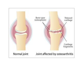
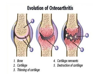
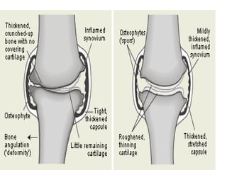
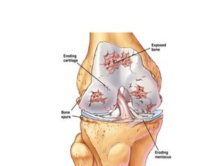
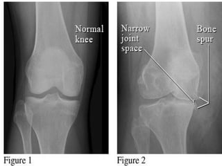
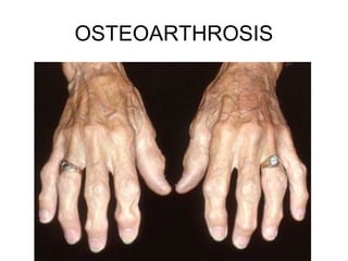
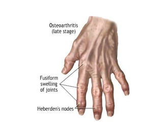
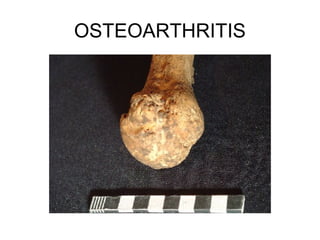
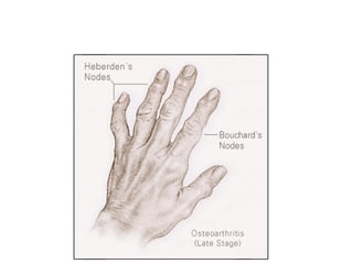
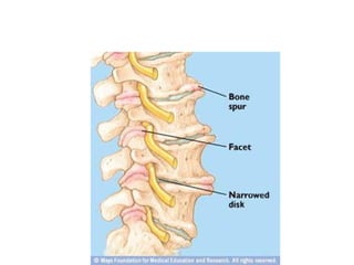
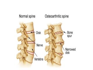
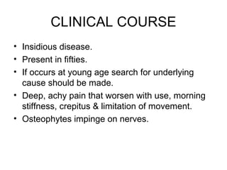
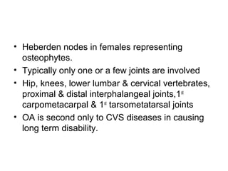
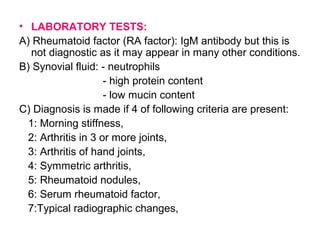
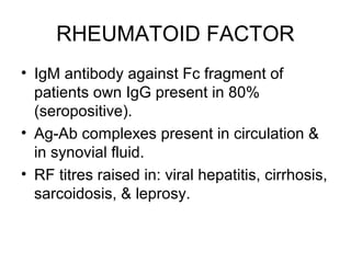
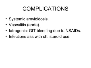
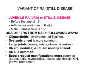
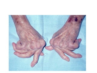
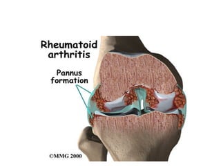
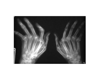
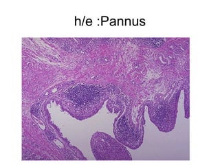
DISEASES OF JOINTS
NORMAL JOINTS
• Provide movement & mechanical support
• 2 types: Synovial & Non-Synovial
1) NON SYNOVIAL JOINTS (SOLID OR
SYNARTHROSES)
• Lack joint space
• Minimal movement
• Connected by fibrous tissue or cartilage
• Fibrous synarthroses:
- include cranial sutures & bonds b/w root of teeth &
jawbone
• Cartilaginous synarthroses:
- are represented by symphyses
2) SYNOVIAL JOINTS
• Have joint space, allow wide range of
movements
• Strengthened by fibrous capsule &
reinforced by ligaments & muscles
• Lined by synovial membrane anchored to
capsule
• Covered by synoviocytes which are
cuboidal cells 1-4 layers deep
• Synovial membrane is not present over
cartilage.
• Lacks BM & merges with underlying CT.
• Synovial fluid is viscus, clear & is plasma
filtrate containing hyaluronic acid, acts as
lubricant & provides nutrition to articulate
cartilage.
• HYALINE CARTILAGE is a unique C.T.
• Serves as elastic shock absorber & wear-
resistant surface.
• Lacks blood supply & has no lymphatics or
nerves.
• Composed of type 2 collagen, water,
proteoglycans & chondrocytes.
• Chondrocytes synthesize the matrix as
well as enzymes which digest it.
OSTEOARTHRITIS (0A)
Or Degenerated Joint Disease or
Osteoarthrosis
• Most common type of joint disease → disablity.
• “Characterized by progressive erosion of
articular cartilage particularly in wt. bearing
joints”.
• Term OS implies an “inflammatory disease”.
• OA is intrinsic disease of articular cartilage in
which biochemical & metabolic alterations result
in its breakdown.
• Main target in this disease is the ARTICULAR
CARTILAGE.
• Idiopathic or Primary OA (95%).
* appears insiduously, without any predis cause as an
aging phenomina.
- Few joints (oligoarthritis).
- Old age.
• Secondary OA: (5%).
* Previous or repeated trauma to a joint.
* Congenital deformity of a jt.
* DM.
* Ochronosis.
* Hemochromatosis.
* Marked obesity.
- Young age.
- One joint or several joints.
• Knees & hands are commonly affected in
women & hip joints in men
PATHOGENESIS
• Normal articular cartilage bathed in
synovial fluid performs 2 functions:
1): Ensures friction free movement in joints.
2): Spreads load across joint surface to
allow underlying bone to absorb shock &
wt. without being crushed.
For this purpose cartilage is elastic & has
unusually high tensile strength
• Two components responsible for these attributes of
articular cartilage are:
1) Type II collagen,
2) Proteoglycans,
Both are secreted by chondrocytes.
• Articular cartilage is not static.
• Turnover occurs in which worn out matrix is
degraded & replaced.
• This balance is maintained by chondrocytes.
• Therefore, healthy chondrocytes are essential for
joint integrity→ 1) synthesize the matrix &
2)secrete matrix degrading enzymes.
• In OS this process is disturbed .
FACTORS AFFECTING OA:
• 1)Age: increasing frequency with advancing
age.
• 2)Mechanical effects:
1) OA is due to wear & tear process.
2) Occur in wt. bearing joints.
3) An increased frequency is seen in conditions
that predisposes joints to abnormal mechanical
stresses like obesity & previous joint
deformity.
• 3) Genetic factors: ↑susceptibility esp. in
cases involving hands & hip.
Specific gene or genes have not been
identified, although linkage to ch.2 & 11
has been suggested.
• 4) Bone density : risk of OA α to bone
density.
• 5)↑Oestrogen levels : ass with ↑risk of
OA.
• OA is characterized by changes in both the
COMPOSITION & MECHANICAL
PROPERTIES of DISEASED CARTILAGE.
1) Increase in its water content.
2) Decrease in proteoglycans.
3) Weakening in collagen network due to
decrease in local synthesis of collagen type 2 &
increase breakdown of pre-existing collagen.
• IL-1, TNF & NO are increased.
• ↑ed APOPTOSIS → ↓ no. of functional
chondrocytes.
• THESE CHANGES TEND TO REDUCE THE
TENSILE STRENGTH & RESILIENCE OF
ARTICULAR CARTILAGE.
• In response to these regressive changes,
chondrocytes in deeper layers proliferate
& attempt to “repair” the damage by
producing new collagen &
proteoglycans.
• These reparative changes are initially able
to keep pace with deterioration of cartilage
but eventually loss & changes in ECM
predominate.
MORPHOLOGY
• Gross picture:
1) Granular, irregular articular cartilage as it
becomes soft due to degradation.
2) Eventually full thickness portions of cartilage
are sloughed exposing subchondral bones
which now become new articular surface.
3) Friction smoothens bony surfaces giving it
appearance of polished ivory (bone
eburnation).
4) Sclerosis of underlying bone.
5) Small fractures of underlying bone are
common & dislodged pieces of cartilage &
bone tumble in joint space forming loose
bodies ( joint mice).
• Fracture gaps allow synovial fluid to be
forced into subchondral regions resulting
in fibrous walled cysts.
• Mushroom shaped osteophytes (bony
outgrowths) develop at margins of
articular cartilage & are capped by
cartilage that gradually ossify.
• Synovium shows minor alterations & is
congested & fibrotic & may have scattered
ch. inflammatory cells.
• In severe cases, fibrous synovial pannus
is formed.
OSTEOARTHROSIS
OSTEOARTHRITIS
CLINICAL COURSE
• Insidious disease.
• Present in fifties.
• If occurs at young age search for underlying
cause should be made.
• Deep, achy pain that worsen with use, morning
stiffness, crepitus & limitation of movement.
• Osteophytes impinge on nerves.
• Heberden nodes in females representing
osteophytes.
• Typically only one or a few joints are involved
• Hip, knees, lower lumbar & cervical vertebrates,
proximal & distal interphalangeal joints,1st
carpometacarpal & 1st
tarsometatarsal joints
• OA is second only to CVS diseases in causing
long term disability.
• 32 SLIDES
RHEUMATOID ARTHRITIS
• Chronic systemic inflammatory disorder
that may affect many tissues & organs-
skin, BVs, heart,lungs & muscles- but
principally attacks the joints, producing
nonsuppurative proliferative &
inflammatory synovitis that often
progresses to destruction of articular
cartilage & ankylosis of joints.
• 1% of world’s population is afflicted by RA.
• Male to female ratio 1:3.
• Age : 4th
– 7th
decade but no age is
immune.
• Cause not known but AUTOIMMUNITY
plays a major role in its pathogenicity.
MORPHOLOGY
• JOINTS:
1): SYNOVIUM (gross) becomes edematous,
thickened, & hyperplastic.
2): Normal smooth contour is transformed→
formation of fronds & villi.
3): (microscopy): Infiltration of synovium by
dense perivascular inflammatory infiltrate
consisting of B cells & CD4+ helper T cells,
plasma cells & macrophages with formation of
lymphoid follicles.
4): Increased vascularity due to vasodilation
& angiogenesis with hemosiderin deposits.
5): Aggregation of organizing fibrin covering
synovium & floating in joint space as rice
bodies.
6): Accumulation of neutrophils in synovial fluid
& superficial synovium.
7): Osteoclastic activity in underlying bone
→synovium penetrating into bone→ juxtra-
articular erosions, subchondrial cysts, &
osteoporosis.
8): Pannus formation.
PANNUS : mass of synovium & synovial stroma
consisting of inflammatory cells, G.T, &
fibroblasts.
- Grows over articular cartilage & causes its
erosion.
- Pannus bridges the apposing bones forming
fibrous ankylosis which ossifies resulting in
bony ankylosis.
- Inflammation in adjacent tendons, ligaments, &
skeletal muscles is common.
• SKIN:
RHEUMATOID NODULES : seen in 25% of pt.
Arise in regions subjected to pressure like ulnar
aspect of forearm, elbows, occiput &
lumbosacral areas.
Also formed in lungs, spleen, pericardium,
myocardium, valves, aorta,
Firm, nontender, round to oval within
subcutaneous tissue
M/E: central zone of fibrinoid necrosis surrounded
by a rim of epithelioid histiocytes, lymphocytes
& plasma cells.
BILAT RH. NODULES
RH.NODULE
Rh. nodule
Rh nodule
• BLOOD VESSELS:
VASCULITIS (is a potentially bad prognostic
indicator of RA).
* Medium to small arteries are involved like PAN
(kidney bv are not involved).
* Vasa nervorum & digital arteries are obstructed
by endarteritis obliterans resulting in neuropathy,
ulcers, & gangrene
* Venulitis produces purpura, ulcers, nail bed
infarction.
• BV are involved in severe disease with
rheumatoid nodules & high levels of RF
• It is potentially catastrophic complication
of RA particularly when it affects vital
organs.
PATHOGENESIS
• Autoimmune disease due to exposure of
genetically susceptible host to an
unknown arthritogenic antigen.
• Therefore, key considerations in
pathogenesis are :-
1) Nature of autoimmune reaction,
2) Mediators of tissue injury,
3) Genetic susceptibility,
4) Arthritogenic antigen,
1) AUTOIMMUNE REACTION:
Consists of ACTIVATED CD4+ T cells & B
LYMPHOCYTES:
• Target antigens & how these lymphocytes are
initiated is not known.
• T-cells stimulate other cells in joint to produce
cytokines.
• Role of B cells is controversial but immune
complex deposition play some role in joint
destruction.
2) MEDIATORS OF JOINT INJURY:
- CYTOKINES play pivotal role & imp. ones are
TNF & IL-1.
- Secreted by macrophages & synovial cells
activated by T cells in the joint.
- TNF & IL-1 in turn, stimulate synovial cells to
proliferate & produce various mediators (PG) &
matrix metalloproteinases (MMPs) causing
cartilage destruction.
• T cells & synovial fibroblasts also
produce RANKL which activate
osteoclasts & promotes bone
destruction.
• Net result is hyperplastic synovium with
inflammatory cells forming pannus→
sustained, irreversible cartilage
destruction & erosion of subchondral
bone.
• Anticytokine therapy (esp against TNF).
3) GENETIC SUSCEPTIBILITY:
- Well defined familial predisposition
- High rate of concordance b/w
monozygotic twins
- Class II HLA locus (HLA DRB1*0401 &
*0404 alleles).
4) ANTIGENS :
- Not known
- Microbial antigens are a possibility but
their role is not confirmed.
CLINICAL COURSE of RA.
• Variable, slow, insidious disease.
• Malaise, fatigue, & generalized musculoskeletal
pain.
• 10% have acute onset.
• Small joints are affected before larger ones.
MCP, PIP,MTP, IP joints followed by wrist,
ankles, & knee.
• Cervical spine also affected.
• Hip jt rarely affected (only late in course of
disease).
• Typically sparing of lumbosacral region.
• Swollen, painful, morning stiffness.
• Disease may be slow or rapid &
fluctuates over period of years with
periods of partial or complete
remission.
• Maximum damage occurs during the
1st
4 -5 yrs.
• X-rays: Juxta-articular osteopenia,
bone erosion with narrowing of joint
space from loss of articular cartilage.
CHARACTERISTIC GROSS
DEFORMITIES:
• Radial deviation of wrist.
• Ulnar deviation of fingers.
• Flexion-hyperextension of fingers (swan
neck).
• Bakers cyst (large synovial cysts) in post
knee due to ↑ intraarticular pressure.
• LABORATORY TESTS:
A) Rheumatoid factor (RA factor): IgM antibody but this is
not diagnostic as it may appear in many other conditions.
B) Synovial fluid: - neutrophils
- high protein content
- low mucin content
C) Diagnosis is made if 4 of following criteria are present:
1: Morning stiffness,
2: Arthritis in 3 or more joints,
3: Arthritis of hand joints,
4: Symmetric arthritis,
5: Rheumatoid nodules,
6: Serum rheumatoid factor,
7:Typical radiographic changes,
RHEUMATOID FACTOR
• IgM antibody against Fc fragment of
patients own IgG present in 80%
(seropositive).
• Ag-Ab complexes present in circulation &
in synovial fluid.
• RF titres raised in: viral hepatitis, cirrhosis,
sarcoidosis, & leprosy.
COMPLICATIONS
• Systemic amyloidosis.
• Vasculitis (aorta).
• Iatrogenic: GIT bleeding due to NSAIDs.
• Infections ass with ch. steroid use.
VARIANT OF RA (STILL DISEASE)
• JUVENILE RA (JRA) or STILL’S DISEASE
- Before the age of 16.
- Arthritis for minimum of 6 wks.
- Male: Female ratio is 1:2.
JRA DIFFERS FROM RA IN FOLLOWING WAYS:
• Oligoarthritis (involvement of 5 joints).
• Systemic onset is more common.
• Large joints (knees, wrists,elbows, & ankles).
• RN (rh. nodules) & RF are usually absent.
• ANA is common.
• Extra-articular manifestations more common
(perocarditis, myocarditis, uveitis, pul fibrosis, GN,
growth retardation)
• FELTY’S SYNDROME:
RA associated with splenomegaly &
hypersplenism & consequently
haematological derangements.
h/e :Pannus
• 70 slides

Osteoarth & Rheumatoid arthritis

  • 1.
  • 2.
    NORMAL JOINTS • Providemovement & mechanical support • 2 types: Synovial & Non-Synovial 1) NON SYNOVIAL JOINTS (SOLID OR SYNARTHROSES) • Lack joint space • Minimal movement • Connected by fibrous tissue or cartilage • Fibrous synarthroses: - include cranial sutures & bonds b/w root of teeth & jawbone • Cartilaginous synarthroses: - are represented by symphyses
  • 3.
    2) SYNOVIAL JOINTS •Have joint space, allow wide range of movements • Strengthened by fibrous capsule & reinforced by ligaments & muscles • Lined by synovial membrane anchored to capsule • Covered by synoviocytes which are cuboidal cells 1-4 layers deep
  • 4.
    • Synovial membraneis not present over cartilage. • Lacks BM & merges with underlying CT. • Synovial fluid is viscus, clear & is plasma filtrate containing hyaluronic acid, acts as lubricant & provides nutrition to articulate cartilage.
  • 5.
    • HYALINE CARTILAGEis a unique C.T. • Serves as elastic shock absorber & wear- resistant surface. • Lacks blood supply & has no lymphatics or nerves. • Composed of type 2 collagen, water, proteoglycans & chondrocytes.
  • 6.
    • Chondrocytes synthesizethe matrix as well as enzymes which digest it.
  • 7.
    OSTEOARTHRITIS (0A) Or DegeneratedJoint Disease or Osteoarthrosis
  • 8.
    • Most commontype of joint disease → disablity. • “Characterized by progressive erosion of articular cartilage particularly in wt. bearing joints”. • Term OS implies an “inflammatory disease”. • OA is intrinsic disease of articular cartilage in which biochemical & metabolic alterations result in its breakdown. • Main target in this disease is the ARTICULAR CARTILAGE.
  • 9.
    • Idiopathic orPrimary OA (95%). * appears insiduously, without any predis cause as an aging phenomina. - Few joints (oligoarthritis). - Old age. • Secondary OA: (5%). * Previous or repeated trauma to a joint. * Congenital deformity of a jt. * DM. * Ochronosis. * Hemochromatosis. * Marked obesity. - Young age. - One joint or several joints.
  • 10.
    • Knees &hands are commonly affected in women & hip joints in men
  • 11.
    PATHOGENESIS • Normal articularcartilage bathed in synovial fluid performs 2 functions: 1): Ensures friction free movement in joints. 2): Spreads load across joint surface to allow underlying bone to absorb shock & wt. without being crushed. For this purpose cartilage is elastic & has unusually high tensile strength
  • 12.
    • Two componentsresponsible for these attributes of articular cartilage are: 1) Type II collagen, 2) Proteoglycans, Both are secreted by chondrocytes. • Articular cartilage is not static. • Turnover occurs in which worn out matrix is degraded & replaced. • This balance is maintained by chondrocytes. • Therefore, healthy chondrocytes are essential for joint integrity→ 1) synthesize the matrix & 2)secrete matrix degrading enzymes. • In OS this process is disturbed .
  • 13.
    FACTORS AFFECTING OA: •1)Age: increasing frequency with advancing age. • 2)Mechanical effects: 1) OA is due to wear & tear process. 2) Occur in wt. bearing joints. 3) An increased frequency is seen in conditions that predisposes joints to abnormal mechanical stresses like obesity & previous joint deformity.
  • 14.
    • 3) Geneticfactors: ↑susceptibility esp. in cases involving hands & hip. Specific gene or genes have not been identified, although linkage to ch.2 & 11 has been suggested. • 4) Bone density : risk of OA α to bone density. • 5)↑Oestrogen levels : ass with ↑risk of OA.
  • 15.
    • OA ischaracterized by changes in both the COMPOSITION & MECHANICAL PROPERTIES of DISEASED CARTILAGE. 1) Increase in its water content. 2) Decrease in proteoglycans. 3) Weakening in collagen network due to decrease in local synthesis of collagen type 2 & increase breakdown of pre-existing collagen. • IL-1, TNF & NO are increased. • ↑ed APOPTOSIS → ↓ no. of functional chondrocytes. • THESE CHANGES TEND TO REDUCE THE TENSILE STRENGTH & RESILIENCE OF ARTICULAR CARTILAGE.
  • 16.
    • In responseto these regressive changes, chondrocytes in deeper layers proliferate & attempt to “repair” the damage by producing new collagen & proteoglycans. • These reparative changes are initially able to keep pace with deterioration of cartilage but eventually loss & changes in ECM predominate.
  • 17.
    MORPHOLOGY • Gross picture: 1)Granular, irregular articular cartilage as it becomes soft due to degradation. 2) Eventually full thickness portions of cartilage are sloughed exposing subchondral bones which now become new articular surface. 3) Friction smoothens bony surfaces giving it appearance of polished ivory (bone eburnation).
  • 18.
    4) Sclerosis ofunderlying bone. 5) Small fractures of underlying bone are common & dislodged pieces of cartilage & bone tumble in joint space forming loose bodies ( joint mice).
  • 19.
    • Fracture gapsallow synovial fluid to be forced into subchondral regions resulting in fibrous walled cysts. • Mushroom shaped osteophytes (bony outgrowths) develop at margins of articular cartilage & are capped by cartilage that gradually ossify. • Synovium shows minor alterations & is congested & fibrotic & may have scattered ch. inflammatory cells. • In severe cases, fibrous synovial pannus is formed.
  • 25.
  • 27.
  • 31.
    CLINICAL COURSE • Insidiousdisease. • Present in fifties. • If occurs at young age search for underlying cause should be made. • Deep, achy pain that worsen with use, morning stiffness, crepitus & limitation of movement. • Osteophytes impinge on nerves.
  • 32.
    • Heberden nodesin females representing osteophytes. • Typically only one or a few joints are involved • Hip, knees, lower lumbar & cervical vertebrates, proximal & distal interphalangeal joints,1st carpometacarpal & 1st tarsometatarsal joints • OA is second only to CVS diseases in causing long term disability.
  • 33.
  • 34.
  • 35.
    RHEUMATOID ARTHRITIS • Chronicsystemic inflammatory disorder that may affect many tissues & organs- skin, BVs, heart,lungs & muscles- but principally attacks the joints, producing nonsuppurative proliferative & inflammatory synovitis that often progresses to destruction of articular cartilage & ankylosis of joints.
  • 36.
    • 1% ofworld’s population is afflicted by RA. • Male to female ratio 1:3. • Age : 4th – 7th decade but no age is immune. • Cause not known but AUTOIMMUNITY plays a major role in its pathogenicity.
  • 37.
    MORPHOLOGY • JOINTS: 1): SYNOVIUM(gross) becomes edematous, thickened, & hyperplastic. 2): Normal smooth contour is transformed→ formation of fronds & villi. 3): (microscopy): Infiltration of synovium by dense perivascular inflammatory infiltrate consisting of B cells & CD4+ helper T cells, plasma cells & macrophages with formation of lymphoid follicles.
  • 38.
    4): Increased vascularitydue to vasodilation & angiogenesis with hemosiderin deposits. 5): Aggregation of organizing fibrin covering synovium & floating in joint space as rice bodies. 6): Accumulation of neutrophils in synovial fluid & superficial synovium. 7): Osteoclastic activity in underlying bone →synovium penetrating into bone→ juxtra- articular erosions, subchondrial cysts, & osteoporosis. 8): Pannus formation.
  • 39.
    PANNUS : massof synovium & synovial stroma consisting of inflammatory cells, G.T, & fibroblasts. - Grows over articular cartilage & causes its erosion. - Pannus bridges the apposing bones forming fibrous ankylosis which ossifies resulting in bony ankylosis. - Inflammation in adjacent tendons, ligaments, & skeletal muscles is common.
  • 41.
    • SKIN: RHEUMATOID NODULES: seen in 25% of pt. Arise in regions subjected to pressure like ulnar aspect of forearm, elbows, occiput & lumbosacral areas. Also formed in lungs, spleen, pericardium, myocardium, valves, aorta, Firm, nontender, round to oval within subcutaneous tissue M/E: central zone of fibrinoid necrosis surrounded by a rim of epithelioid histiocytes, lymphocytes & plasma cells.
  • 42.
  • 43.
  • 44.
  • 45.
  • 46.
    • BLOOD VESSELS: VASCULITIS(is a potentially bad prognostic indicator of RA). * Medium to small arteries are involved like PAN (kidney bv are not involved). * Vasa nervorum & digital arteries are obstructed by endarteritis obliterans resulting in neuropathy, ulcers, & gangrene * Venulitis produces purpura, ulcers, nail bed infarction.
  • 47.
    • BV areinvolved in severe disease with rheumatoid nodules & high levels of RF • It is potentially catastrophic complication of RA particularly when it affects vital organs.
  • 48.
    PATHOGENESIS • Autoimmune diseasedue to exposure of genetically susceptible host to an unknown arthritogenic antigen. • Therefore, key considerations in pathogenesis are :- 1) Nature of autoimmune reaction, 2) Mediators of tissue injury, 3) Genetic susceptibility, 4) Arthritogenic antigen,
  • 49.
    1) AUTOIMMUNE REACTION: Consistsof ACTIVATED CD4+ T cells & B LYMPHOCYTES: • Target antigens & how these lymphocytes are initiated is not known. • T-cells stimulate other cells in joint to produce cytokines. • Role of B cells is controversial but immune complex deposition play some role in joint destruction.
  • 50.
    2) MEDIATORS OFJOINT INJURY: - CYTOKINES play pivotal role & imp. ones are TNF & IL-1. - Secreted by macrophages & synovial cells activated by T cells in the joint. - TNF & IL-1 in turn, stimulate synovial cells to proliferate & produce various mediators (PG) & matrix metalloproteinases (MMPs) causing cartilage destruction.
  • 51.
    • T cells& synovial fibroblasts also produce RANKL which activate osteoclasts & promotes bone destruction. • Net result is hyperplastic synovium with inflammatory cells forming pannus→ sustained, irreversible cartilage destruction & erosion of subchondral bone. • Anticytokine therapy (esp against TNF).
  • 52.
    3) GENETIC SUSCEPTIBILITY: -Well defined familial predisposition - High rate of concordance b/w monozygotic twins - Class II HLA locus (HLA DRB1*0401 & *0404 alleles).
  • 53.
    4) ANTIGENS : -Not known - Microbial antigens are a possibility but their role is not confirmed.
  • 54.
    CLINICAL COURSE ofRA. • Variable, slow, insidious disease. • Malaise, fatigue, & generalized musculoskeletal pain. • 10% have acute onset. • Small joints are affected before larger ones. MCP, PIP,MTP, IP joints followed by wrist, ankles, & knee. • Cervical spine also affected. • Hip jt rarely affected (only late in course of disease). • Typically sparing of lumbosacral region.
  • 55.
    • Swollen, painful,morning stiffness. • Disease may be slow or rapid & fluctuates over period of years with periods of partial or complete remission. • Maximum damage occurs during the 1st 4 -5 yrs. • X-rays: Juxta-articular osteopenia, bone erosion with narrowing of joint space from loss of articular cartilage.
  • 56.
    CHARACTERISTIC GROSS DEFORMITIES: • Radialdeviation of wrist. • Ulnar deviation of fingers. • Flexion-hyperextension of fingers (swan neck). • Bakers cyst (large synovial cysts) in post knee due to ↑ intraarticular pressure.
  • 57.
    • LABORATORY TESTS: A)Rheumatoid factor (RA factor): IgM antibody but this is not diagnostic as it may appear in many other conditions. B) Synovial fluid: - neutrophils - high protein content - low mucin content C) Diagnosis is made if 4 of following criteria are present: 1: Morning stiffness, 2: Arthritis in 3 or more joints, 3: Arthritis of hand joints, 4: Symmetric arthritis, 5: Rheumatoid nodules, 6: Serum rheumatoid factor, 7:Typical radiographic changes,
  • 58.
    RHEUMATOID FACTOR • IgMantibody against Fc fragment of patients own IgG present in 80% (seropositive). • Ag-Ab complexes present in circulation & in synovial fluid. • RF titres raised in: viral hepatitis, cirrhosis, sarcoidosis, & leprosy.
  • 59.
    COMPLICATIONS • Systemic amyloidosis. •Vasculitis (aorta). • Iatrogenic: GIT bleeding due to NSAIDs. • Infections ass with ch. steroid use.
  • 60.
    VARIANT OF RA(STILL DISEASE) • JUVENILE RA (JRA) or STILL’S DISEASE - Before the age of 16. - Arthritis for minimum of 6 wks. - Male: Female ratio is 1:2. JRA DIFFERS FROM RA IN FOLLOWING WAYS: • Oligoarthritis (involvement of 5 joints). • Systemic onset is more common. • Large joints (knees, wrists,elbows, & ankles). • RN (rh. nodules) & RF are usually absent. • ANA is common. • Extra-articular manifestations more common (perocarditis, myocarditis, uveitis, pul fibrosis, GN, growth retardation)
  • 61.
    • FELTY’S SYNDROME: RAassociated with splenomegaly & hypersplenism & consequently haematological derangements.
  • 70.
  • 71.