This document provides an overview of the major joints in the lower extremity, including the hip, knee, ankle, and foot. It describes the tissues, motions, biomechanics, configurations, ligaments, and common pathologies of each joint. The document is authored by Kylie Bauman, Jessie Brown, Sivan Fogel, Mariah Granzella, Michael Kaspin, Kelsey Poos-Benson, Megan Smith, and Allie Stone as part of an arthrology guide for the lower extremity.


![Lower Extremity Arthrology Guide 31
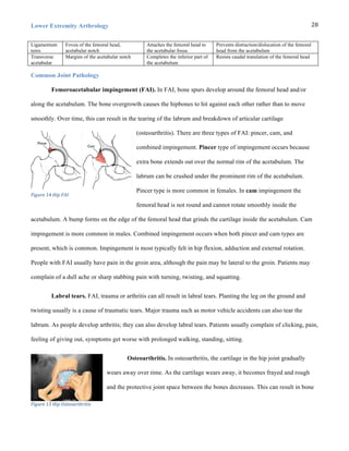
Muscles of the Knee Joint Complex
Muscles Proximal attachment Distal attachment Action Segmental
Innervation
Peripheral
innervation
Sartorius anterior superior iliac
spine
medial aspect of the proximal
tibia
flexes and assists internal
rotation of the knee
(L2-3 [4]) Femoral nerve
Rectus
femoris
anterior inferior iliac spine
and groove superior to the
acetabulum
the base of the patella extends knee (L2-3-4) Femoral nerve
Vastus
intermedius
anterior aspect of the
proximal 2/3rds of the
femoral shaft
lateral border of the patella
actions- extends knee
Extends knee (L2-3-4) Femoral nerve
Vastus
lateralis
Intertrochanteric line,
greater trochanter, gluteal
tuberosity and linea aspera
Base and lateral border of the
patella
Extends knee (L2-3-4) Femoral nerve
Vastus
medialis
Intertrochanteric line,
spiral line, linea aspera
and medial supracondylar
line
Base and medial border of the
patella
Extends knee (L2-3-4) Femoral nerve
Tensor
fasciae latae
ASIS & external lip iliac
crest
iliotibial tract assists in maintaining
knee extension
(L4-5-S1) Superior
gluteal nerve
Gracilis body of the pubis &
inferior pubic ramus
medial surface of tibia, distal
to condyle, proximal
to insertion of semitendinosus,
lateral to insertion of sartorius
flexes & medially rotates
the knee
(L2-3-4) Obturator
nerve
Biceps
femoris
ischial tuberosity &
sacrotuberous lig. (long
head) ; lateral lip of linea
aspera & lateral
supracondylar line (short
head)
lateral side of fibular head Both heads: Flex knee
Long Head: Extends hip
Long head:
(L5-S1-2-3)
Short head:
(L5-S1-2)
Long head:
tibial branch of
sciatic nerve
Short head:
Fibular branch
of sciatic nerve
Semimembr
anosus
Posterior aspect of the
medial tibial condyle
posterior aspect of the medial
tibial condyle
Ischial tuberosity (L4-5-S1-2) Tibial division
of the sciatic
Semitendin
osus
ischial tuberosity proximal, medial tibia flexes & medially rotates
knee
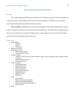
(L4-5-S1-2) Tibial division
of the sciatic
Gastrocnem
ius
posterior aspect of the
condyles and joint capsule
Posterior calcaneal surface flexes knee (S1-2) Tibial nerve
Popliteus lateral femoral condyle
and oblique popliteal
ligament
Soleal line of the tibia In NWB, IR of tibia and
knee flexion; in WB
insertion is fixed:
ER of femur and knee
flexion; unlocks the knee
from extension into early
flexion
(L4-5-S1) Tibial nerve
Articularis
Genu
Distal anterior shaft of
femur
Proximal portion of synovial
membrane of knee joint
Pulls articular capsule
proximally
(L2-3-4) Femoral](https://image.slidesharecdn.com/finallearthrologyguidetable25-151030233803-lva1-app6891/85/Final-le-arthrology-guide-table-25-31-320.jpg)


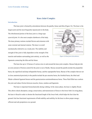
![Lower Extremity Arthrology Guide 49
Muscles of the Ankle Joint Complex
Muscle Proximal Attachment Distal Attachment Innervation Action
Anterior Crural Muscles
Extensor
Digitorum
Longus
Lateral tibial condyle,
proximal ¾ of the fibula and
interosseus mem
Dorsal digital
expansions of toes 2-
5
Deep fibular nerve (L4-
5-S1)
Dorsiflexes ankle, and extends
digits 2-5 (IP and MP)
Extensor
Hallucis
Longus
Middle ½ of fibular surface &
interosseous mem
Distal phalangeal
base of the 1st toe
Deep fibular nerve (L4-
5-S1)
Dorsiflexes ankle and extends
great toe (MP and IP)
Fibularis
Tertius
Distal fibula and interosseus
membrane
Base of the 5th
metatarsal
Deep fibular nerve (L4-
5-S1)
Dorsiflexes ankle and everts
foot
Tibialis
Anterior
Lateral condyle and proximal
2/3 of the tibia’s lateral
surface and interosseus
membrane
Medial cuneiform
and adjacent 1st
metatarsal
Deep fibular nerve (L4-
5-S1)
Dorsiflexes ankle and inverts
foot
Lateral Crural Muscles
Fibularis
Longus
Head and proximal 2/3 of the
fibula
Lateral aspects of the
1st metatarsal and
adjacent medial
cuneiform
Superficial fibular
(fibular) nerve (L4-5-S1)
Everts foot, plantarflexes
ankle and depresses 1st
metatarsal head
Fibularis
Brevis
Distal 2/3 of the fibula Lateral base of the
5th metatarsal
Superficial fibular
(fibular) nerve (L4-5-S1)
Everts foot and plantarflexes
ankle
Posterior Crural Muscles
Flexor
Digitorum
Longus
Posterior tibia distal to the
soleal line
Plantar surfaces of
the distal phalangeal
bases
Tibial nerve (L5-S1 [2]) Plantarflexes ankle and flexes
digits 2-5 (MP and IP)
Flexor
Hallucis
Longus
Distal 2/3 of the posterior
fibular surface and interosseus
membrane
Plantar aspect of the
distal phalangeal base
of the 1st toe
Tibial nerve (L5-S1-2) Plantarflexes ankle and flexes
great toe (MP and IP)
Gastrocnemius Posterior aspect of the
femoral condyles and joint
capsule
Posterior calcaneal
surface
Tibial nerve (S1-2) Flexes knee and plantarflexes
ankle
Plantaris Lateral supracondylar line Posterior calcaneal
surface
Tibial nerve (L4-5-S1
[2])
Flexes knee and plantarflexes
ankle
Popliteus Lateral femoral condyle and
oblique popliteal ligament
Soleal line of the
tibia
Tibial nerve (L4-5-S1) In NWB, IR of tibia and knee
flexion; In WB insertion is
fixed: ER of femur and knee
flexion; Unlocks knee
extension to early flexion
Soleus Post aspect of the head &
proximal ¼ of the fibula and
tibial soleal line
Posterior calcaneal
surface
Tibial nerve (L5-S1-2) Plantarflexes ankle
Tibialis
Posterior
Interosseous membrane,
lateral tibial surface and
medial fibular surface
Navicular,
intermediate
cuneiform and bases
of metatarsals 2-4
Tibial nerve ([L4]L5-S1) Inverts foot and plantarflexes
ankle](https://image.slidesharecdn.com/finallearthrologyguidetable25-151030233803-lva1-app6891/85/Final-le-arthrology-guide-table-25-49-320.jpg)


![Lower Extremity Arthrology Guide 103
Reference List
Adams E, Madden C. Cuboid subluxation: a case study and review of the literature. Current sports medicine
reports. Nov-Dec 2009;8(6):300-307.
Adams, T., Band-Entrup, D., Kuhn, S., Legere, L., Mace, K., Paggi, A. and Penney, M. (2013). Physical Therapy
Management of Knee Osteoarthritis in the Middle-aged Athlete. Sports Medicine and Arthroscopy
Review, 21 (1), p.2–10. [Online]. Available at: doi:10.1097/jsa.0b013e318272f530.
Becker I, Woodley SJ, Stringer MD. The adult human pubic symphysis: a systematic review. Journal of anatomy.
Nov 2010;217(5):475-487.
Bookstein, N. 2014. Lower Extremity Study Guide
Brunnstrom S, Lehmkuhl LD, Smith LK. Brunnstrom's Clinical kinesiology. 4th ed. Philadelphia: F.A. Davis Co.;
1983.
Christiansen, C. (2015). Knee Joint Functional Anatomy.
Christiansen, C. (2015). Fundamentals of Gait.
Cook, Orthopedic Manual Therapy: An Evidence-Based Approach, 2/E © 2012 by Pearson Education, Inc.,
Upper Saddle River, NJ
Davies MS, Saxby TS. Intercuneiform instability and the "gap" sign. Foot & ankle international. Sep
1999;20(9):606-609.
DiDomenico LA, Cross D. Tarsometatarsal/Lisfranc joint. Clinics in podiatric medicine and surgery. Apr
2012;29(2):221-242, vii-viii.
Foley BS, Buschbacher RM. Sacroiliac joint pain: anatomy, biomechanics, diagnosis, and treatment. Am J Phys
Med Rehabil. 2006;85(12):997-1006.
Forst SL, Wheeler MT, Fortin JD, Vilensky JA. The sacroiliac joint: anatomy, physiology and clinical
significance. Pain Physician. 2006;9(1):61-7.
Gilroy AM, MacPherson BR, Ross LM et al. Atlas of Anatomy. Thieme Medical Pub; 2012: 406-423.
Glasoe WM, Yack HJ, Saltzman CL. Anatomy and biomechanics of the first ray. Phys Ther. Sep 1999;79(9):854-
859.
Goldstein Y, Gold A, Chechik O, Drexler M. Dislocation of the proximal tibiofibular joint: a rare sports-related
injury. The Israel Medical Association journal : IMAJ. 2011;13(1):62-63.
Grant’s Atlas of Anatomy, Thirteenth Edition.
Ibrahim A, El-Sherbini A (1961) The different ligaments of the
symphysis
pubis
in
the
pregnant
and the non-
pregnant state.
J Obstet Gynaecol Br Emp 68, 592–596.](https://image.slidesharecdn.com/finallearthrologyguidetable25-151030233803-lva1-app6891/85/Final-le-arthrology-guide-table-25-103-320.jpg)
![Lower Extremity Arthrology
104
J K Loudon SLB. The foot and ankle: an overview of arthrokinematics and selected joint techniques. Journal of
athletic training. 1996;31(2):173-178.
James, D. (2015). Knee Soft Tissue Pathology 2015
Kendall, FP. Muscles: testing and function with posture and pain. 5th ed. Baltimore, MD: Lippincott Williams &
Wilkins; 2005.
Kura H, Luo ZP, Kitaoka HB, Smutz WP, An KN. Mechanical behavior of the Lisfranc and dorsal
cuneometatarsal ligaments: in vitro biomechanical study. Journal of orthopaedic trauma. Feb
2001;15(2):107-110.
Lee, K., Park., Y, Jegal, H., Young, K., Kim, J., Lim, S. Osteoarthritis of the second metatarsophalangeal joint
associated with hallux valgus deformity. Foot Ankle Int. 2014 35(12):1329-33.
Maceira E, Monteagudo M. Subtalar Anatomy and Mechanics. Foot Ankle Clin. 2015. 20(2):195-221.
Mintken, P. (2015). Knee Ligamentous Injuries.
Mintken, P. (2015). Meniscal Injuries
Moore, K. L., Dally, A. F. and Agur, A. M. R. (2010). Clinically Oriented Anatomy. 6th edition. Montalbano, J.
(ed.). Lippincott Williams & Wilkins.
Moore, K., Dalley, A., Agur, A. Clinically Oriented Anatomy. Seventh Edition.
Morelli, V., Bright, C. and Fields, A. (2013). Ligamentous Injuries of the Knee. Primary Care: Clinics in Office
Practice, 40 (2), p.335–356. [Online]. Available at: doi:10.1016/j.pop.2013.02.004
Neumann, D. A. and Kelly, E. R. (2010). Kinesiology of the Musculoskeletal System: Foundations for
Rehabilitation. 2nd ed. United States: Elsevier Health Sciences.
Netter FH. Atlas of human anatomy. 5th ed. Philadelphia, PA: Saunders/Elsevier; 2010.
Norkin CC, White DJ. Measurement of Joint Motion, A Guide to Goniometry. F A Davis Company; 2009: 241-
242.
Scott SH, Winter DA. Biomechanical model of the human foot: kinematics and kinetics during the stance phase of
walking. Journal of biomechanics. Sep 1993;26(9):1091-1104.
Solan MC, Moorman CT, 3rd, Miyamoto RG, Jasper LE, Belkoff SM. Ligamentous restraints of the second
tarsometatarsal joint: a biomechanical evaluation. Foot & ankle international. Aug 2001;22(8):637-641.
Vleeming A, Schuenke MD, Masi AT, Carreiro JE, Danneels L, Willard FH. The sacroiliac joint: an overview of
its anatomy, function and potential clinical implications. J Anat. 2012;221(6):537-67.](https://image.slidesharecdn.com/finallearthrologyguidetable25-151030233803-lva1-app6891/85/Final-le-arthrology-guide-table-25-104-320.jpg)


![Gcse workbook[1]](https://cdn.slidesharecdn.com/ss_thumbnails/gcseworkbook1-120509024110-phpapp02-thumbnail.jpg?width=640&height=640&fit=bounds)


