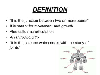
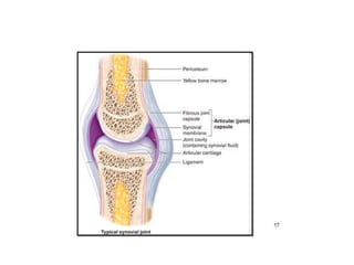
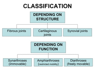
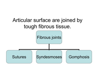
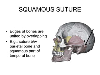
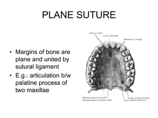
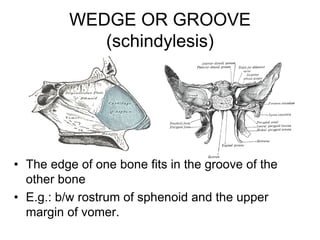
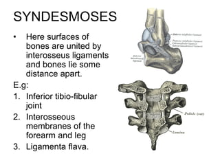
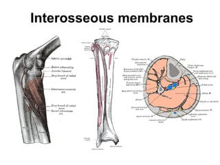
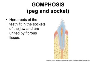
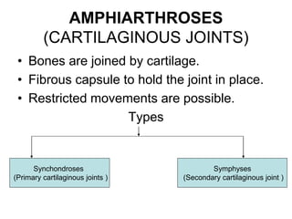
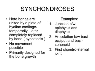
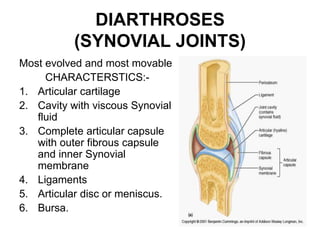
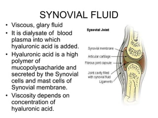
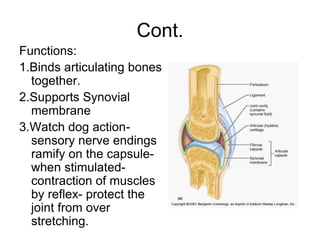
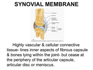
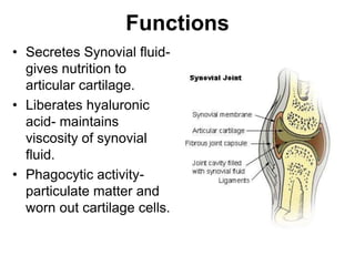
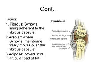
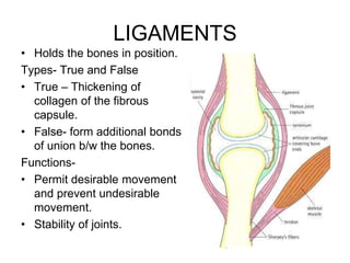
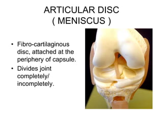
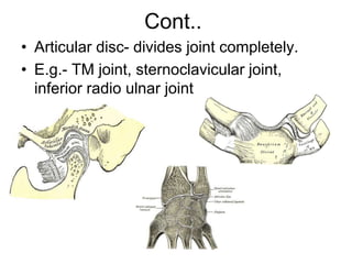
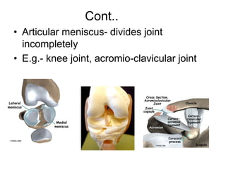
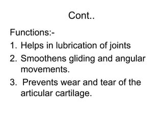
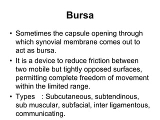
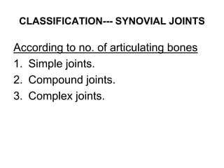
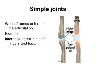
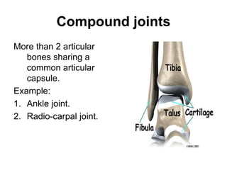
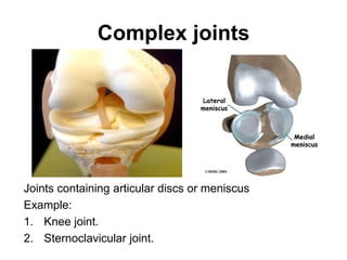
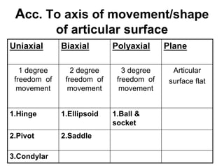
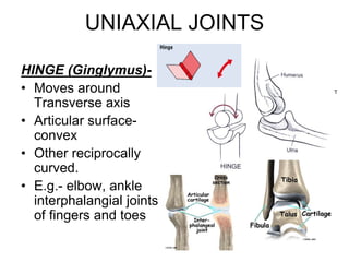
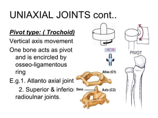
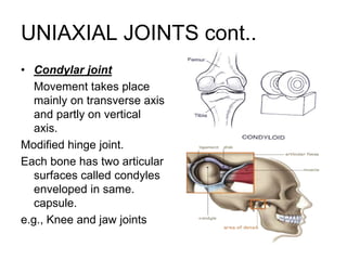
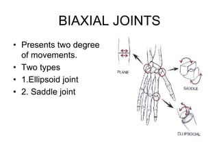
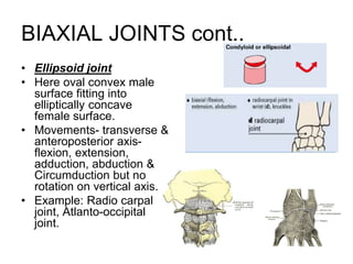
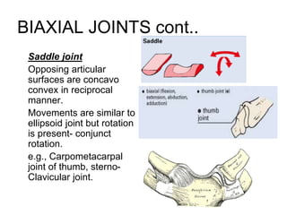
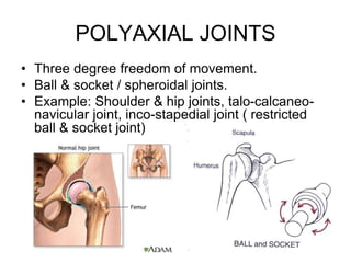
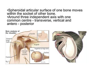
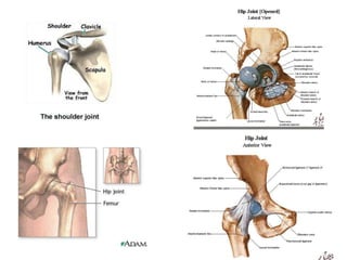
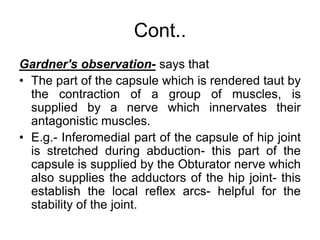
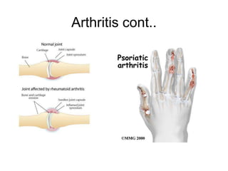
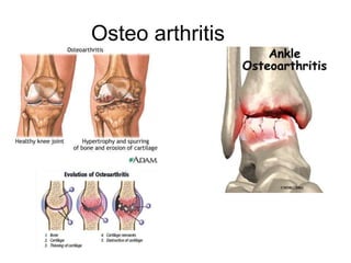
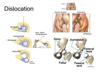
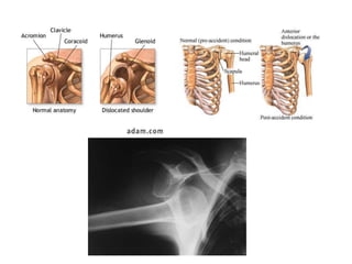
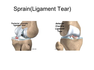
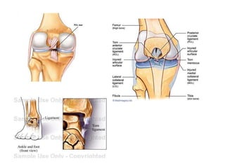
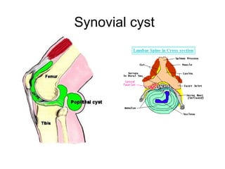
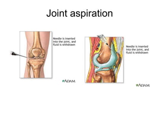
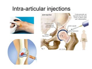
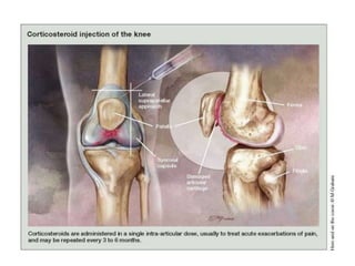
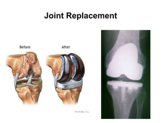
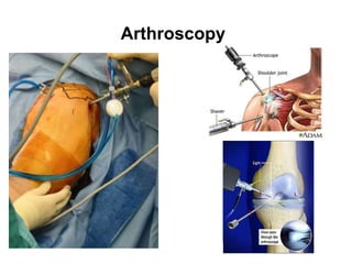
JOINTS
JOINTS
• DEFINITION
• CLASSIFICATION
• SYNOVIAL JOINTS
• BLOOD SUPPLY
• NERVE SUPPLY
• KINESIOLOGY
• APPLIED ASPECTS
DEFINITION
• “It is the junction between two or more bones”
• It is meant for movement and growth.
• Also called as articulation
• ARTHROLOGY:-
• “It is the science which deals with the study of
joints”
CLASSIFICATION
DEPENDING ON
STRUCTURE
Cartilaginous
joints
Fibrous joints Synovial joints
Amphiarthroses
(restricted mobility)
Synarthroses
(immovable)
Diarthroses
(freely movable)
DEPENDING ON
FUNCTION
Articular surface are joined by
tough fibrous tissue.
Fibrous joints
Sutures Syndesmoses Gomphosis
SUTURES
• Most of the Joints of the skull
• Appear b/w the bones which ossify in membranes except
clavicle
Suture membranes (Ligaments) present between opposed
margins
They binds periosteum and promote bone growth.
Ossification till twenties, then resulting in synostosis.
SERRATE SUTURE
• Edges of the bones present saw toothed
appearance
• E.g.: sagittal suture
DENTICULATE SUTURE
• The margins looks
like teeth, with the tips
being broader than
roots.
• E.g.: Lambdoid suture
SQUAMOUS SUTURE
• Edges of bones are
united by overlapping
• E.g.: suture b/w
parietal bone and
squamous part of
temporal bone
PLANE SUTURE
• Margins of bone are
plane and united by
sutural ligament
• E.g.: articulation b/w
palatine process of
two maxillae
WEDGE OR GROOVE
(schindylesis)
• The edge of one bone fits in the groove of the
other bone
• E.g.: b/w rostrum of sphenoid and the upper
margin of vomer.
SYNDESMOSES
• Here surfaces of
bones are united by
interosseus ligaments
and bones lie some
distance apart.
E.g:
1. Inferior tibio-fibular
joint
2. Interosseous
membranes of the
forearm and leg
3. Ligamenta flava.
Interosseous membranes
GOMPHOSIS
(peg and socket)
• Here roots of the
teeth fit in the sockets
of the jaw and are
united by fibrous
tissue.
AMPHIARTHROSES
(CARTILAGINOUS JOINTS)
• Bones are joined by cartilage.
• Fibrous capsule to hold the joint in place.
• Restricted movements are possible.
Types
Synchondroses
(Primary cartilaginous joints )
Symphyses
(Secondary cartilaginous joint )
SYNCHONDROSES
• Here bones are
united by a plate of
hyaline cartilage-
temporarily –later
completely replaced
by bone ( synostosis )
• No movement
possible
• Primarily designed for
the bone growth
Examples:
1. Junction b/w
epiphysis and
diaphysis
2. Articulation b/w basi-
occiput and basi-
sphenoid
3. First chondro-sternal
joint
Junction b/w epiphysis
and diaphysis
Articulation b/w basi-occiput and
basi-sphenoid
• Synostosis at 25
years
• Early Synostosis
before eruption of
permanent teeth
leads to dental
malocclution (short
maxilla)
First chondro-sternal joint
SYMPHYSES
• Articular surface covered by hyaline cartilage
and are united by a plate of fibro cartilage.
• Some times the joints are enveloped by
incomplete fibrous capsule
• They persists through out life and are in median
plane of the body
• Limited movement is possible due to
compression of fibro cartilage.
• Thickness of fibro cartilage is related to range of
movement.
• Represents intermediate stage in evolution of
synovial joints.
1. Intervertebral discs
• Structure
• Annulus fibrosus- concentric layer of fibers.
• Nucleus pulposus- gelatinous- water
• cartilage cells, multinucleated notochordal cells
• Functions: shock-absorber, resistance to
compression
• Present in anterior arch of pelvis in between body
of two pubic bone.
• When medial thrusts of femoral head are
transmitted through the anterior arch- interpubic
disc resists the force by acting as a shock
absorber.
2. Symphysis pubis
3. Manubrio-sternal joint
• Pump handle movement
responsible for increase
in anteroposterior
diameter.
• After 60 years partly or
completely replaced by
bone.
DIARTHROSES
(SYNOVIAL JOINTS)
Most evolved and most movable
CHARACTERSTICS:-
1. Articular cartilage
2. Cavity with viscous Synovial
fluid
3. Complete articular capsule
with outer fibrous capsule
and inner Synovial
membrane
4. Ligaments
5. Articular disc or meniscus.
6. Bursa.
ARTICULAR CARTILAGE
• Hyaline cartilage
• Avascular. Non-nervous,
elastic.
• Damaged- cannot be
replaced by hyaline
cartilage but by fibrous
tissue - indispensable.
• On convex articular
surface.
• On concave articular
surface.
Cont..
Functions:
• Smooth gliding surface and reduce the
force of compression or muscle action.
• Coefficient of friction is equal to ice on ice.
• The surface shows undulations - filled with
Synovial fluid - i.e...., it is extremely porous
and absorbs Synovial fluid in resting
condition – on compression fluid is
squeezed out.
• Regulates the epiphysial growth.
Cont..
Nutrition:
1. From Synovial fluid
2. By diffusion from the capillaries at the
periphery of the articular cartilage
3. By diffusion from the adjacent epiphysial
blood vessels.
SYNOVIAL FLUID
• Viscous, glary fluid
• It is dialysate of blood
plasma into which
hyaluronic acid is added.
• Hyaluronic acid is a high
polymer of
mucopolysacharide and
secreted by the Synovial
cells and mast cells of
Synovial membrane.
• Viscosity depends on
concentration of
hyaluronic acid.
Cont..
Alkaline
Cellular contents:
• Monocytes,
lymphocytes,
macrophages,
Synovial cells and
few leucocytes.
• Proteins traces.
Cont..
Functions:
1.Nutrition to articular capsule
2.Lubrication of joint cavity to prevent wear and
tear:- this is helped by,
A) Incongruous articular surfaces- this provides
space for the flushing of fluid- Synovial fluid
spread as an elastic fluid film over moving
articular surface.
B) Viscosity of fluid maintains the lubrication, dep on
concentration of hyaluronic acid.
C) More movements of the joint increase the
lubrication.
Joint cracks
ARTICULAR CAPSULE
• Outer- fibrous capsule
• Inner- Synovial
membrane.
FIBROUS CAPSULE-
• Completely invests joint
• Attached on the bones
close to articular cartilage
continuously.
• Formed by bundles of
collagen fibers-irregular
spirals-
• Sensitive to changes of
position of joint.
Cont.
Functions:
1.Binds articulating bones
together.
2.Supports Synovial
membrane
3.Watch dog action-
sensory nerve endings
ramify on the capsule-
when stimulated-
contraction of muscles
by reflex- protect the
joint from over
stretching.
SYNOVIAL MEMBRANE
Highly vascular & cellular connective
tissue- lines inner aspects of fibrous capsule
& bones lying within the joint- but cease at
the periphery of the articular capsule,
articular disc or meniscus.
Functions
• Secretes Synovial fluid-
gives nutrition to
articular cartilage.
• Liberates hyaluronic
acid- maintains
viscosity of synovial
fluid.
• Phagocytic activity-
particulate matter and
worn out cartilage cells.
Cont..
Types:
1. Fibrous: Synovial
lining adherent to the
fibrous capsule
2.Areolar: where
Synovial membrane
freely moves over the
fibrous capsule
3.Adipose: covers intra
articular pad of fat.
LIGAMENTS
• Holds the bones in position.
Types- True and False
• True – Thickening of
collagen of the fibrous
capsule.
• False- form additional bonds
of union b/w the bones.
Functions-
• Permit desirable movement
and prevent undesirable
movement.
• Stability of joints.
ARTICULAR DISC
( MENISCUS )
• Fibro-cartilaginous
disc, attached at the
periphery of capsule.
• Divides joint
completely/
incompletely.
Cont..
• Articular disc- divides joint completely.
• E.g.- TM joint, sternoclavicular joint,
inferior radio ulnar joint
Cont..
• Articular meniscus- divides joint
incompletely
• E.g.- knee joint, acromio-clavicular joint
Cont..
Functions:-
1. Helps in lubrication of joints
2. Smoothens gliding and angular
movements.
3. Prevents wear and tear of the
articular cartilage.
Bursa
• Sometimes the capsule opening through
which synovial membrane comes out to
act as bursa.
• It is a device to reduce friction between
two mobile but tightly opposed surfaces,
permitting complete freedom of movement
within the limited range.
• Types : Subcutaneous, subtendinous,
sub muscular, subfacial, inter ligamentous,
communicating.
CLASSIFICATION--- SYNOVIAL JOINTS
According to no. of articulating bones
1. Simple joints.
2. Compound joints.
3. Complex joints.
Simple joints
When 2 bones enters in
the articulation.
Example:
Interphalangeal joints of
fingers and toes.
Compound joints
More than 2 articular
bones sharing a
common articular
capsule.
Example:
1. Ankle joint.
2. Radio-carpal joint.
Complex joints
Joints containing articular discs or meniscus
Example:
1. Knee joint.
2. Sternoclavicular joint.
Acc. To axis of movement/shape
of articular surface
Uniaxial Biaxial Polyaxial Plane
1 degree
freedom of
movement
2 degree
freedom of
movement
3 degree
freedom of
movement
Articular
surface flat
1.Hinge 1.Ellipsoid 1.Ball &
socket
2.Pivot 2.Saddle
3.Condylar
UNIAXIAL JOINTS
HINGE (Ginglymus)-
• Moves around
Transverse axis
• Articular surface-
convex
• Other reciprocally
curved.
• E.g.- elbow, ankle
interphalangial joints
of fingers and toes
UNIAXIAL JOINTS cont..
Pivot type: ( Trochoid)
Vertical axis movement
One bone acts as pivot
and is encircled by
osseo-ligamentous
ring
E.g.1. Atlanto axial joint
2. Superior & inferior
radioulnar joints.
UNIAXIAL JOINTS cont..
• Condylar joint
Movement takes place
mainly on transverse axis
and partly on vertical
axis.
Modified hinge joint.
Each bone has two articular
surfaces called condyles
enveloped in same.
capsule.
e.g., Knee and jaw joints
BIAXIAL JOINTS
• Presents two degree
of movements.
• Two types
• 1.Ellipsoid joint
• 2. Saddle joint
BIAXIAL JOINTS cont..
• Ellipsoid joint
• Here oval convex male
surface fitting into
elliptically concave
female surface.
• Movements- transverse &
anteroposterior axis-
flexion, extension,
adduction, abduction &
Circumduction but no
rotation on vertical axis.
• Example: Radio carpal
joint, Atlanto-occipital
joint.
BIAXIAL JOINTS cont..
Saddle joint
Opposing articular
surfaces are concavo
convex in reciprocal
manner.
Movements are similar to
ellipsoid joint but rotation
is present- conjunct
rotation.
e.g., Carpometacarpal
joint of thumb, sterno-
Clavicular joint.
POLYAXIAL JOINTS
• Three degree freedom of movement.
• Ball & socket / spheroidal joints.
• Example: Shoulder & hip joints, talo-calcaneo-
navicular joint, inco-stapedial joint ( restricted
ball & socket joint)
•Spheroidal articular surface of one bone moves
within the socket of other bone.
•Around three independent axis with one
common centre - transverse, vertical and
antero - posterior
Blood supply
• Epiphyseal vessels- Enter the long bone at or
near the attachment of the fibrous capsule- give
articular branches- form rich capillary plexus in
Synovial membrane- circulus vasculosus
• These end around the articular margin in a
fringe of looped anastomosis.
Nerve supply
• Articular capsule and ligaments have rich nerve
supply.
1.Sensory 2.Autonomic ( vasomotor)
• Sensory- form Ruffini endings and paccinian
corpuscles- convey proprioceptive sensation-
concerned with control of posture, locomotion
and perception of position and movement.
• Some sensory fibers form free nerve endings-
pain sensation.
Cont..
Hiltons law- says that,
• Nerves which supply the joint also give
branches to the muscles regulating the
movements of the joint and skin over the
joint.
• Irritation of the nerve in joint disease-
reflex spasm of the muscles and fix the
joint in the position of comfort and the pain
referred to the skin.
Cont..
Gardner's observation- says that
• The part of the capsule which is rendered taut by
the contraction of a group of muscles, is
supplied by a nerve which innervates their
antagonistic muscles.
• E.g.- Inferomedial part of the capsule of hip joint
is stretched during abduction- this part of the
capsule is supplied by the Obturator nerve which
also supplies the adductors of the hip joint- this
establish the local reflex arcs- helpful for the
stability of the joint.
• LAST’S FORMULATION ( R.J LAST)
1.Four contiguous spinal segments regulate
movements of a particular joint : upper two
segments control one movement, lower
two segments regulate opposite
movement.
2.For a joint one segment more distal in the
limb, the Centers lie en block, one
segment lower in the cord.
APPLIED ASPECT
• ARTHRITIS
• ARTHRALGIA
• OSTEOARTHRITIS
• DISLOCATION
• INTRA ARTICULAR FRACTURE
• LIGAMENT TEARS
• SYNOVIAL CYST
• JOINT ASPIRATION
• INTRA ARTICULAR INJECTIONS
• JOINT REPLACEMENT
• ARTHROSCOPY
Arthritis
Arthritis cont..
Osteo arthritis
Dislocation
Intra articular fracture
Sprain(Ligament Tear)
Synovial cyst
Joint aspiration
Intra-articular injections
Joint Replacement
Arthroscopy
JOINTS   .ppt

JOINTS .ppt

  • 1.
  • 2.
    JOINTS • DEFINITION • CLASSIFICATION •SYNOVIAL JOINTS • BLOOD SUPPLY • NERVE SUPPLY • KINESIOLOGY • APPLIED ASPECTS
  • 3.
    DEFINITION • “It isthe junction between two or more bones” • It is meant for movement and growth. • Also called as articulation • ARTHROLOGY:- • “It is the science which deals with the study of joints”
  • 5.
    CLASSIFICATION DEPENDING ON STRUCTURE Cartilaginous joints Fibrous jointsSynovial joints Amphiarthroses (restricted mobility) Synarthroses (immovable) Diarthroses (freely movable) DEPENDING ON FUNCTION
  • 6.
    Articular surface arejoined by tough fibrous tissue. Fibrous joints Sutures Syndesmoses Gomphosis
  • 7.
    SUTURES • Most ofthe Joints of the skull • Appear b/w the bones which ossify in membranes except clavicle Suture membranes (Ligaments) present between opposed margins They binds periosteum and promote bone growth. Ossification till twenties, then resulting in synostosis.
  • 8.
    SERRATE SUTURE • Edgesof the bones present saw toothed appearance • E.g.: sagittal suture
  • 9.
    DENTICULATE SUTURE • Themargins looks like teeth, with the tips being broader than roots. • E.g.: Lambdoid suture
  • 10.
    SQUAMOUS SUTURE • Edgesof bones are united by overlapping • E.g.: suture b/w parietal bone and squamous part of temporal bone
  • 11.
    PLANE SUTURE • Marginsof bone are plane and united by sutural ligament • E.g.: articulation b/w palatine process of two maxillae
  • 12.
    WEDGE OR GROOVE (schindylesis) •The edge of one bone fits in the groove of the other bone • E.g.: b/w rostrum of sphenoid and the upper margin of vomer.
  • 13.
    SYNDESMOSES • Here surfacesof bones are united by interosseus ligaments and bones lie some distance apart. E.g: 1. Inferior tibio-fibular joint 2. Interosseous membranes of the forearm and leg 3. Ligamenta flava.
  • 14.
  • 15.
    GOMPHOSIS (peg and socket) •Here roots of the teeth fit in the sockets of the jaw and are united by fibrous tissue.
  • 16.
    AMPHIARTHROSES (CARTILAGINOUS JOINTS) • Bonesare joined by cartilage. • Fibrous capsule to hold the joint in place. • Restricted movements are possible. Types Synchondroses (Primary cartilaginous joints ) Symphyses (Secondary cartilaginous joint )
  • 17.
    SYNCHONDROSES • Here bonesare united by a plate of hyaline cartilage- temporarily –later completely replaced by bone ( synostosis ) • No movement possible • Primarily designed for the bone growth Examples: 1. Junction b/w epiphysis and diaphysis 2. Articulation b/w basi- occiput and basi- sphenoid 3. First chondro-sternal joint
  • 18.
  • 19.
    Articulation b/w basi-occiputand basi-sphenoid • Synostosis at 25 years • Early Synostosis before eruption of permanent teeth leads to dental malocclution (short maxilla)
  • 20.
  • 21.
    SYMPHYSES • Articular surfacecovered by hyaline cartilage and are united by a plate of fibro cartilage. • Some times the joints are enveloped by incomplete fibrous capsule • They persists through out life and are in median plane of the body • Limited movement is possible due to compression of fibro cartilage. • Thickness of fibro cartilage is related to range of movement. • Represents intermediate stage in evolution of synovial joints.
  • 22.
    1. Intervertebral discs •Structure • Annulus fibrosus- concentric layer of fibers. • Nucleus pulposus- gelatinous- water • cartilage cells, multinucleated notochordal cells • Functions: shock-absorber, resistance to compression
  • 24.
    • Present inanterior arch of pelvis in between body of two pubic bone. • When medial thrusts of femoral head are transmitted through the anterior arch- interpubic disc resists the force by acting as a shock absorber. 2. Symphysis pubis
  • 25.
    3. Manubrio-sternal joint •Pump handle movement responsible for increase in anteroposterior diameter. • After 60 years partly or completely replaced by bone.
  • 26.
    DIARTHROSES (SYNOVIAL JOINTS) Most evolvedand most movable CHARACTERSTICS:- 1. Articular cartilage 2. Cavity with viscous Synovial fluid 3. Complete articular capsule with outer fibrous capsule and inner Synovial membrane 4. Ligaments 5. Articular disc or meniscus. 6. Bursa.
  • 27.
    ARTICULAR CARTILAGE • Hyalinecartilage • Avascular. Non-nervous, elastic. • Damaged- cannot be replaced by hyaline cartilage but by fibrous tissue - indispensable. • On convex articular surface. • On concave articular surface.
  • 28.
    Cont.. Functions: • Smooth glidingsurface and reduce the force of compression or muscle action. • Coefficient of friction is equal to ice on ice. • The surface shows undulations - filled with Synovial fluid - i.e...., it is extremely porous and absorbs Synovial fluid in resting condition – on compression fluid is squeezed out. • Regulates the epiphysial growth.
  • 29.
    Cont.. Nutrition: 1. From Synovialfluid 2. By diffusion from the capillaries at the periphery of the articular cartilage 3. By diffusion from the adjacent epiphysial blood vessels.
  • 30.
    SYNOVIAL FLUID • Viscous,glary fluid • It is dialysate of blood plasma into which hyaluronic acid is added. • Hyaluronic acid is a high polymer of mucopolysacharide and secreted by the Synovial cells and mast cells of Synovial membrane. • Viscosity depends on concentration of hyaluronic acid.
  • 31.
  • 32.
    Cont.. Functions: 1.Nutrition to articularcapsule 2.Lubrication of joint cavity to prevent wear and tear:- this is helped by, A) Incongruous articular surfaces- this provides space for the flushing of fluid- Synovial fluid spread as an elastic fluid film over moving articular surface. B) Viscosity of fluid maintains the lubrication, dep on concentration of hyaluronic acid. C) More movements of the joint increase the lubrication. Joint cracks
  • 33.
    ARTICULAR CAPSULE • Outer-fibrous capsule • Inner- Synovial membrane. FIBROUS CAPSULE- • Completely invests joint • Attached on the bones close to articular cartilage continuously. • Formed by bundles of collagen fibers-irregular spirals- • Sensitive to changes of position of joint.
  • 34.
    Cont. Functions: 1.Binds articulating bones together. 2.SupportsSynovial membrane 3.Watch dog action- sensory nerve endings ramify on the capsule- when stimulated- contraction of muscles by reflex- protect the joint from over stretching.
  • 35.
    SYNOVIAL MEMBRANE Highly vascular& cellular connective tissue- lines inner aspects of fibrous capsule & bones lying within the joint- but cease at the periphery of the articular capsule, articular disc or meniscus.
  • 36.
    Functions • Secretes Synovialfluid- gives nutrition to articular cartilage. • Liberates hyaluronic acid- maintains viscosity of synovial fluid. • Phagocytic activity- particulate matter and worn out cartilage cells.
  • 37.
    Cont.. Types: 1. Fibrous: Synovial liningadherent to the fibrous capsule 2.Areolar: where Synovial membrane freely moves over the fibrous capsule 3.Adipose: covers intra articular pad of fat.
  • 38.
    LIGAMENTS • Holds thebones in position. Types- True and False • True – Thickening of collagen of the fibrous capsule. • False- form additional bonds of union b/w the bones. Functions- • Permit desirable movement and prevent undesirable movement. • Stability of joints.
  • 39.
    ARTICULAR DISC ( MENISCUS) • Fibro-cartilaginous disc, attached at the periphery of capsule. • Divides joint completely/ incompletely.
  • 40.
    Cont.. • Articular disc-divides joint completely. • E.g.- TM joint, sternoclavicular joint, inferior radio ulnar joint
  • 41.
    Cont.. • Articular meniscus-divides joint incompletely • E.g.- knee joint, acromio-clavicular joint
  • 42.
    Cont.. Functions:- 1. Helps inlubrication of joints 2. Smoothens gliding and angular movements. 3. Prevents wear and tear of the articular cartilage.
  • 43.
    Bursa • Sometimes thecapsule opening through which synovial membrane comes out to act as bursa. • It is a device to reduce friction between two mobile but tightly opposed surfaces, permitting complete freedom of movement within the limited range. • Types : Subcutaneous, subtendinous, sub muscular, subfacial, inter ligamentous, communicating.
  • 44.
    CLASSIFICATION--- SYNOVIAL JOINTS Accordingto no. of articulating bones 1. Simple joints. 2. Compound joints. 3. Complex joints.
  • 45.
    Simple joints When 2bones enters in the articulation. Example: Interphalangeal joints of fingers and toes.
  • 46.
    Compound joints More than2 articular bones sharing a common articular capsule. Example: 1. Ankle joint. 2. Radio-carpal joint.
  • 47.
    Complex joints Joints containingarticular discs or meniscus Example: 1. Knee joint. 2. Sternoclavicular joint.
  • 48.
    Acc. To axisof movement/shape of articular surface Uniaxial Biaxial Polyaxial Plane 1 degree freedom of movement 2 degree freedom of movement 3 degree freedom of movement Articular surface flat 1.Hinge 1.Ellipsoid 1.Ball & socket 2.Pivot 2.Saddle 3.Condylar
  • 49.
    UNIAXIAL JOINTS HINGE (Ginglymus)- •Moves around Transverse axis • Articular surface- convex • Other reciprocally curved. • E.g.- elbow, ankle interphalangial joints of fingers and toes
  • 50.
    UNIAXIAL JOINTS cont.. Pivottype: ( Trochoid) Vertical axis movement One bone acts as pivot and is encircled by osseo-ligamentous ring E.g.1. Atlanto axial joint 2. Superior & inferior radioulnar joints.
  • 51.
    UNIAXIAL JOINTS cont.. •Condylar joint Movement takes place mainly on transverse axis and partly on vertical axis. Modified hinge joint. Each bone has two articular surfaces called condyles enveloped in same. capsule. e.g., Knee and jaw joints
  • 52.
    BIAXIAL JOINTS • Presentstwo degree of movements. • Two types • 1.Ellipsoid joint • 2. Saddle joint
  • 53.
    BIAXIAL JOINTS cont.. •Ellipsoid joint • Here oval convex male surface fitting into elliptically concave female surface. • Movements- transverse & anteroposterior axis- flexion, extension, adduction, abduction & Circumduction but no rotation on vertical axis. • Example: Radio carpal joint, Atlanto-occipital joint.
  • 54.
    BIAXIAL JOINTS cont.. Saddlejoint Opposing articular surfaces are concavo convex in reciprocal manner. Movements are similar to ellipsoid joint but rotation is present- conjunct rotation. e.g., Carpometacarpal joint of thumb, sterno- Clavicular joint.
  • 55.
    POLYAXIAL JOINTS • Threedegree freedom of movement. • Ball & socket / spheroidal joints. • Example: Shoulder & hip joints, talo-calcaneo- navicular joint, inco-stapedial joint ( restricted ball & socket joint)
  • 56.
    •Spheroidal articular surfaceof one bone moves within the socket of other bone. •Around three independent axis with one common centre - transverse, vertical and antero - posterior
  • 59.
    Blood supply • Epiphysealvessels- Enter the long bone at or near the attachment of the fibrous capsule- give articular branches- form rich capillary plexus in Synovial membrane- circulus vasculosus • These end around the articular margin in a fringe of looped anastomosis.
  • 60.
    Nerve supply • Articularcapsule and ligaments have rich nerve supply. 1.Sensory 2.Autonomic ( vasomotor) • Sensory- form Ruffini endings and paccinian corpuscles- convey proprioceptive sensation- concerned with control of posture, locomotion and perception of position and movement. • Some sensory fibers form free nerve endings- pain sensation.
  • 61.
    Cont.. Hiltons law- saysthat, • Nerves which supply the joint also give branches to the muscles regulating the movements of the joint and skin over the joint. • Irritation of the nerve in joint disease- reflex spasm of the muscles and fix the joint in the position of comfort and the pain referred to the skin.
  • 62.
    Cont.. Gardner's observation- saysthat • The part of the capsule which is rendered taut by the contraction of a group of muscles, is supplied by a nerve which innervates their antagonistic muscles. • E.g.- Inferomedial part of the capsule of hip joint is stretched during abduction- this part of the capsule is supplied by the Obturator nerve which also supplies the adductors of the hip joint- this establish the local reflex arcs- helpful for the stability of the joint.
  • 63.
    • LAST’S FORMULATION( R.J LAST) 1.Four contiguous spinal segments regulate movements of a particular joint : upper two segments control one movement, lower two segments regulate opposite movement. 2.For a joint one segment more distal in the limb, the Centers lie en block, one segment lower in the cord.
  • 64.
    APPLIED ASPECT • ARTHRITIS •ARTHRALGIA • OSTEOARTHRITIS • DISLOCATION • INTRA ARTICULAR FRACTURE • LIGAMENT TEARS • SYNOVIAL CYST • JOINT ASPIRATION • INTRA ARTICULAR INJECTIONS • JOINT REPLACEMENT • ARTHROSCOPY
  • 65.
  • 66.
  • 67.
  • 68.
  • 70.
  • 72.
  • 74.
  • 75.
  • 77.
  • 79.
  • 80.