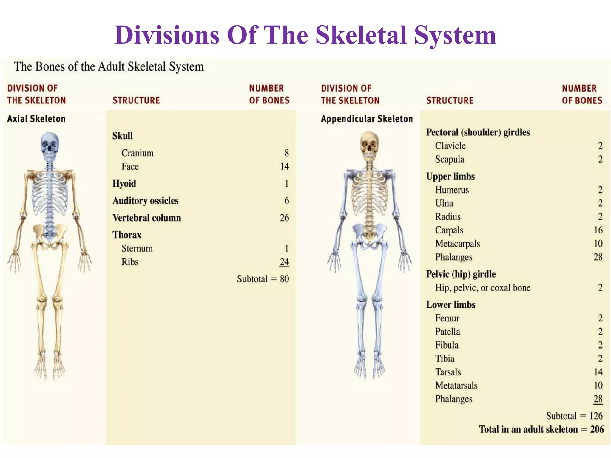
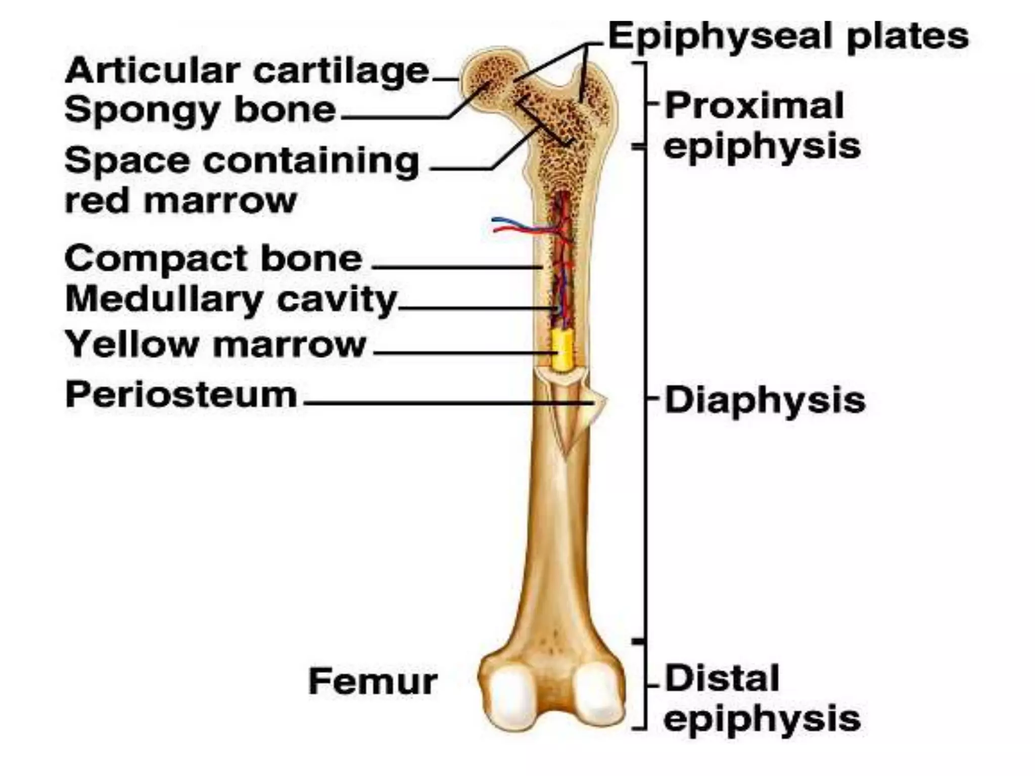
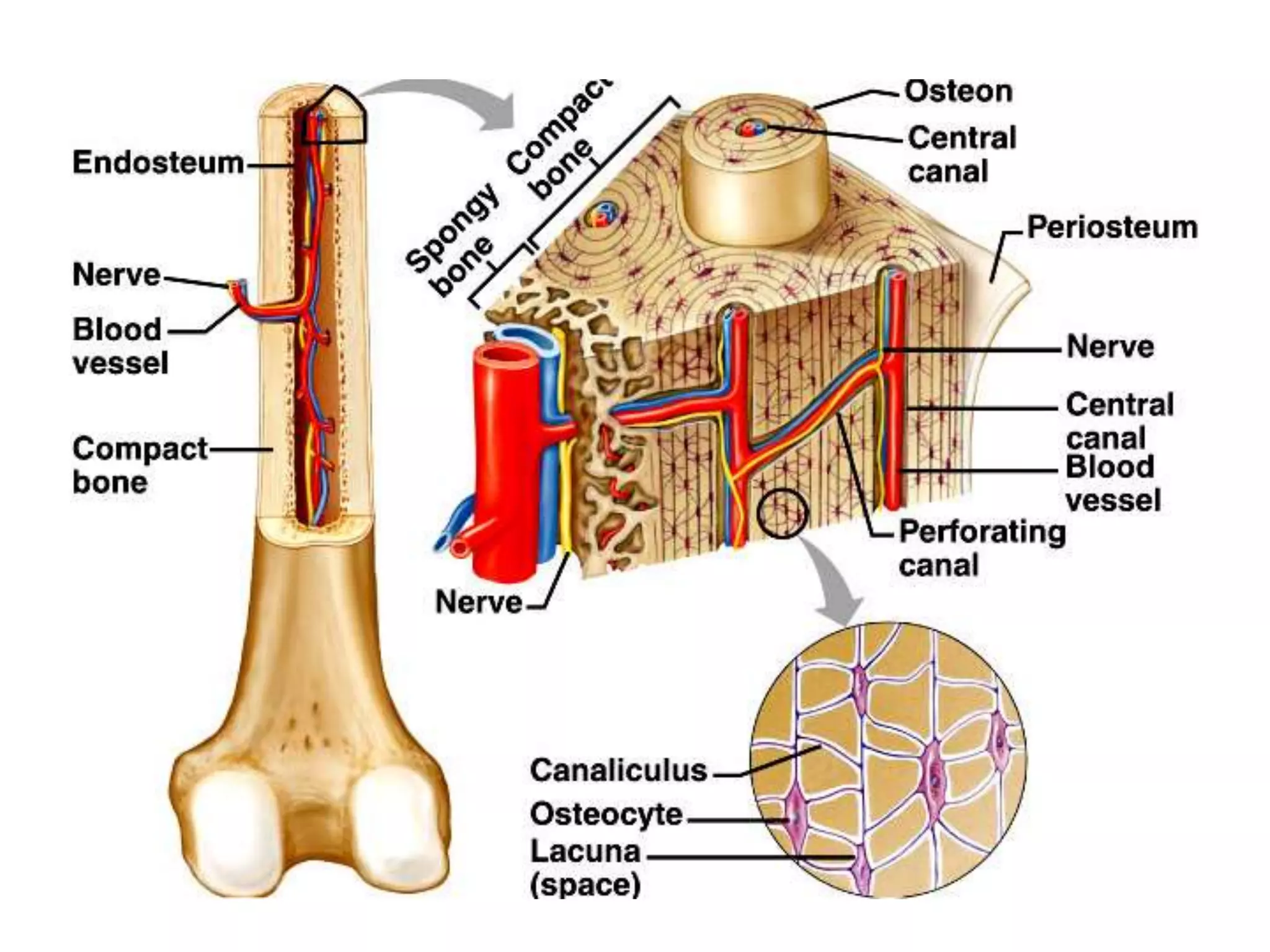
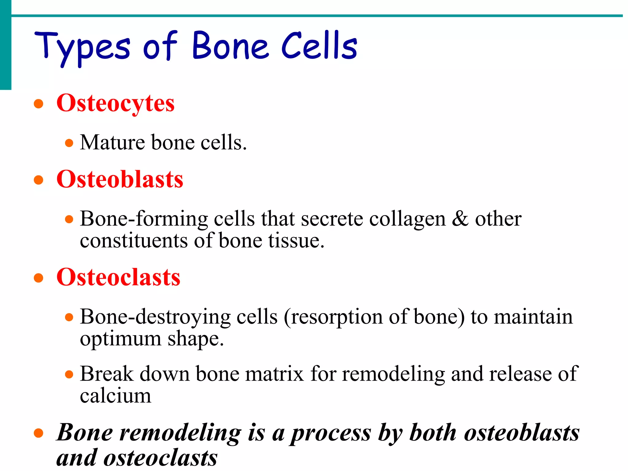
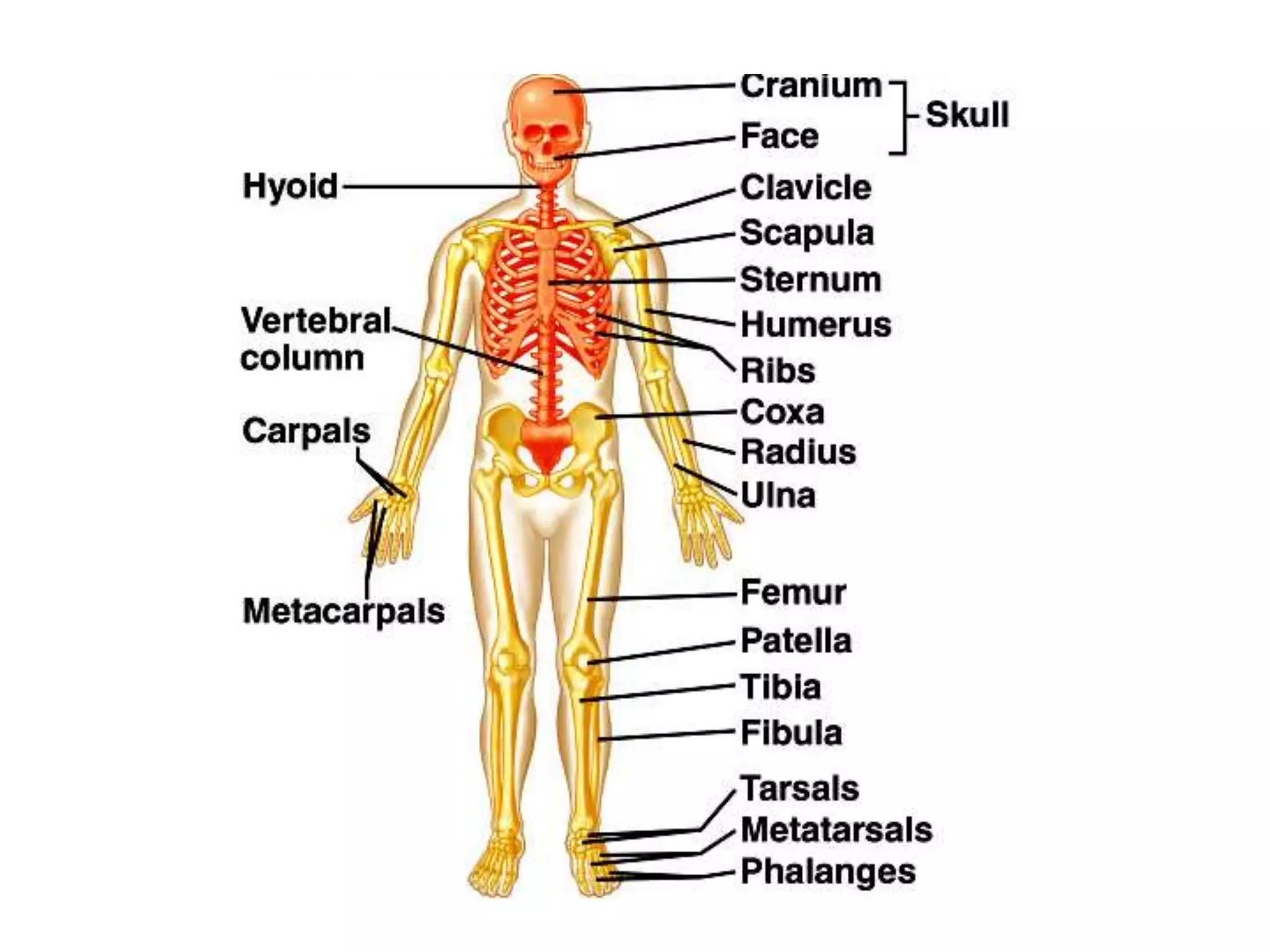
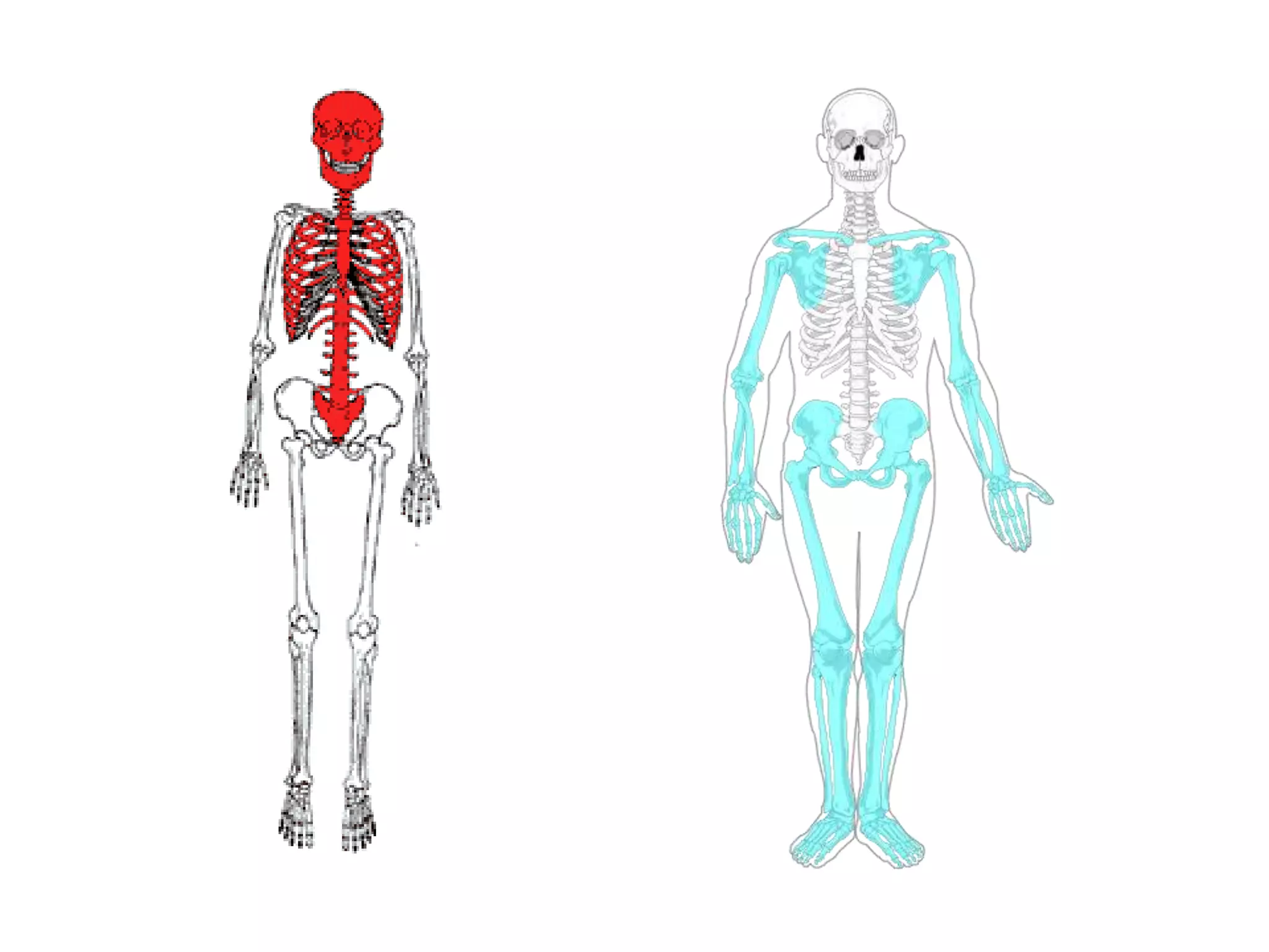
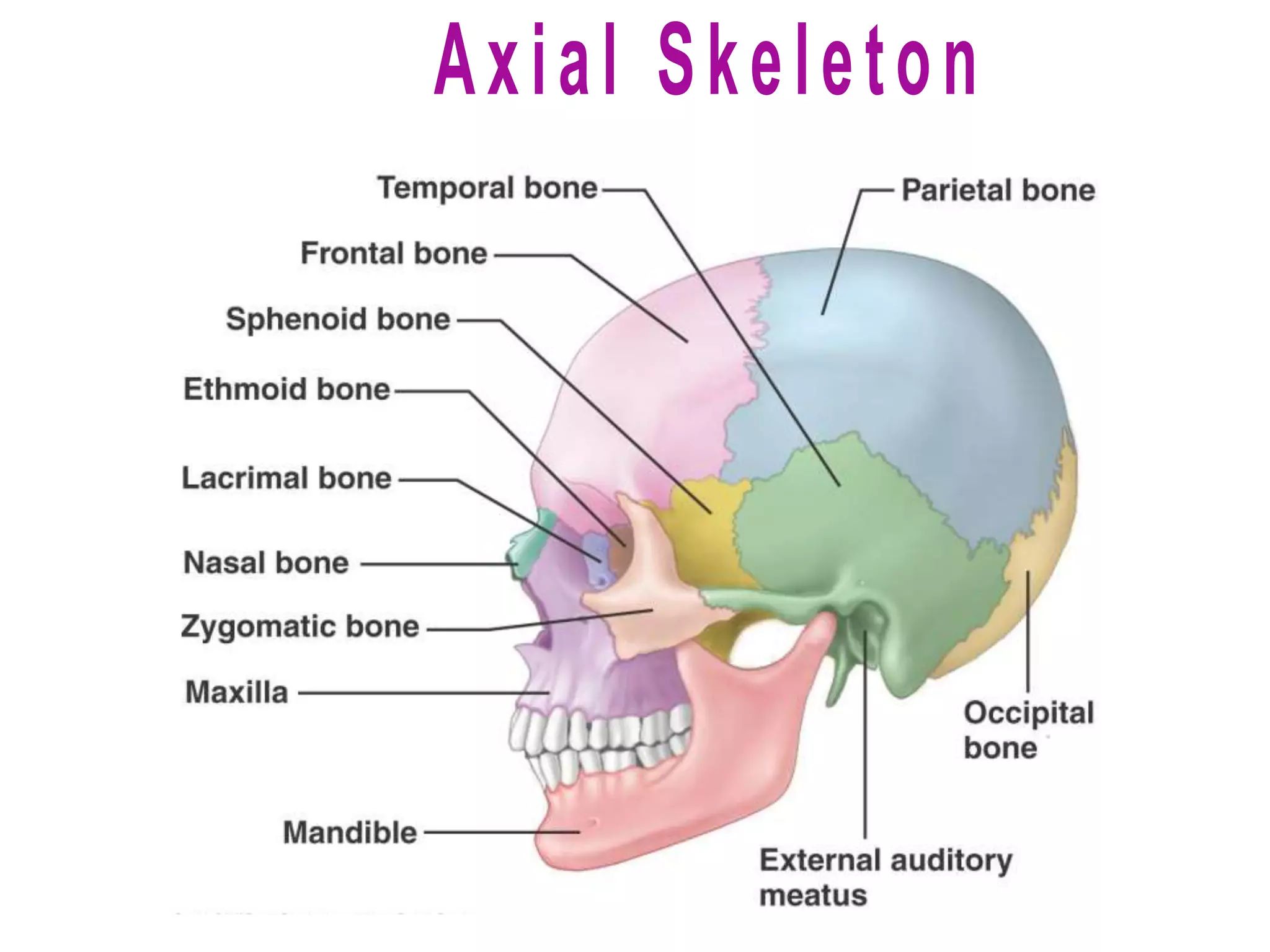
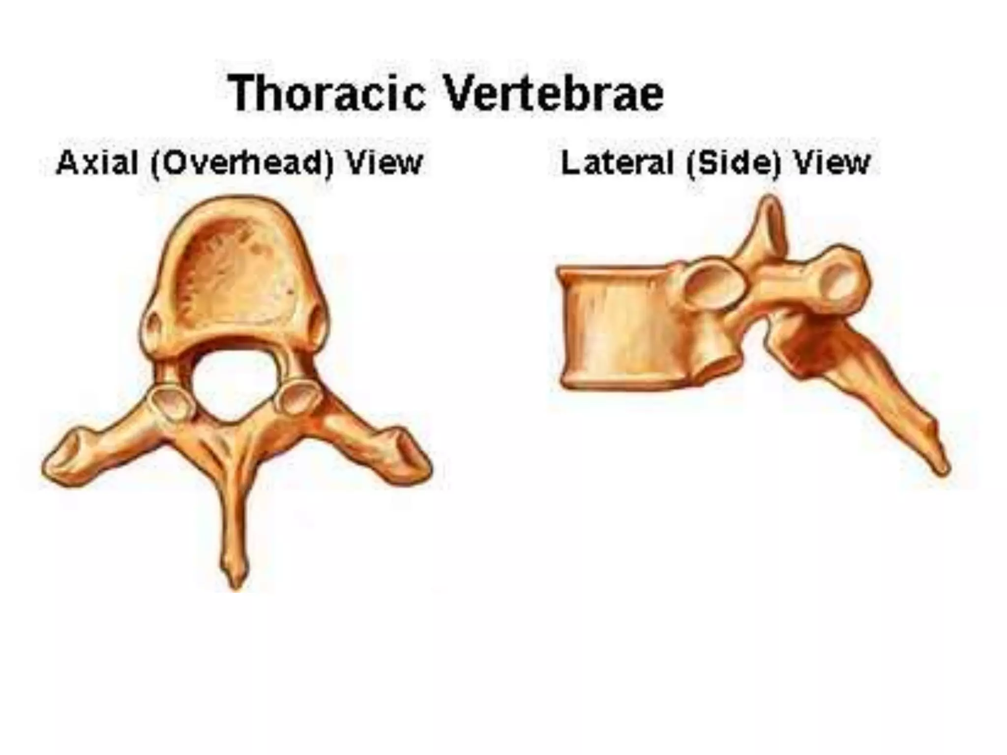
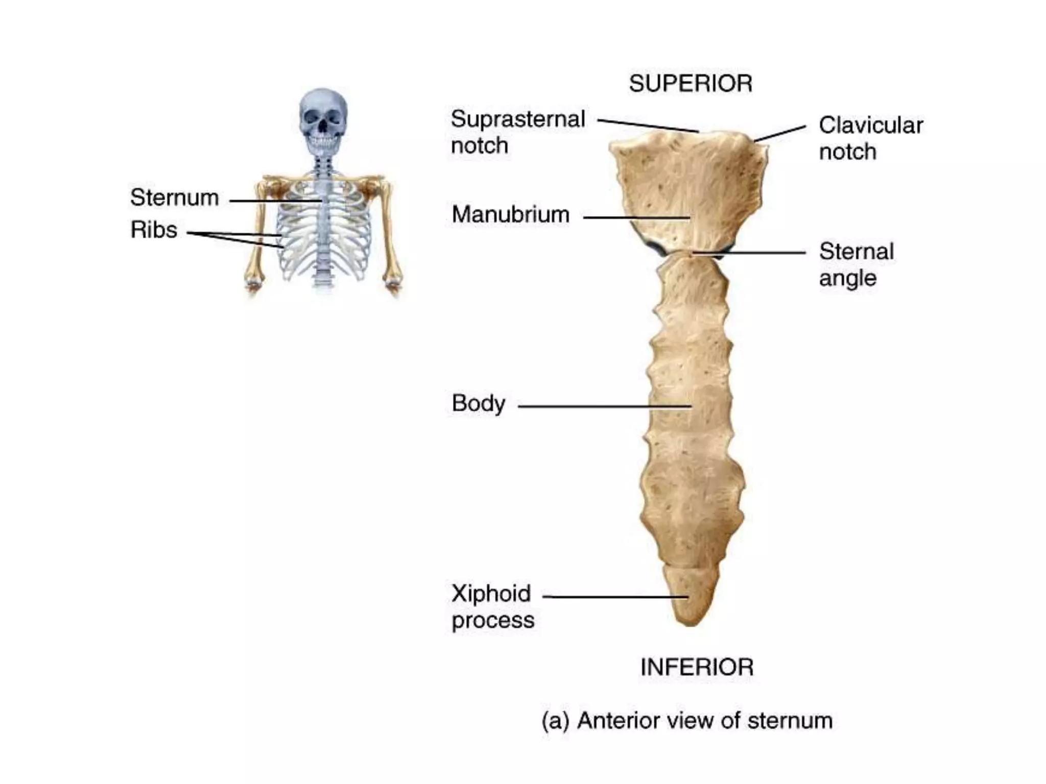
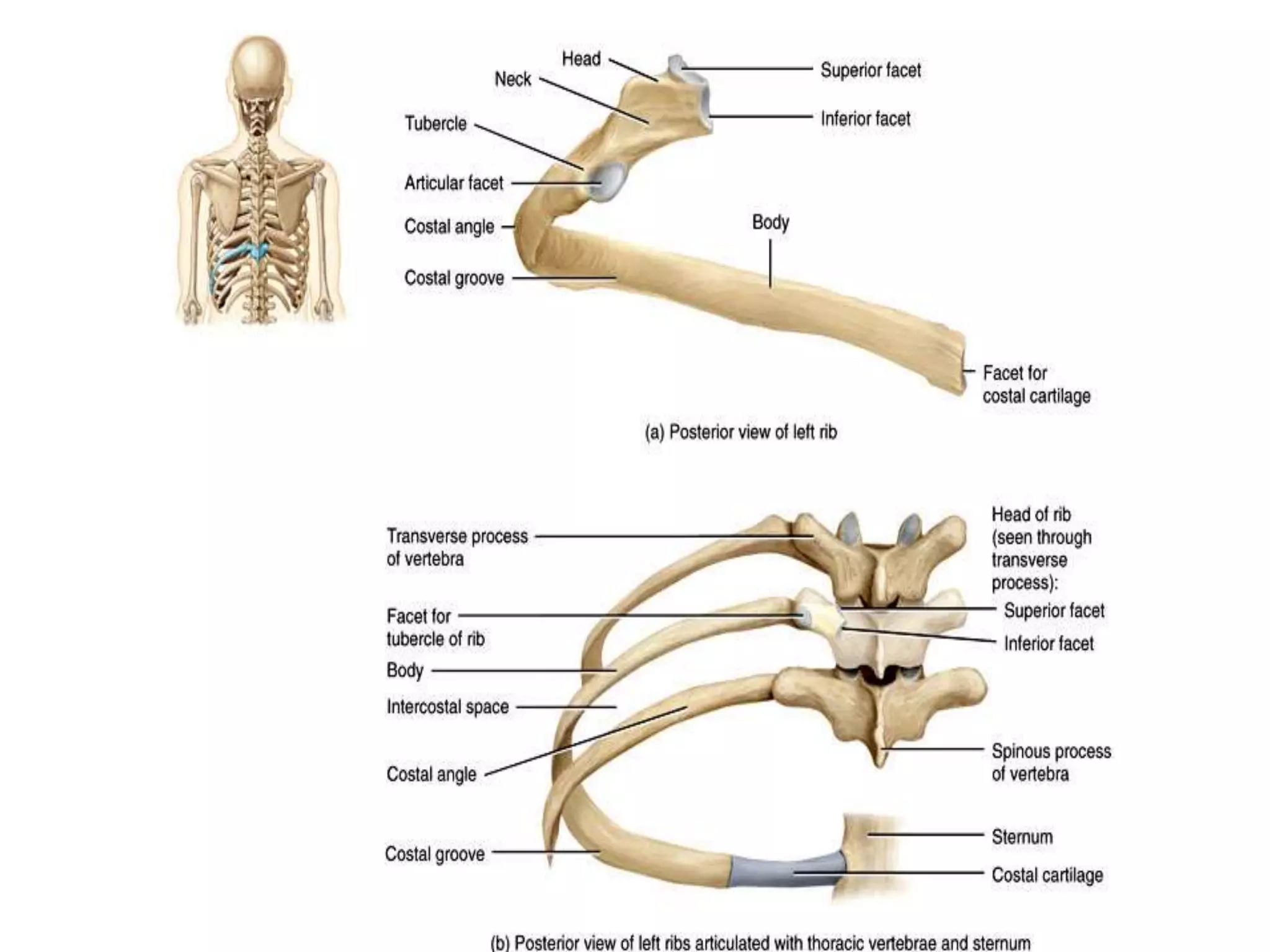
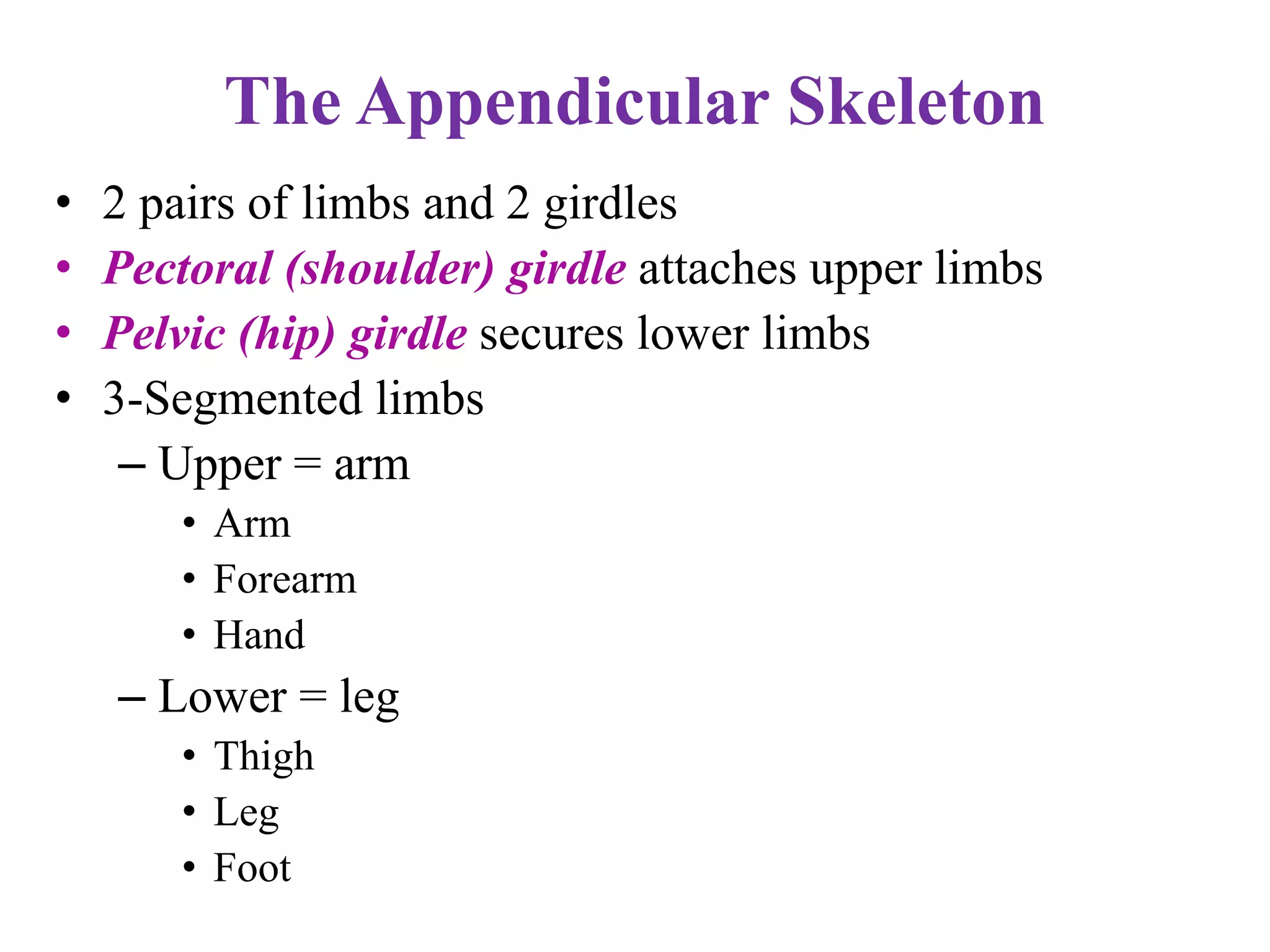
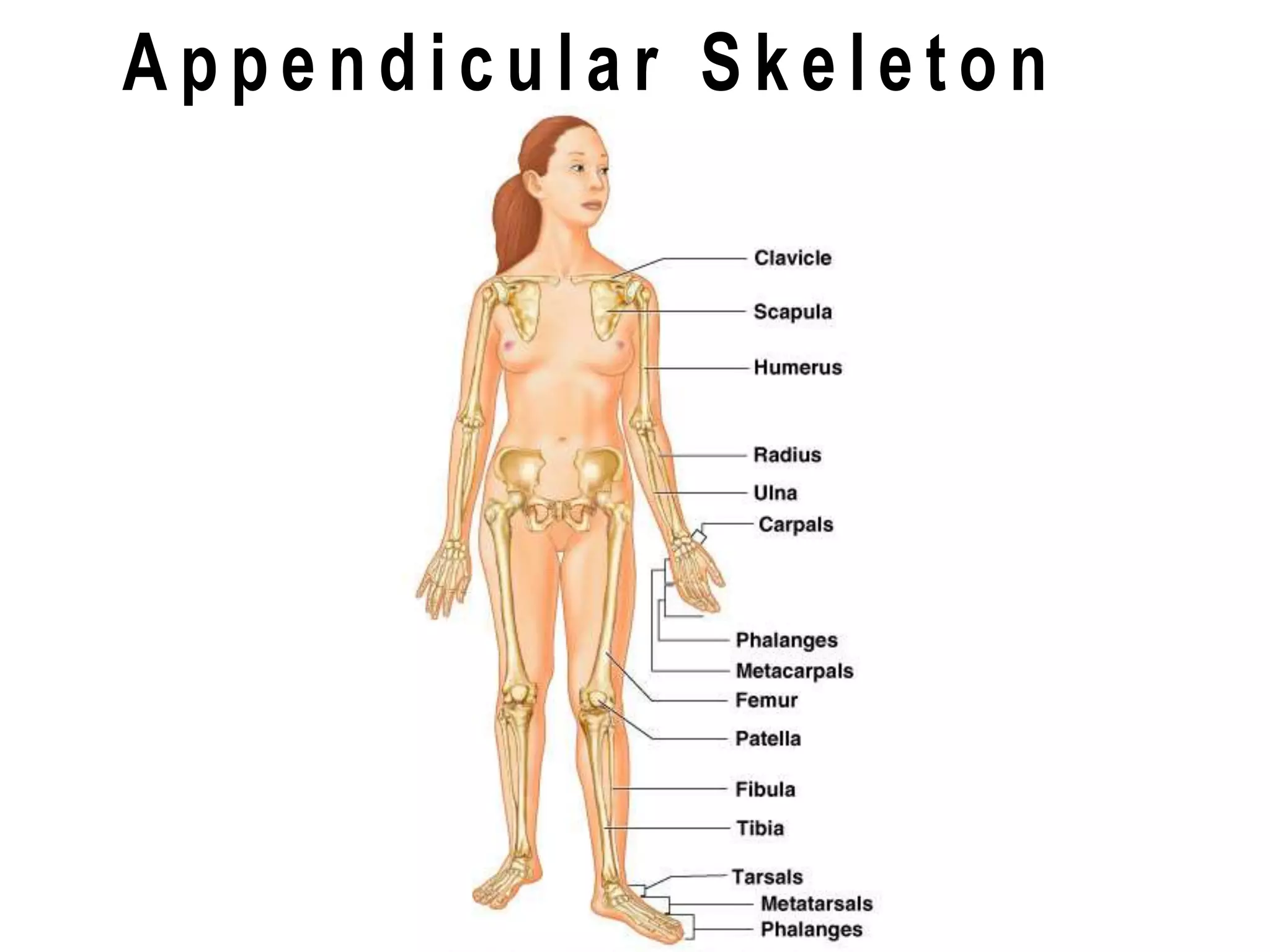
Mr Rahul Sharma
RN, Nursing Tutor
IGSCON, Amethi
Bone
• Bone is a strong and durable type of
connective tissue, it consists of :
• Water (25%)
• Organic constituents including osteoid (the
carbon containing part of the matrix) and
bone cells (25%)
• Inorganic constituents, mainly calcium
phosphate (50%)
• Support- framework that supports body and
cradles its soft organs
• Protection- for delicate organs, heart, lungs,
brain
• Movement- bones act as levers for muscles
• Mineral storage- calcium & phosphate
• Blood cell formation- hematopoesis
 Parts of the skeletal system
 Bones (skeleton)
 Joints
 Cartilages
 Ligaments (bone to bone)(tendon=bone to muscle)
 Divided into two divisions
 Axial skeleton- skull, spinal column
 Appendicular skeleton – limbs and girdle
Divisions of the Skeletal System
• The human skeleton consists of 206 named bones
• Bones of the skeleton are grouped into two principal
divisions:
– Axial skeleton
• Consists of the bones that lie around the longitudinal axis
of the human body
• Skull bones, auditory ossicles (ear bones), hyoid bone, ribs,
sternum (breastbone), and bones of the vertebral column
– Appendicular skeleton
• Consists of the bones of the upper and lower limbs
(extremities), plus the bones forming the girdles that
connect the limbs to the axial skeleton
Divisions Of The Skeletal System
• Long Bones- consists of a shaft & two
extremities. Femur, humerus, ulna, radius,
tibia, fibula
• Short Bones- carpals, tarsals
• Flat Bones- ribs, scapula, skull, sternum
• Irregular Bones- vertebrae, some facial bones
• Sesamoid- patella (knee cap)
22 bones in skull
6 in middle ears
1 hyoid bone
26 in vertebral column
25 in thoracic cage
4 in pectoral girdle
60 in upper limbs
60 in lower limbs
2 in pelvic girdle
206 bones in all
Distal
epiphysis
Proximal
epiphysis
diaphysis
yellow marrow
epiphyseal line
periosteum
compact bone
spongy bone
Endosteum
hyaline cartilage
Sharpey’s fibers
Bone enclosed in periosteum, which is continuous with
tendons and ligaments, blood vessels in periosteum
Epiphysis- ends, spongy bone contains red marrow,
compact bone, articular cartilage
Diaphysis- middle, compact bone, medullary cavity-
contains yellow marrow (fat)lined with endosteum
(squamous epithelium)
Compact bone- osteocytes within lacunae arranged in
concentric circles called lamellae
This surround a central canal; complex is called
Haversian system
Canaliculi connect osteocytes to central canal and to each
other
Microscopic Structure of Bone
 The skeleton has 206 bones
 Two basic types of bone tissue
Compact bone
 Homogeneous
Spongy bone
 Small needle-like
pieces of bone
 Many open spaces
Types of Bone Cells
 Osteocytes
 Mature bone cells.
 Osteoblasts
 Bone-forming cells that secrete collagen & other
constituents of bone tissue.
 Osteoclasts
 Bone-destroying cells (resorption of bone) to maintain
optimum shape.
 Break down bone matrix for remodeling and release of
calcium
 Bone remodeling is a process by both osteoblasts
and osteoclasts
Stages of development of
a long bone
Break
Axial skeleton
• This part consists of the skull, vertebral
column, ribs & sternum.
• Together the bones forming these structures
constitute the central bony core of the body,
the axis
Axial Skeleton- Skull
The skull rests on the upper end of the vertebral column and is
divided into the cranium and the face.
Cranium
The cranium is formed by a number of flat and irregular bones
that provides a bony protection for the brain.
It has a base upon which the brain rests and a vault that covers
and surround it.
 the periosteum inside the skull bones consists of the outer layer
of dura matter.
In mature skull the joints between the bones(sutures) are
immovable.
The bones have numerous perforations through which nerves,
blood vessels and lymph vessels pass.
Skull
• Skull (cranium)
• Consists of 22 bones
• Bones of the skull are grouped into two categories:
– Cranial bones
• Eight cranial bones form the cranial cavity
– Frontal bone, two parietal bones, two temporal bones, the occipital
bone, the sphenoid bone, ethmoid bone
– Facial bones
• Fourteen facial bones form the face
– Two nasal bones, two maxillae, two zygomatic bones, the mandible,
two lacrimal bones, two palatine bones, two inferior nasal conchae,
vomer
Skull
• The cranial and facial bones protect and support special
sense organs and the brain
• Besides forming the large cranial cavity, the skull also
forms several smaller cavities
– Nasal cavity
– Orbits (eye sockets)
– Paranasal sinuses
– Small cavities which house organs involved in
hearing and equilibrium
Skull
• Immovable joints called sutures fuse most of the
skull bones together
• The skull provides large areas of attachment for
muscles that move various parts of the head
• Skull and facial bones provide attachment for
muscles that produce facial expressions
• The facial bones form the framework of the face
and provide support for the entrances to the
digestive and respiratory systems
Skull (Cranial Bones)
• Frontal Bone
– Forms the forehead
• Parietal Bones
– Form the sides and roof of the cranial cavity
• Temporal Bones
– Form the lateral aspects and floor of the cranium
• Occipital Bone
– Forms the posterior part and most of the base of the cranium
• Sphenoid Bone
– Lies at the middle part of the base of the skull
• Ethmoid Bone
– Located on the midline in the anterior part of the cranial floor medial to the
orbits
– A major superior supporting structure of the nasal cavity
– Contain thin projections called conchae which are lined by mucous
membranes
– Increased surface area in the nasal cavity helps to humidify inhaled air
trapping inhaled particles
The bones of the cranium are
1 frontal bone
2 parietal bone
2 temporal bone
1 occipital bone
1 sphenoid bone
1 ethmoid bone
Posterior View
• Warm and moisten air
• Lighten the skull
• Enhance voice resonance
Cervical Vertebrae (7)
Thoracic Vertebrae (12)
Lumbar Vertberae (5)
Sacrum
Coccyx
The Vertebral Column
Cervical Vertebrae
Sternum
True Ribs (7)
False Ribs (3)
Floating Ribs (2)
The Thoracic Cage
Sacrum & Coccyx
Break
The Appendicular Skeleton
• 2 pairs of limbs and 2 girdles
• Pectoral (shoulder) girdle attaches upper limbs
• Pelvic (hip) girdle secures lower limbs
• 3-Segmented limbs
– Upper = arm
• Arm
• Forearm
• Hand
– Lower = leg
• Thigh
• Leg
• Foot
Pectoral
Girdle
(Shoulder Girdle)
• Clavicle – anterior: collar bone
– Sternal end attaches to the manubrium medially
– Acromial end articulates with the scapula laterally
• Scapula – posterior: shoulder blade
Clavicle
• Extend horizontally across the superior thorax
• Sternal end articulates with the manubrium
• Acromial end articulates with scapula
• Provide attachment for muscles
• Hold the scapulae and arms laterally
• Transmit compression forces from the upper
limbs to the axial skeleton
Scapulae
• Lie on the dorsal surface of the rib cage
• Located between ribs 2 – 7
• Have three borders
– Superior
– Medial (vertebral)
– Lateral (axillary)
• Have three angles
– Lateral, superior, and inferior
Structures of the Scapula
The Upper Limb
• 30 bones form each upper limb
• Grouped into bones of the
– Arm
– Forearm
– Hand
Humerus
Ulna
Radius
8 Carpals
14 Phalanges
5 Metacarpals
Arm
• Region of the upper limb between the shoulder and elbow
• Humerus
– The only bone of the arm
– Longest and strongest bone of the upper limb
– Articulates with the scapula at the shoulder
– Articulates with the radius and ulna at the elbow
– Many structures of the humerus provide sites for
muscle attachment
– Other structures of the humerus provide articulation
sites for other bones
Structures of the Humerus of the
Right Arm
Forearm
• Formed from the radius and ulna
• Proximal ends articulate with the humerus
• Distal ends articulate with carpals
• Radius and ulna articulate with each other
– At the proximal and distal radioulnar joints
• The interosseous membrane
– Interconnects radius and ulna
• In anatomical position
– The radius is lateral and the ulna is medial
Copyright © 2008 Pearson
Education, Inc., publishing
as Benjamin Cummings
Details of Arm and Forearm
Figure 8.5a
Ulna
• Main bone responsible for forming the elbow
joint with the humerus
• Hinge joint allows forearm to bend on arm
• Distal end is separated from carpals by
fibrocartilage
• Plays little to no role in hand movement
Proximal Part of the Ulna
Radius and Ulna
Radius
• Superior surface of the head of the radius
articulates with the capitulum
• Medially – the head of the radius articulates
with the radial notch of the ulna
• Contributes heavily to the wrist joint
– Distal radius articulates with carpal bones
– When radius moves, the hand moves with it
Distal Ends of the Radius and Ulna
Pelvis
Ischium
Ilium
Acetabulum
Pubis
Ischium
Obturator
foramen
Pelvis (lateral view)
Female Vs Male Pelvis
Patella
The Lower Limb
(Legs)
Femur
Tibia
Fibula
5 Metatarsals
14 Phalanges
7 Tarsals
metatarsals
phelangies
tarsals
metatarsals
phalenges
tarsals
Immovable Joints
(synarthrosis)
suture
pubis symphisis
Slightly Movable Joint
(ampharthrosis)
femur
ligaments
pelvis
(diarthrosis)- freely moveable
femur
pelvis
hyaline
cartilage synovial
cavity
joint
capsule
Abduction
Extension
Rotation
Flexion
Adduction
Synovial Joint Movement
Osteoblast
Osteocyte
Osteoclast
Eats bone
Builds new bone
Mature bone cell
hematoma
callus bony callus
bone
remodeling
Diseases of the Skeletal System:
Osteoporosis- bone reabsorption
outpaces bone deposit; bones
become lighter and fracture easier
Factors:
• age, gender (more in women)
• estrogen and testosterone decrease
• insufficient exercise (or too much)
• diet poor in Ca++ and protein
• abnormal vitamin D receptors
• smoking
Rickets- vitamin D deficiency
Osteomalacia- soft bones, inadequate
mineralization in bones, lack of vitamin D
Pagets Disease- spotty weakening in the
bones, excessive and abnormal bone
remodeling
Rheumatoid arthritis- autoimmune
reaction
Diseases of the Skeletal System:

Skeletal System

  • 1.
    Mr Rahul Sharma RN,Nursing Tutor IGSCON, Amethi
  • 2.
    Bone • Bone isa strong and durable type of connective tissue, it consists of : • Water (25%) • Organic constituents including osteoid (the carbon containing part of the matrix) and bone cells (25%) • Inorganic constituents, mainly calcium phosphate (50%)
  • 3.
    • Support- frameworkthat supports body and cradles its soft organs • Protection- for delicate organs, heart, lungs, brain • Movement- bones act as levers for muscles • Mineral storage- calcium & phosphate • Blood cell formation- hematopoesis
  • 4.
     Parts ofthe skeletal system  Bones (skeleton)  Joints  Cartilages  Ligaments (bone to bone)(tendon=bone to muscle)  Divided into two divisions  Axial skeleton- skull, spinal column  Appendicular skeleton – limbs and girdle
  • 5.
    Divisions of theSkeletal System • The human skeleton consists of 206 named bones • Bones of the skeleton are grouped into two principal divisions: – Axial skeleton • Consists of the bones that lie around the longitudinal axis of the human body • Skull bones, auditory ossicles (ear bones), hyoid bone, ribs, sternum (breastbone), and bones of the vertebral column – Appendicular skeleton • Consists of the bones of the upper and lower limbs (extremities), plus the bones forming the girdles that connect the limbs to the axial skeleton
  • 6.
    Divisions Of TheSkeletal System
  • 7.
    • Long Bones-consists of a shaft & two extremities. Femur, humerus, ulna, radius, tibia, fibula • Short Bones- carpals, tarsals • Flat Bones- ribs, scapula, skull, sternum • Irregular Bones- vertebrae, some facial bones • Sesamoid- patella (knee cap)
  • 9.
    22 bones inskull 6 in middle ears 1 hyoid bone 26 in vertebral column 25 in thoracic cage 4 in pectoral girdle 60 in upper limbs 60 in lower limbs 2 in pelvic girdle 206 bones in all
  • 10.
    Distal epiphysis Proximal epiphysis diaphysis yellow marrow epiphyseal line periosteum compactbone spongy bone Endosteum hyaline cartilage Sharpey’s fibers
  • 11.
    Bone enclosed inperiosteum, which is continuous with tendons and ligaments, blood vessels in periosteum Epiphysis- ends, spongy bone contains red marrow, compact bone, articular cartilage Diaphysis- middle, compact bone, medullary cavity- contains yellow marrow (fat)lined with endosteum (squamous epithelium) Compact bone- osteocytes within lacunae arranged in concentric circles called lamellae This surround a central canal; complex is called Haversian system Canaliculi connect osteocytes to central canal and to each other
  • 13.
    Microscopic Structure ofBone  The skeleton has 206 bones  Two basic types of bone tissue Compact bone  Homogeneous Spongy bone  Small needle-like pieces of bone  Many open spaces
  • 15.
    Types of BoneCells  Osteocytes  Mature bone cells.  Osteoblasts  Bone-forming cells that secrete collagen & other constituents of bone tissue.  Osteoclasts  Bone-destroying cells (resorption of bone) to maintain optimum shape.  Break down bone matrix for remodeling and release of calcium  Bone remodeling is a process by both osteoblasts and osteoclasts
  • 16.
    Stages of developmentof a long bone
  • 17.
  • 20.
    Axial skeleton • Thispart consists of the skull, vertebral column, ribs & sternum. • Together the bones forming these structures constitute the central bony core of the body, the axis
  • 22.
    Axial Skeleton- Skull Theskull rests on the upper end of the vertebral column and is divided into the cranium and the face. Cranium The cranium is formed by a number of flat and irregular bones that provides a bony protection for the brain. It has a base upon which the brain rests and a vault that covers and surround it.  the periosteum inside the skull bones consists of the outer layer of dura matter. In mature skull the joints between the bones(sutures) are immovable. The bones have numerous perforations through which nerves, blood vessels and lymph vessels pass.
  • 23.
    Skull • Skull (cranium) •Consists of 22 bones • Bones of the skull are grouped into two categories: – Cranial bones • Eight cranial bones form the cranial cavity – Frontal bone, two parietal bones, two temporal bones, the occipital bone, the sphenoid bone, ethmoid bone – Facial bones • Fourteen facial bones form the face – Two nasal bones, two maxillae, two zygomatic bones, the mandible, two lacrimal bones, two palatine bones, two inferior nasal conchae, vomer
  • 24.
    Skull • The cranialand facial bones protect and support special sense organs and the brain • Besides forming the large cranial cavity, the skull also forms several smaller cavities – Nasal cavity – Orbits (eye sockets) – Paranasal sinuses – Small cavities which house organs involved in hearing and equilibrium
  • 25.
    Skull • Immovable jointscalled sutures fuse most of the skull bones together • The skull provides large areas of attachment for muscles that move various parts of the head • Skull and facial bones provide attachment for muscles that produce facial expressions • The facial bones form the framework of the face and provide support for the entrances to the digestive and respiratory systems
  • 26.
    Skull (Cranial Bones) •Frontal Bone – Forms the forehead • Parietal Bones – Form the sides and roof of the cranial cavity • Temporal Bones – Form the lateral aspects and floor of the cranium • Occipital Bone – Forms the posterior part and most of the base of the cranium • Sphenoid Bone – Lies at the middle part of the base of the skull • Ethmoid Bone – Located on the midline in the anterior part of the cranial floor medial to the orbits – A major superior supporting structure of the nasal cavity – Contain thin projections called conchae which are lined by mucous membranes – Increased surface area in the nasal cavity helps to humidify inhaled air trapping inhaled particles
  • 27.
    The bones ofthe cranium are 1 frontal bone 2 parietal bone 2 temporal bone 1 occipital bone 1 sphenoid bone 1 ethmoid bone
  • 33.
  • 35.
    • Warm andmoisten air • Lighten the skull • Enhance voice resonance
  • 36.
    Cervical Vertebrae (7) ThoracicVertebrae (12) Lumbar Vertberae (5) Sacrum Coccyx The Vertebral Column
  • 38.
  • 41.
    Sternum True Ribs (7) FalseRibs (3) Floating Ribs (2) The Thoracic Cage
  • 45.
  • 46.
  • 47.
    The Appendicular Skeleton •2 pairs of limbs and 2 girdles • Pectoral (shoulder) girdle attaches upper limbs • Pelvic (hip) girdle secures lower limbs • 3-Segmented limbs – Upper = arm • Arm • Forearm • Hand – Lower = leg • Thigh • Leg • Foot
  • 49.
    Pectoral Girdle (Shoulder Girdle) • Clavicle– anterior: collar bone – Sternal end attaches to the manubrium medially – Acromial end articulates with the scapula laterally • Scapula – posterior: shoulder blade
  • 50.
    Clavicle • Extend horizontallyacross the superior thorax • Sternal end articulates with the manubrium • Acromial end articulates with scapula • Provide attachment for muscles • Hold the scapulae and arms laterally • Transmit compression forces from the upper limbs to the axial skeleton
  • 52.
    Scapulae • Lie onthe dorsal surface of the rib cage • Located between ribs 2 – 7 • Have three borders – Superior – Medial (vertebral) – Lateral (axillary) • Have three angles – Lateral, superior, and inferior
  • 53.
  • 54.
  • 55.
  • 56.
    The Upper Limb •30 bones form each upper limb • Grouped into bones of the – Arm – Forearm – Hand
  • 57.
  • 58.
    Arm • Region ofthe upper limb between the shoulder and elbow • Humerus – The only bone of the arm – Longest and strongest bone of the upper limb – Articulates with the scapula at the shoulder – Articulates with the radius and ulna at the elbow – Many structures of the humerus provide sites for muscle attachment – Other structures of the humerus provide articulation sites for other bones
  • 59.
    Structures of theHumerus of the Right Arm
  • 60.
    Forearm • Formed fromthe radius and ulna • Proximal ends articulate with the humerus • Distal ends articulate with carpals • Radius and ulna articulate with each other – At the proximal and distal radioulnar joints • The interosseous membrane – Interconnects radius and ulna • In anatomical position – The radius is lateral and the ulna is medial
  • 61.
    Copyright © 2008Pearson Education, Inc., publishing as Benjamin Cummings Details of Arm and Forearm Figure 8.5a
  • 62.
    Ulna • Main boneresponsible for forming the elbow joint with the humerus • Hinge joint allows forearm to bend on arm • Distal end is separated from carpals by fibrocartilage • Plays little to no role in hand movement
  • 63.
  • 64.
  • 65.
    Radius • Superior surfaceof the head of the radius articulates with the capitulum • Medially – the head of the radius articulates with the radial notch of the ulna • Contributes heavily to the wrist joint – Distal radius articulates with carpal bones – When radius moves, the hand moves with it
  • 66.
    Distal Ends ofthe Radius and Ulna
  • 68.
  • 69.
  • 70.
  • 72.
    Patella The Lower Limb (Legs) Femur Tibia Fibula 5Metatarsals 14 Phalanges 7 Tarsals
  • 76.
  • 77.
  • 78.
  • 79.
  • 80.
  • 82.
  • 83.
  • 84.
  • 85.
    Diseases of theSkeletal System: Osteoporosis- bone reabsorption outpaces bone deposit; bones become lighter and fracture easier Factors: • age, gender (more in women) • estrogen and testosterone decrease • insufficient exercise (or too much) • diet poor in Ca++ and protein • abnormal vitamin D receptors • smoking
  • 86.
    Rickets- vitamin Ddeficiency Osteomalacia- soft bones, inadequate mineralization in bones, lack of vitamin D Pagets Disease- spotty weakening in the bones, excessive and abnormal bone remodeling Rheumatoid arthritis- autoimmune reaction Diseases of the Skeletal System: