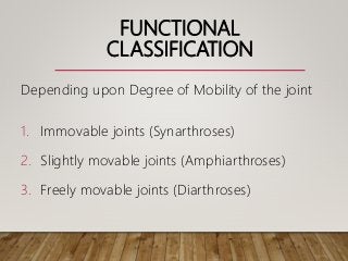
JOINTS
DR. SUNDIP CHARMODE
ASSOCIATE PROFESSOR
DEPARTMENT OF ANATOMY
AIIMS RAJKOT
OBJECTIVES
• Definition of Joint
• Classification of Joints
• Features of various Joints
• Movements of various Joints
• Blood supply of Joints
• Clinical Correlates of Joints
INTRODUCTION
• It means a place where two things are
joined together.
• Articulation
• Arthrosis
• Arthrology
• Arthritis
TYPES
1. Depending on the Function
2. Depending on the Structure
FUNCTIONAL
CLASSIFICATION
Depending upon Degree of Mobility of the joint
1. Immovable joints (Synarthroses)
2. Slightly movable joints (Amphiarthroses)
3. Freely movable joints (Diarthroses)
SYNARTHROSES
• Solid Joints
• Without any cavity
• No mobility
• E.g. : Fibrous joints, Primary Cartilaginous joints.
AMPHIARTHROSES
• Without any cavity
• Restricted movements take place
• E.g. : Secondary Cartilaginous joints
DIARTHROSES
• Possess joint cavity filled with Synovial fluid.
• Permit free movements
• Examples : Synovial joints
STRUCTURAL
CLASSIFICATION
Depending upon :
• Type of Connective Tissue
• Presence and absence of Joint Cavity.
1. Fibrous
2. Cartilaginous
3. Synovial
FIBROUS JOINTS
1. United by Fibrous Connective Tissue ?
2. No movement or only slight movement is
permitted.
Three types :
I. Sutures
II. Syndesmoses
III. Gomphoses
SUTURES
• Immovable confined to Skull.
• Appear between Bones which ossify in
membrane
• Connected by thin layer of connective tissue
(Sutural ligament)
SUTURAL LIGAMENT
• Unossified part of Mesenchyme
sheet
• Connects
• Provides
• Binds
• Ossification in Sutural ligament
SUTURAL LIGAMENT / MEMBRANE
TYPES OF SUTURES
Depending upon : Shape of Articular Surface
and
Margins of Articulating Bones
1. Plane suture
2. Serrate / Limbus suture
3. Squamous suture / Lap suture
4. Denticulate suture
5. Schindylesis (Wedge and Groove suture)
SYNOSTOSIS
• In Elderly, most of the sutures of the
skull undergo ossification leading to
rigid bony union.
• It can begin in early twenties.
SYNDESMOSES
• It is a type of fibrous joint where the surfaces of
bones are united by Inter-osseous ligaments
and the bones lie some distance apart.
• These ligaments persist for lifetime.
• Slight amount of movement is possible.
SYNDESMOSES
• Examples :
• Inter-osseous membranes of forearm and leg
• Inferior tibio-fibular joint
• Ligamentum flava
• Tympano-stapedial joints
GOMPHOSES
• Peg and Socket joint.
• It’s a special type of Fibrous joint
restricted to fixation of root of teeth in
alveolar sockets of mandible and
maxilla united by fibrous tissue.
• Perio-dontal ligament.
PERIODONTITIS
• Inflammation and degeneration of Periodontal
ligament.
• Tooth become loose and fall.
CARTILAGINOUS JOINTS
• These are those joints in which the bones
forming the joint are united by means of either
Hyaline cartilage or Fibrocartilage.
• They also lack joint cavity.
• Two types :
• Primary cartilaginous joint
• Secondary cartilaginous joint
PRIMARY CARTILAGINOUS
JOINTS
• The bones or part of the bones forming the joint
are joined by plate of Hyaline cartilage (non-
articular).
• They also lack joint cavity.
• These are immovable
• Mostly temporary in nature
• Also called as ‘Synchondroses’
SECONDARY CARTILAGINOUS JOINTS
• The articular surfaces of bones forming the joint
are covered by thin plate of Hyaline cartilage
(articular), connected by a disc or pad of
Fibrocartilage.
• They also lack joint cavity.
• These are slightly movable and permanent in
nature.
• Always occur in midline.
• Also called as ‘Symphyses’
Primary Cartilaginous joint Secondary Cartilaginous
joint
Bones are connected by Hyaline
cartilage
Bones are covered by Hyaline
cartilage and connected by a plate
of fibrocartilage.
Immovable Slightly movable
Temporary joints Permanent joints
Rarely occur in midline Always occur in midline
Synchondroses Symphyses
E.g. Joint between epiphysis and
diaphysis, Spheno-occipital joint,
First Sterno-chondral joint, Costo-
chondral joint.
E.g. Symphyses pubis, Manubrio-
sternal joint, Symphyses Menti,
Xiphi-sternal joint.
SYNOVIAL JOINTS
1. Possess cavity
2. Articular ends of bones are enclosed
within a Fibrous Capsule.
3. Articular cavity containing synovial
fluid is present between articulating
ends.
CHARACTERISTIC
FEATURES
1. Articular cartilage
2. Synovial fluid
3. Outer fibrous capsule and Inner
synovial membrane
4. Ligaments
5. Articular disc/ Meniscus
COMPONENTS
1. Articular
cartilage
2. Synovial fluid
3. Fibrous capsule
4. Synovial
membrane
5. Ligaments
6. Articular disc or
meniscus
7. Bursae
8. Fat pads
ARTICULAR CARTILAGE
• Made up of Hyaline cartilage except ?
• Resists compressive forces
• Provides a smooth gliding surface
• Reduces forces of compression during
weight bearing and muscle action.
• Regulates the growth of epiphysis
ARTICULAR CARTILAGE
• Extremely porous and absorbs
synovial fluid in resting condition.
• Nutrition from synovial fluid, diffusion
from capillaries and adjacent
epiphyseal blood vessels.
• Changes due to senility
SYNOVIAL FLUID
• Viscous, glary and alkaline fluid
• Dialysate of blood plasma along with
Hyaluronic acid
• Hyaluronic acid - High polymer of
polysaccharide secreted by synovial cells
and mast cells
• Viscosity depends on concentration of
HA
SYNOVIAL FLUID
• Maintain nutrition of articular cartilage
• Lubrication of joint cavity
FIBROUS CAPSULE
• It completely invests the joint
• It consists of longitudinal and
interlacing bundles of white connective
tissue fibres.
• It stabilizes the joint by permitting free
movements and resisting dislocation.
• Watch –dog action of the capsule.
FIBROUS CAPSULE
• It is sensitive to the changes of
position of the joints
• It is pierced by the blood vessels and
nerves
• It binds the articulating bones together
• It supports the synovial membrane on
the inner surface
SYNOVIAL MEMBRANE
• It is highly vascular and cellular
connective tissue, lining the inner
aspect of fibrous capsule and bones
lying within the capsule except at the
periphery of the articular cartilage,
articular disc or meniscus.
SYNOVIAL MEMBRANE
• Secretes synovial fluid and provides
nutrition
• Liberates hyaluronic acid and
maintains viscosity
• Removes particulate matter and
worn-out cartilage cells by
phagocytosis.
LIGAMENTS
• Two types:
• True
• False
• True ligaments are thickening of
collagen fibres of fibrous capsule.
• False ligaments are additional
bonds of union between the bones.
LIGAMENTS
• Permit desirable movements and
prevent undesirable ones
• Maintain stability of the joints
ARTICULAR DISC / MENISCUS
• They are pads of fibrocartilage
• Divide the cavity completely or incompletely
• Attached at the periphery of the fibrous
capsule
• Examples: Temporo-mandibular joint,
Sterno-clavicular joint, Inferior radio-ulnar
joint, Knee joint and Acromio-clavicular
joint.
ARTICULAR DISC / MENISCUS
• Facilitates lubrication of the joints.
• Appears in the joints where gliding movement
is associated with angular movements.
• Acts as a ligament to modify certain joint
movements
• Prevents wear and tear of cartilages by
cushioning effect.
BURSAE
• Pouch like sacs of connective tissue filled
with synovial fluid.
• Commonly located between:
• Tendon and bone
• Muscle and bone
• Skin and bone
• Tendon and skin
BURSAE AROUND KNEE JOINT
BURSAE
• They reduce friction of one structure
moving over another.
• They cushion certain muscles, particularly
those that cross the wrist and ankle joints.
FAT PADS
• Pads of fat placed between synovial
membrane and capsule or between synovial
membrane and bone.
• E.g. Acetabular fat, Infra-patellar Fat-pad,
etc.
CLASSIFICATION
 According to the Shape of Articular surface :
1. Plane joints
2. Hinge joints / Ginglymus joints
3. Pivot joints / Trochiod joints
4. Condylar joints
5. Ellipsoidal joints
6. Saddle or Sellar joints
ACCORDING TO THE SHAPE OF
ARTICULAR SURFACE
7. Ball and socket joints
PLANE JOINT
• Articular surfaces are nearly flat.
• Permit gliding movements in various directions
• Side to side
• Back and forth
PLANE JOINT
• Examples:
• Inter-carpal joints
• Inter-tarsal joints
• Inter-metacarpal joints
• Inter-metatarsal joints
• Joints between articular facets of adjacent
vertebra
HINGE JOINT
• One articular surface is convex like a cylinder
and other is reciprocally curved.
• Movements are possible only in one plane
around a transverse axis.
• These joints have strong collateral ligaments to
prevent other ligaments.
HINGE JOINT
• Examples:
• Elbow joint (Humero-Ulnar joint)
• Inter phalangeal joints
• Knee joint
• Ankle joint
PIVOT JOINT
• One articular surface is rounded which fits into
the concavity of another bone.
• The rounded part is surrounded by an osseo-
ligamentous ring.
• “Rod surrounded by a ring”
• Movement is limited rotation around a vertical
axis.
PIVOT JOINT
• Examples:
• Superior Radio-ulnar joint
• Median Atlanto-axial joint
CONDYLAR JOINT
• One articular surface is rounded which fits into
the socket like articular surface of another bone.
• The end of bone bearing round articular surface
is called ‘condyle’
• Movements are permitted mainly along a
transverse axis and partly along a vertical axis.
• Movements are permitted in two directions
(biaxial) up and down and side to side.
CONDYLAR JOINT
• Examples:
• Right and left Temporo-mandibular joint
• Knee joints
ELLIPSOID JOINT
• The elliptical convex surface of one bone
articulates with the elliptical concave
surface of another bone.
• The movements takes place around
transverse and antero-posterior axes.
ELLIPSOID JOINT
• Movements are permitted in two
directions (biaxial)
• Flexion and extension around transverse axis
• Abduction and adduction around Antero-
posterior axis
• Combination of these movements produces
‘Circumduction’
• Typical rotation around a vertical axis does
not take place.
ELLIPSOID JOINT
• Examples:
• Radio-carpal joint (wrist joint)
• Atlanto-occipital joint
• Metacarpo-phalangeal joint
• Metatarso-phalangeal joint
SADDLE / SELLAR JOINT
• The opposing articular surfaces are
concavo-convex in reciprocal manner.
• These permit movements like ellipsoid
joints.
• Some rotation is also associated called
as ‘Conjunct rotation’.
SADDLE / SELLAR JOINT
• Examples:
• First Carpo-metacarpal joint
• Sterno-clavicular joint
• Calcaneo-cuboid joint
• Incudo-malleolar joint
BALL AND SOCKET JOINT
• Rounded convex surface of one bone
fits into cup like socket of another bone.
• Greatest range of movements of all
synovial joints around three
independent axes (transverse, antero-
posterior and vertical) which have one
common centre.
BALL AND SOCKET JOINT
• Flexion, extension, adduction, abduction,
medial rotation, lateral rotation and
circumduction.
• Examples:
• Hip joint (largest ball and socket joint)
• Shoulder joint
• Incudo-stapedial joint (smallest ball and
socket joint)
ACCORDING TO THE AXIS OF
MOVEMENT
1. Uniaxial Joints – Hinge and Pivot
joints
2. Biaxial Joints – Condylar, Ellipsoidal,
Saddle joints
3. Multiaxial / Polyaxial Joints: Ball and
socket joint
4. Gliding type of movement: Plane
joints
ACCORDING TO NUMBER OF
ARTICULATING BONES
1. Simple Joints
2. Compound Joints
3. Complex joints
FACTORS LIMITING THE RANGE
OF MOVEMENT
1. Shape of articulating bones
2. Tension of ligaments
3. Tension of antagonistic muscles
4. The approximation of soft parts
FACTORS MAINTAINING STABILITY
OF JOINTS
1. Bony configuration
2. Ligaments
3. Muscles
4. Atmospheric pressure and force of
cohesion
BLOOD SUPPLY
• Epiphyseal vessels
• Articular branches
• Rich capillary plexus in synovial
membrane
• This periarterial plexus – Circulus
vasculosus
• Arterio-venous anastomoses do exist
in joints but function unknown
NERVE SUPPLY
• Articular capsule and ligaments – rich
nerve supply
• Articular nerves contain:
• Sensory fibers –
• Proprioceptive function from Ruffini’s endings
and Paccinian corpuscles of joint capsule
• Pain sensations from fibrous capsule from
free nerve endings
• Autonomic fibers – vasomotor function
CLINICAL CORRELATION
• Hilton’s law
• Gardner’s observation

Joints.pptx

  • 1.
    JOINTS DR. SUNDIP CHARMODE ASSOCIATEPROFESSOR DEPARTMENT OF ANATOMY AIIMS RAJKOT
  • 2.
    OBJECTIVES • Definition ofJoint • Classification of Joints • Features of various Joints • Movements of various Joints • Blood supply of Joints • Clinical Correlates of Joints
  • 3.
    INTRODUCTION • It meansa place where two things are joined together. • Articulation • Arthrosis • Arthrology • Arthritis
  • 4.
    TYPES 1. Depending onthe Function 2. Depending on the Structure
  • 5.
    FUNCTIONAL CLASSIFICATION Depending upon Degreeof Mobility of the joint 1. Immovable joints (Synarthroses) 2. Slightly movable joints (Amphiarthroses) 3. Freely movable joints (Diarthroses)
  • 6.
    SYNARTHROSES • Solid Joints •Without any cavity • No mobility • E.g. : Fibrous joints, Primary Cartilaginous joints.
  • 7.
    AMPHIARTHROSES • Without anycavity • Restricted movements take place • E.g. : Secondary Cartilaginous joints
  • 8.
    DIARTHROSES • Possess jointcavity filled with Synovial fluid. • Permit free movements • Examples : Synovial joints
  • 9.
    STRUCTURAL CLASSIFICATION Depending upon : •Type of Connective Tissue • Presence and absence of Joint Cavity. 1. Fibrous 2. Cartilaginous 3. Synovial
  • 10.
    FIBROUS JOINTS 1. Unitedby Fibrous Connective Tissue ? 2. No movement or only slight movement is permitted. Three types : I. Sutures II. Syndesmoses III. Gomphoses
  • 12.
    SUTURES • Immovable confinedto Skull. • Appear between Bones which ossify in membrane • Connected by thin layer of connective tissue (Sutural ligament)
  • 13.
    SUTURAL LIGAMENT • Unossifiedpart of Mesenchyme sheet • Connects • Provides • Binds • Ossification in Sutural ligament
  • 14.
  • 15.
    TYPES OF SUTURES Dependingupon : Shape of Articular Surface and Margins of Articulating Bones 1. Plane suture 2. Serrate / Limbus suture 3. Squamous suture / Lap suture 4. Denticulate suture 5. Schindylesis (Wedge and Groove suture)
  • 20.
    SYNOSTOSIS • In Elderly,most of the sutures of the skull undergo ossification leading to rigid bony union. • It can begin in early twenties.
  • 24.
    SYNDESMOSES • It isa type of fibrous joint where the surfaces of bones are united by Inter-osseous ligaments and the bones lie some distance apart. • These ligaments persist for lifetime. • Slight amount of movement is possible.
  • 25.
    SYNDESMOSES • Examples : •Inter-osseous membranes of forearm and leg • Inferior tibio-fibular joint • Ligamentum flava • Tympano-stapedial joints
  • 29.
    GOMPHOSES • Peg andSocket joint. • It’s a special type of Fibrous joint restricted to fixation of root of teeth in alveolar sockets of mandible and maxilla united by fibrous tissue. • Perio-dontal ligament.
  • 31.
    PERIODONTITIS • Inflammation anddegeneration of Periodontal ligament. • Tooth become loose and fall.
  • 32.
    CARTILAGINOUS JOINTS • Theseare those joints in which the bones forming the joint are united by means of either Hyaline cartilage or Fibrocartilage. • They also lack joint cavity. • Two types : • Primary cartilaginous joint • Secondary cartilaginous joint
  • 33.
    PRIMARY CARTILAGINOUS JOINTS • Thebones or part of the bones forming the joint are joined by plate of Hyaline cartilage (non- articular). • They also lack joint cavity. • These are immovable • Mostly temporary in nature • Also called as ‘Synchondroses’
  • 38.
    SECONDARY CARTILAGINOUS JOINTS •The articular surfaces of bones forming the joint are covered by thin plate of Hyaline cartilage (articular), connected by a disc or pad of Fibrocartilage. • They also lack joint cavity. • These are slightly movable and permanent in nature. • Always occur in midline. • Also called as ‘Symphyses’
  • 43.
    Primary Cartilaginous jointSecondary Cartilaginous joint Bones are connected by Hyaline cartilage Bones are covered by Hyaline cartilage and connected by a plate of fibrocartilage. Immovable Slightly movable Temporary joints Permanent joints Rarely occur in midline Always occur in midline Synchondroses Symphyses E.g. Joint between epiphysis and diaphysis, Spheno-occipital joint, First Sterno-chondral joint, Costo- chondral joint. E.g. Symphyses pubis, Manubrio- sternal joint, Symphyses Menti, Xiphi-sternal joint.
  • 44.
    SYNOVIAL JOINTS 1. Possesscavity 2. Articular ends of bones are enclosed within a Fibrous Capsule. 3. Articular cavity containing synovial fluid is present between articulating ends.
  • 46.
    CHARACTERISTIC FEATURES 1. Articular cartilage 2.Synovial fluid 3. Outer fibrous capsule and Inner synovial membrane 4. Ligaments 5. Articular disc/ Meniscus
  • 47.
    COMPONENTS 1. Articular cartilage 2. Synovialfluid 3. Fibrous capsule 4. Synovial membrane 5. Ligaments 6. Articular disc or meniscus 7. Bursae 8. Fat pads
  • 48.
    ARTICULAR CARTILAGE • Madeup of Hyaline cartilage except ? • Resists compressive forces • Provides a smooth gliding surface • Reduces forces of compression during weight bearing and muscle action. • Regulates the growth of epiphysis
  • 49.
    ARTICULAR CARTILAGE • Extremelyporous and absorbs synovial fluid in resting condition. • Nutrition from synovial fluid, diffusion from capillaries and adjacent epiphyseal blood vessels. • Changes due to senility
  • 51.
    SYNOVIAL FLUID • Viscous,glary and alkaline fluid • Dialysate of blood plasma along with Hyaluronic acid • Hyaluronic acid - High polymer of polysaccharide secreted by synovial cells and mast cells • Viscosity depends on concentration of HA
  • 52.
    SYNOVIAL FLUID • Maintainnutrition of articular cartilage • Lubrication of joint cavity
  • 53.
    FIBROUS CAPSULE • Itcompletely invests the joint • It consists of longitudinal and interlacing bundles of white connective tissue fibres. • It stabilizes the joint by permitting free movements and resisting dislocation. • Watch –dog action of the capsule.
  • 54.
    FIBROUS CAPSULE • Itis sensitive to the changes of position of the joints • It is pierced by the blood vessels and nerves • It binds the articulating bones together • It supports the synovial membrane on the inner surface
  • 55.
    SYNOVIAL MEMBRANE • Itis highly vascular and cellular connective tissue, lining the inner aspect of fibrous capsule and bones lying within the capsule except at the periphery of the articular cartilage, articular disc or meniscus.
  • 56.
    SYNOVIAL MEMBRANE • Secretessynovial fluid and provides nutrition • Liberates hyaluronic acid and maintains viscosity • Removes particulate matter and worn-out cartilage cells by phagocytosis.
  • 57.
    LIGAMENTS • Two types: •True • False • True ligaments are thickening of collagen fibres of fibrous capsule. • False ligaments are additional bonds of union between the bones.
  • 58.
    LIGAMENTS • Permit desirablemovements and prevent undesirable ones • Maintain stability of the joints
  • 59.
    ARTICULAR DISC /MENISCUS • They are pads of fibrocartilage • Divide the cavity completely or incompletely • Attached at the periphery of the fibrous capsule • Examples: Temporo-mandibular joint, Sterno-clavicular joint, Inferior radio-ulnar joint, Knee joint and Acromio-clavicular joint.
  • 60.
    ARTICULAR DISC /MENISCUS • Facilitates lubrication of the joints. • Appears in the joints where gliding movement is associated with angular movements. • Acts as a ligament to modify certain joint movements • Prevents wear and tear of cartilages by cushioning effect.
  • 61.
    BURSAE • Pouch likesacs of connective tissue filled with synovial fluid. • Commonly located between: • Tendon and bone • Muscle and bone • Skin and bone • Tendon and skin
  • 62.
  • 63.
    BURSAE • They reducefriction of one structure moving over another. • They cushion certain muscles, particularly those that cross the wrist and ankle joints.
  • 64.
    FAT PADS • Padsof fat placed between synovial membrane and capsule or between synovial membrane and bone. • E.g. Acetabular fat, Infra-patellar Fat-pad, etc.
  • 65.
    CLASSIFICATION  According tothe Shape of Articular surface : 1. Plane joints 2. Hinge joints / Ginglymus joints 3. Pivot joints / Trochiod joints 4. Condylar joints 5. Ellipsoidal joints 6. Saddle or Sellar joints
  • 66.
    ACCORDING TO THESHAPE OF ARTICULAR SURFACE 7. Ball and socket joints
  • 67.
    PLANE JOINT • Articularsurfaces are nearly flat. • Permit gliding movements in various directions • Side to side • Back and forth
  • 68.
    PLANE JOINT • Examples: •Inter-carpal joints • Inter-tarsal joints • Inter-metacarpal joints • Inter-metatarsal joints • Joints between articular facets of adjacent vertebra
  • 70.
    HINGE JOINT • Onearticular surface is convex like a cylinder and other is reciprocally curved. • Movements are possible only in one plane around a transverse axis. • These joints have strong collateral ligaments to prevent other ligaments.
  • 71.
    HINGE JOINT • Examples: •Elbow joint (Humero-Ulnar joint) • Inter phalangeal joints • Knee joint • Ankle joint
  • 75.
    PIVOT JOINT • Onearticular surface is rounded which fits into the concavity of another bone. • The rounded part is surrounded by an osseo- ligamentous ring. • “Rod surrounded by a ring” • Movement is limited rotation around a vertical axis.
  • 76.
    PIVOT JOINT • Examples: •Superior Radio-ulnar joint • Median Atlanto-axial joint
  • 79.
    CONDYLAR JOINT • Onearticular surface is rounded which fits into the socket like articular surface of another bone. • The end of bone bearing round articular surface is called ‘condyle’ • Movements are permitted mainly along a transverse axis and partly along a vertical axis. • Movements are permitted in two directions (biaxial) up and down and side to side.
  • 80.
    CONDYLAR JOINT • Examples: •Right and left Temporo-mandibular joint • Knee joints
  • 82.
    ELLIPSOID JOINT • Theelliptical convex surface of one bone articulates with the elliptical concave surface of another bone. • The movements takes place around transverse and antero-posterior axes.
  • 83.
    ELLIPSOID JOINT • Movementsare permitted in two directions (biaxial) • Flexion and extension around transverse axis • Abduction and adduction around Antero- posterior axis • Combination of these movements produces ‘Circumduction’ • Typical rotation around a vertical axis does not take place.
  • 84.
    ELLIPSOID JOINT • Examples: •Radio-carpal joint (wrist joint) • Atlanto-occipital joint • Metacarpo-phalangeal joint • Metatarso-phalangeal joint
  • 87.
    SADDLE / SELLARJOINT • The opposing articular surfaces are concavo-convex in reciprocal manner. • These permit movements like ellipsoid joints. • Some rotation is also associated called as ‘Conjunct rotation’.
  • 88.
    SADDLE / SELLARJOINT • Examples: • First Carpo-metacarpal joint • Sterno-clavicular joint • Calcaneo-cuboid joint • Incudo-malleolar joint
  • 91.
    BALL AND SOCKETJOINT • Rounded convex surface of one bone fits into cup like socket of another bone. • Greatest range of movements of all synovial joints around three independent axes (transverse, antero- posterior and vertical) which have one common centre.
  • 92.
    BALL AND SOCKETJOINT • Flexion, extension, adduction, abduction, medial rotation, lateral rotation and circumduction. • Examples: • Hip joint (largest ball and socket joint) • Shoulder joint • Incudo-stapedial joint (smallest ball and socket joint)
  • 96.
    ACCORDING TO THEAXIS OF MOVEMENT 1. Uniaxial Joints – Hinge and Pivot joints 2. Biaxial Joints – Condylar, Ellipsoidal, Saddle joints 3. Multiaxial / Polyaxial Joints: Ball and socket joint 4. Gliding type of movement: Plane joints
  • 97.
    ACCORDING TO NUMBEROF ARTICULATING BONES 1. Simple Joints 2. Compound Joints 3. Complex joints
  • 98.
    FACTORS LIMITING THERANGE OF MOVEMENT 1. Shape of articulating bones 2. Tension of ligaments 3. Tension of antagonistic muscles 4. The approximation of soft parts
  • 99.
    FACTORS MAINTAINING STABILITY OFJOINTS 1. Bony configuration 2. Ligaments 3. Muscles 4. Atmospheric pressure and force of cohesion
  • 100.
    BLOOD SUPPLY • Epiphysealvessels • Articular branches • Rich capillary plexus in synovial membrane • This periarterial plexus – Circulus vasculosus • Arterio-venous anastomoses do exist in joints but function unknown
  • 101.
    NERVE SUPPLY • Articularcapsule and ligaments – rich nerve supply • Articular nerves contain: • Sensory fibers – • Proprioceptive function from Ruffini’s endings and Paccinian corpuscles of joint capsule • Pain sensations from fibrous capsule from free nerve endings • Autonomic fibers – vasomotor function
  • 102.
    CLINICAL CORRELATION • Hilton’slaw • Gardner’s observation

Editor's Notes

  • #39 Primarily designed by bone growth
  • #49 Membranous bones have fibrocartilage Hyaline cartilage is avascular, non nervous and elastic
  • #50 Membranous bones have fibrocartilage Hyaline cartilage is avascular, non nervous and elastic
  • #54 Numerous nerve endings ramify on the capsule These nerves when stimulated produce contractions of the muscles by reflexes and thereby protect the joints This is watch dog action of the capsule
  • #56 The cells of the membrane are arranged in two zones- Inner and outer Synovial cells are of two types A and B A cells – more numerous, present filopodia, contain pinocytic vesicles and Golgi apparatus- secrete hyaluronic acid and phagocytose particulate matter B cells – rich in Endoplasmic reticulum and resemble the fibroblasts – secrete proteins in the synovial fluid
  • #102 Proprioceptive function means they are concerned with reflex control of posture, locomotion and the perception of position and movement.