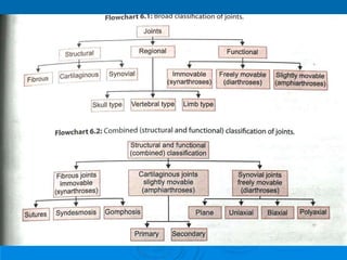
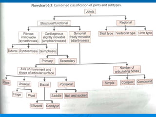
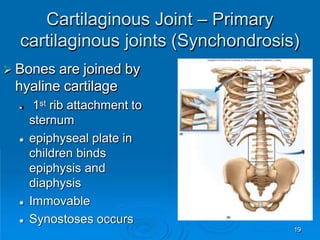
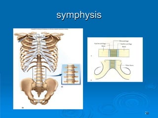
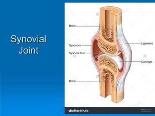
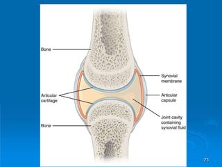
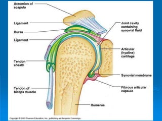
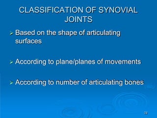
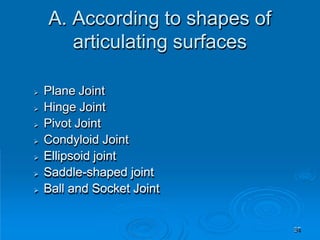
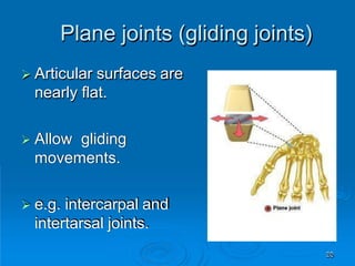
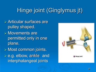
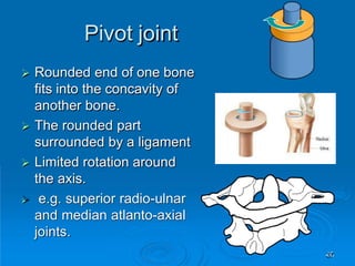
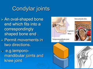
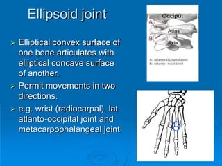
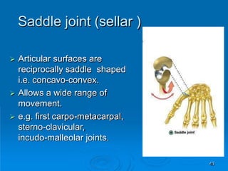
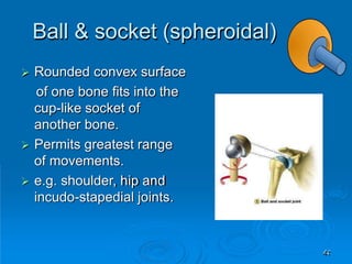
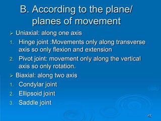
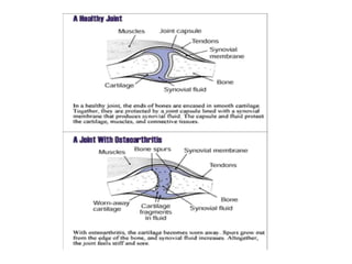
JOINTS
 Definition of joint
 Classification of joints
 General features of synovial joint
 Hilton’s laws
 Bursa
 Clinical case with anatomical explanation
Learning objectives
2
 regions of the skeleton where two or more
bones meet and articulate.
 supported by a variety of soft tissue
structures
 prime functions : facilitate growth or to
transmit forces between bones, thereby
enabling movement or weight to be
transferred.
JOINTS
3
 Arthrology = study of the joints
 Kinesiology = study of musculoskeletal
movement
 Classified by freedom of movement
(functional)
 diarthrosis (freely movable)
 amphiarthrosis (slightly movable)
 synarthrosis (little or no movement)
Joints and Their Classification
6
 Classified how adjacent bones
are joined (structural)
 Fibrous
 Cartilaginous
 Synovial
7
Fibrous Joints (Synarthrosis)
 The bones forming the joint are united
by fibrous connective tissue. These
are either immovable or permit only
slight degree of movement..
 Sutures
 Gomphoses and
 Syndesmoses
8
 Cranial and facial bones
 In a suture, the two bones are separated by
a layer of membrane-derived connective
tissue.
 This connective tissue decreases with age
so that the osteogenic surfaces become
apposed.
 On completion of growth, many sutures
synostose and are obliterated.
Fibrous Joint -- Sutures
9
10
Types of Sutures
11
• Plane
• Serrate
• Denticulate
• Squamous
• Schindylesis
12
13
14
15
Schindylesis
16
Fibrous Joint -- Gomphoses
 Attachment of a tooth
to its socket
 Held in place by
fibrous
periodontal ligament
 collagen fibers attach
tooth to jawbone
 Some movement
while chewing
17
15
 Two bones bound by
CT called the interosseus
membrane.
 Most movable of fibrous
joints
 Interosseus membranes
unite radius to ulna and
tibia to fibula
Fibrous Joint -- Syndesmosis
Cartilaginous Joint – Primary
cartilaginous joints (Synchondrosis)
19
 Bones are joined by
hyaline cartilage
 1st rib attachment to
sternum
 epiphyseal plate in
children binds
epiphysis and
diaphysis
 Immovable
 Synostoses occurs
Cartilaginous Joint – secondary
cartilaginous joints (Symphysis)
They are defined by the presence of an
intervening pad or disc of fibrocartilage
interposed between the articular hyaline
cartilage that covers the ends of two bones
a)pubic symphysis and
b)intervertebral discs (between body of vertebra)
Only slight amount of movement is possible
Mostly all midline joints are symphysis except
symphysis menti
20
symphysis
21
Synovial
Joint
22
23
1. FIBROUS CAPSULE
invests the joint like a sleeve and encloses synovial cavity
(except where there are synovial protusions)
Function :
a)stabilizes the joint in such a way that it permits
movements but resists dislocation.
b)numerous sensory nerve endings ramify on the
capsule. The stimulation of the nerves produce reflex
contraction of muscle acting on the joint.
Components Of Synovial Joints And
Their Functional Significance
24
2. SYNOVIAL MEMBRANE :
• It is a thin vascular membrane of connective tissue
lining inside of the fibrous capsule.
• The articular cartilages are not covered by it.
• Also absent from intra-articular discs and menisci
• FUNCTION: produces synovial fluid in sufficient
quantity to keep the surface lubricated.
3. ARTICULAR CARTILAGE: hyaline cartilage
covering the joint surfaces mostly . It provides smooth
friction free movements and resists compression
forces.
25
4.ARTICULAR DISCS AND MENISCI: these are pads
of fibrocartilage interposed between the articular
surface of some joints eg. Jaw (temporomandibular
joint) , wrist (radioulnar joint), sternoclavicular and
knee joints
FUNCTION:
a) Helps in lubrication of joints by maintaining an
interval between the articular surface.
b) Divides the joint into two compartments.
c) Prevents wear and tear of articular cartilage by
providing a cushioning effect.
26
24
Articular disc
Menisci
5. . LIGAMENT : thickened band of connective
tissue that attaches bone to bone
Two types:
a) True ligaments (intrinsic ligaments):
• local thickenings of fiber bundles of the capsule,
• Stabilize the joint and permit movement in one
plane. Eg. Medial and lateral collateral ligaments.
28
b) Accessory ligaments: these are separate from
the fibrous capsule. Stabilizes the joint and limits
the range of movements
Two types
i)Extracapsular: stylomandibular and
sphenomandibular
ii) Intracapsular. Cruciate ligaments of knee joint
29
6. Bursa (synovial flexor sheaths)
7. Fat Pads
8. Tendon
30
31
32
CLINICAL CORRELATION: osteoarthritis
Blood supply of synovial joint: from the periarticular
network of arteries that surround the joint. Supplies
capsule, synovial membrane and epiphysis.
(articular cartilages are avascular)
Nerve supply of synovial joint:
a) Sensory nerves conveying pain
b) Sensory nerves conveying proprioception.
c) Autonomic fibers which have vasomotor effects.
33
 Segmental innervation of joints.
 HILTON’S LAW: it states that the nerves
supplying the joint also supply the muscles
regulating the movements of joint and skin
over the joint.
34
Factors maintaining the stability
of synovial joint
35
 BONES
 LIGAMENTS
 MUSCLES
Clinical correlation: Dislocation of joint
CLASSIFICATION OF SYNOVIAL
JOINTS
36
 Based on the shape of articulating
surfaces
 According to plane/planes of movements
 According to number of articulating bones
A. According to shapes of
articulating surfaces
37
 Plane Joint
 Hinge Joint
 Pivot Joint
 Condyloid Joint
 Ellipsoid joint
 Saddle-shaped joint
 Ball and Socket Joint
Plane joints (gliding joints)
38
 Articular surfaces are
nearly flat.
 Allow gliding
movements.
 e.g. intercarpal and
intertarsal joints.
Hinge joint (Ginglymus jt)
39
 Articular surfaces are
pulley shaped.
 Movements are
permitted only in one
plane.
 Most common joints.
 e.g. elbow, ankle and
interphalangeal joints
Pivot joint
40
 Rounded end of one bone
fits into the concavity of
another bone.
 The rounded part
surrounded by a ligament
 Limited rotation around
the axis.
 e.g. superior radio-ulnar
and median atlanto-axial
joints.
Condylar joints
41
 An oval-shaped bone
end which fits into a
correspondingly
shaped bone end..
 Permit movements in
two directions.
 e.g.temporo-
mandibular joints and
knee joint
Ellipsoid joint
42
 Elliptical convex surface of
one bone articulates with
elliptical concave surface
of another.
 Permit movements in two
directions.
 e.g. wrist (radiocarpal), lat
atlanto-occipital joint and
metacarpophalangeal joint
Saddle joint (sellar )
43
 Articular surfaces are
reciprocally saddle shaped
i.e. concavo-convex.
 Allows a wide range of
movement.
 e.g. first carpo-metacarpal,
sterno-clavicular,
incudo-malleolar joints.
Ball & socket (spheroidal)
44
 Rounded convex surface
of one bone fits into the
cup-like socket of
another bone.
 Permits greatest range
of movements.
 e.g. shoulder, hip and
incudo-stapedial joints.
B. According to the plane/
planes of movement
45
 Uniaxial: along one axis
1. Hinge joint :Movements only along transverse
axis so only flexion and extension
2. Pivot joint: movement only along the vertical
axis so only rotation.
 Biaxial: along two axis
1. Condylar joint
2. Ellipsoid joint
3. Saddle joint
Multiaxial: along many axis
Eg. Ball and socket
Movements occur in three planes or axis. So
all kind of movements possible.
circumduction
46
Axis of Rotation
Shoulder joint has 3 degrees of freedom : multiaxial jo44int
 Simple joints: only two bones take part in
formation of joint. Eg. Interphalangeal
joints of fingers and toes.
 Compound joints: more than two bones
take part in formation of joint.
a) Ankle joint: tibia , fibula and talus
b) Knee joint: femur, tibia and patella
C. According to the number of
articulating bones
48
Movements of synovial joints
49
 Gliding or slipping
 Angular movements
 Rotary or circular movements
Synovial joints – sporting
examples
50
During the butterfly stroke, the ball
and socket joint of the shoulder
allows the swimmer’s arm to
rotate.
You might head a football using the pivot
joint in your neck, which allows your head
to rotate.
What type of joint allows a handball player’s
fingers to spread apart so that they can control
the ball with one hand?
Answer:
The joints between the metacarpals and phalanges.
Components of a Lever
51
 A lever is a rigid object that rotates around a
fixed point called a fulcrum
 Rotation occurs when effort overcomes
resistance
 resistance arm and effort arm are described
relative to fulcrum
Mechanical Advantage of a Lever
52
 Two effects of levers
 lever that helps increase output of force
• human moving a heavy object with help of crowbar
 lever move object further and faster
• movement of row boat with paddle
 Types of levers produce either increase in
speed or force
50
First-Class Lever
 Has fulcrum in the middle between effort and
resistance
 Atlantooccipital joint lies between the muscles on the
back of the neck and the weight of the face
Second-Class Lever
54
 Resistance between fulcrum and effort
 Resistance from the muscle tone of the temporalis
muscle lies between the jaw joint and the pull of the
diagastric muscle on the chin as it opens the mouth
quickly
Third-Class Lever
55
 Effort between the resistance and the fulcrum
 most joints of the body
 The effort applied by the biceps muscle is applied to
the forearm between the elbow joint and the weight of
the hand and the forearm
Range of Motion
56
 Degrees through which a joint can move
 Determined by
 structure of the articular surfaces
 strength and tautness of ligaments, tendons and
capsule
• stretching of ligaments increases range of motion
 action of the muscles and tendons
• nervous system monitors joint position and muscle
tone
Position of joints
57
 Closed packed position
 Loose packed position
Close packed position
58
 it is the position in which the articular
surfaces are fully congruent and have
maximum area of contact.
 The joint is firm and rigid all the ligaments
in relation to it are mostly taut.
 No dislocation but if does occur then the
most damage occurs
 Eg. Shoulder joint abducted and laterally
rotated, elbow fully extended.
Loose packed position
59
 In this the articular surfaces are
incongruent.
 The joint space is freely mobile in this
position and prone to disclocation.
 Eg. Shoulder joint semiabducted
Flexion, Extension and Hyperextension
60
 Flexion decreases the
angle of a joint
 Extension straightens
and returns to the
anatomical position
 Hyperextension =
extension beyond 180
degrees
Flexion, Extension and Hyperextension
61
Abduction and Adduction
62
 Abduction is movement of a part away from the
midline
 hyperabduction – raise arm over back or front of head
 Adduction is movement towards the midline
 hyperadduction – crossing fingers
Elevation and Depression
63
 Elevation is a movement that raises a bone vertically
 mandibles are elevated during biting and clavicles during
a shrug
 Depression is lowering the mandible or the
shoulders
Protraction and Retraction
64
 Protraction = movement
anteriorly on horizontal
plane
 thrusting the jaw
forward, shoulders or
pelvis forward
 Retraction is movement
posteriorly
Circumduction
65
 Movement in which one end
of an appendage remains
stationary while the other end
makes a circular motion
 Sequence of flexion,
abduction, extension and
adduction movements
 baseball player winding up for
a pitch
Rotation
66
 Movement on
longitudinal axis
 rotation of trunk,
thigh, head or arm
 Medial rotation
turns the bone
inwards
 Lateral rotation
turns the bone
outwards
64
Supination and Pronation
 In the forearm and foot
 Supination
 rotation of forearm so that the
palm faces forward
 inversion and abduction of foot
(raising the medial edge of the
foot)
 Pronation
 rotation of forearm so the palm
faces to the rear
 eversion and abduction of foot
(raising the lateral edge of the
foot)
65
Movements of Head and Trunk
 Flexion, hyperextension and lateral flexion of
vertebral column
66
Rotation of Trunk and Head
 Right rotation of trunk; rotation of head
67
Movements of Mandible
 Lateral excursion = sideways movement
 Medial excursion = movement back to the
midline
 side-to-side grinding during chewing
 Protraction – retraction of mandible
68
Movement of Hand and Digits
 Radial and ulnar
flexion
 Abduction of fingers
and thumb
 Opposition is
movement of the
thumb to approach
or touch the
fingertips
 Reposition is
movement back to
the anatomical
position
69
Movements of the Foot
 Dorsiflexion is raising of the toes as when you swing the
foot forward to take a step (heel strike)
 Plantarflexion is extension of the foot so that the toes point
downward as in standing on tiptoe
 Inversion is a movement in which the soles are turned
medially
 Eversion is a turning of the soles to face laterally
 Bursitis – inflammation of a bursa usually
caused by a blow or friction
 Tendonitis – inflammation of tendon
sheaths
 Arthritis – inflammatory or degenerative
diseases of joints
Inflammatory Conditions
Associated with Joints
70
71
Rheumatoid Arthritis
75
Gouty Arthritis
76
Joint Prostheses
77
Types of
cartilage
78
 Hyaline
 Fibrocartilage
 Elastic
79
80

joints-ppt.pptx

  • 1.
  • 2.
     Definition ofjoint  Classification of joints  General features of synovial joint  Hilton’s laws  Bursa  Clinical case with anatomical explanation Learning objectives 2
  • 3.
     regions ofthe skeleton where two or more bones meet and articulate.  supported by a variety of soft tissue structures  prime functions : facilitate growth or to transmit forces between bones, thereby enabling movement or weight to be transferred. JOINTS 3
  • 6.
     Arthrology =study of the joints  Kinesiology = study of musculoskeletal movement  Classified by freedom of movement (functional)  diarthrosis (freely movable)  amphiarthrosis (slightly movable)  synarthrosis (little or no movement) Joints and Their Classification 6
  • 7.
     Classified howadjacent bones are joined (structural)  Fibrous  Cartilaginous  Synovial 7
  • 8.
    Fibrous Joints (Synarthrosis) The bones forming the joint are united by fibrous connective tissue. These are either immovable or permit only slight degree of movement..  Sutures  Gomphoses and  Syndesmoses 8
  • 9.
     Cranial andfacial bones  In a suture, the two bones are separated by a layer of membrane-derived connective tissue.  This connective tissue decreases with age so that the osteogenic surfaces become apposed.  On completion of growth, many sutures synostose and are obliterated. Fibrous Joint -- Sutures 9
  • 10.
  • 11.
    Types of Sutures 11 •Plane • Serrate • Denticulate • Squamous • Schindylesis
  • 12.
  • 13.
  • 14.
  • 15.
  • 16.
  • 17.
    Fibrous Joint --Gomphoses  Attachment of a tooth to its socket  Held in place by fibrous periodontal ligament  collagen fibers attach tooth to jawbone  Some movement while chewing 17
  • 18.
    15  Two bonesbound by CT called the interosseus membrane.  Most movable of fibrous joints  Interosseus membranes unite radius to ulna and tibia to fibula Fibrous Joint -- Syndesmosis
  • 19.
    Cartilaginous Joint –Primary cartilaginous joints (Synchondrosis) 19  Bones are joined by hyaline cartilage  1st rib attachment to sternum  epiphyseal plate in children binds epiphysis and diaphysis  Immovable  Synostoses occurs
  • 20.
    Cartilaginous Joint –secondary cartilaginous joints (Symphysis) They are defined by the presence of an intervening pad or disc of fibrocartilage interposed between the articular hyaline cartilage that covers the ends of two bones a)pubic symphysis and b)intervertebral discs (between body of vertebra) Only slight amount of movement is possible Mostly all midline joints are symphysis except symphysis menti 20
  • 21.
  • 22.
  • 23.
  • 24.
    1. FIBROUS CAPSULE investsthe joint like a sleeve and encloses synovial cavity (except where there are synovial protusions) Function : a)stabilizes the joint in such a way that it permits movements but resists dislocation. b)numerous sensory nerve endings ramify on the capsule. The stimulation of the nerves produce reflex contraction of muscle acting on the joint. Components Of Synovial Joints And Their Functional Significance 24
  • 25.
    2. SYNOVIAL MEMBRANE: • It is a thin vascular membrane of connective tissue lining inside of the fibrous capsule. • The articular cartilages are not covered by it. • Also absent from intra-articular discs and menisci • FUNCTION: produces synovial fluid in sufficient quantity to keep the surface lubricated. 3. ARTICULAR CARTILAGE: hyaline cartilage covering the joint surfaces mostly . It provides smooth friction free movements and resists compression forces. 25
  • 26.
    4.ARTICULAR DISCS ANDMENISCI: these are pads of fibrocartilage interposed between the articular surface of some joints eg. Jaw (temporomandibular joint) , wrist (radioulnar joint), sternoclavicular and knee joints FUNCTION: a) Helps in lubrication of joints by maintaining an interval between the articular surface. b) Divides the joint into two compartments. c) Prevents wear and tear of articular cartilage by providing a cushioning effect. 26
  • 27.
  • 28.
    5. . LIGAMENT: thickened band of connective tissue that attaches bone to bone Two types: a) True ligaments (intrinsic ligaments): • local thickenings of fiber bundles of the capsule, • Stabilize the joint and permit movement in one plane. Eg. Medial and lateral collateral ligaments. 28
  • 29.
    b) Accessory ligaments:these are separate from the fibrous capsule. Stabilizes the joint and limits the range of movements Two types i)Extracapsular: stylomandibular and sphenomandibular ii) Intracapsular. Cruciate ligaments of knee joint 29
  • 30.
    6. Bursa (synovialflexor sheaths) 7. Fat Pads 8. Tendon 30
  • 31.
  • 32.
  • 33.
    CLINICAL CORRELATION: osteoarthritis Bloodsupply of synovial joint: from the periarticular network of arteries that surround the joint. Supplies capsule, synovial membrane and epiphysis. (articular cartilages are avascular) Nerve supply of synovial joint: a) Sensory nerves conveying pain b) Sensory nerves conveying proprioception. c) Autonomic fibers which have vasomotor effects. 33
  • 34.
     Segmental innervationof joints.  HILTON’S LAW: it states that the nerves supplying the joint also supply the muscles regulating the movements of joint and skin over the joint. 34
  • 35.
    Factors maintaining thestability of synovial joint 35  BONES  LIGAMENTS  MUSCLES Clinical correlation: Dislocation of joint
  • 36.
    CLASSIFICATION OF SYNOVIAL JOINTS 36 Based on the shape of articulating surfaces  According to plane/planes of movements  According to number of articulating bones
  • 37.
    A. According toshapes of articulating surfaces 37  Plane Joint  Hinge Joint  Pivot Joint  Condyloid Joint  Ellipsoid joint  Saddle-shaped joint  Ball and Socket Joint
  • 38.
    Plane joints (glidingjoints) 38  Articular surfaces are nearly flat.  Allow gliding movements.  e.g. intercarpal and intertarsal joints.
  • 39.
    Hinge joint (Ginglymusjt) 39  Articular surfaces are pulley shaped.  Movements are permitted only in one plane.  Most common joints.  e.g. elbow, ankle and interphalangeal joints
  • 40.
    Pivot joint 40  Roundedend of one bone fits into the concavity of another bone.  The rounded part surrounded by a ligament  Limited rotation around the axis.  e.g. superior radio-ulnar and median atlanto-axial joints.
  • 41.
    Condylar joints 41  Anoval-shaped bone end which fits into a correspondingly shaped bone end..  Permit movements in two directions.  e.g.temporo- mandibular joints and knee joint
  • 42.
    Ellipsoid joint 42  Ellipticalconvex surface of one bone articulates with elliptical concave surface of another.  Permit movements in two directions.  e.g. wrist (radiocarpal), lat atlanto-occipital joint and metacarpophalangeal joint
  • 43.
    Saddle joint (sellar) 43  Articular surfaces are reciprocally saddle shaped i.e. concavo-convex.  Allows a wide range of movement.  e.g. first carpo-metacarpal, sterno-clavicular, incudo-malleolar joints.
  • 44.
    Ball & socket(spheroidal) 44  Rounded convex surface of one bone fits into the cup-like socket of another bone.  Permits greatest range of movements.  e.g. shoulder, hip and incudo-stapedial joints.
  • 45.
    B. According tothe plane/ planes of movement 45  Uniaxial: along one axis 1. Hinge joint :Movements only along transverse axis so only flexion and extension 2. Pivot joint: movement only along the vertical axis so only rotation.  Biaxial: along two axis 1. Condylar joint 2. Ellipsoid joint 3. Saddle joint
  • 46.
    Multiaxial: along manyaxis Eg. Ball and socket Movements occur in three planes or axis. So all kind of movements possible. circumduction 46
  • 47.
    Axis of Rotation Shoulderjoint has 3 degrees of freedom : multiaxial jo44int
  • 48.
     Simple joints:only two bones take part in formation of joint. Eg. Interphalangeal joints of fingers and toes.  Compound joints: more than two bones take part in formation of joint. a) Ankle joint: tibia , fibula and talus b) Knee joint: femur, tibia and patella C. According to the number of articulating bones 48
  • 49.
    Movements of synovialjoints 49  Gliding or slipping  Angular movements  Rotary or circular movements
  • 50.
    Synovial joints –sporting examples 50 During the butterfly stroke, the ball and socket joint of the shoulder allows the swimmer’s arm to rotate. You might head a football using the pivot joint in your neck, which allows your head to rotate. What type of joint allows a handball player’s fingers to spread apart so that they can control the ball with one hand? Answer: The joints between the metacarpals and phalanges.
  • 51.
    Components of aLever 51  A lever is a rigid object that rotates around a fixed point called a fulcrum  Rotation occurs when effort overcomes resistance  resistance arm and effort arm are described relative to fulcrum
  • 52.
    Mechanical Advantage ofa Lever 52  Two effects of levers  lever that helps increase output of force • human moving a heavy object with help of crowbar  lever move object further and faster • movement of row boat with paddle  Types of levers produce either increase in speed or force
  • 53.
    50 First-Class Lever  Hasfulcrum in the middle between effort and resistance  Atlantooccipital joint lies between the muscles on the back of the neck and the weight of the face
  • 54.
    Second-Class Lever 54  Resistancebetween fulcrum and effort  Resistance from the muscle tone of the temporalis muscle lies between the jaw joint and the pull of the diagastric muscle on the chin as it opens the mouth quickly
  • 55.
    Third-Class Lever 55  Effortbetween the resistance and the fulcrum  most joints of the body  The effort applied by the biceps muscle is applied to the forearm between the elbow joint and the weight of the hand and the forearm
  • 56.
    Range of Motion 56 Degrees through which a joint can move  Determined by  structure of the articular surfaces  strength and tautness of ligaments, tendons and capsule • stretching of ligaments increases range of motion  action of the muscles and tendons • nervous system monitors joint position and muscle tone
  • 57.
    Position of joints 57 Closed packed position  Loose packed position
  • 58.
    Close packed position 58 it is the position in which the articular surfaces are fully congruent and have maximum area of contact.  The joint is firm and rigid all the ligaments in relation to it are mostly taut.  No dislocation but if does occur then the most damage occurs  Eg. Shoulder joint abducted and laterally rotated, elbow fully extended.
  • 59.
    Loose packed position 59 In this the articular surfaces are incongruent.  The joint space is freely mobile in this position and prone to disclocation.  Eg. Shoulder joint semiabducted
  • 60.
    Flexion, Extension andHyperextension 60  Flexion decreases the angle of a joint  Extension straightens and returns to the anatomical position  Hyperextension = extension beyond 180 degrees
  • 61.
    Flexion, Extension andHyperextension 61
  • 62.
    Abduction and Adduction 62 Abduction is movement of a part away from the midline  hyperabduction – raise arm over back or front of head  Adduction is movement towards the midline  hyperadduction – crossing fingers
  • 63.
    Elevation and Depression 63 Elevation is a movement that raises a bone vertically  mandibles are elevated during biting and clavicles during a shrug  Depression is lowering the mandible or the shoulders
  • 64.
    Protraction and Retraction 64 Protraction = movement anteriorly on horizontal plane  thrusting the jaw forward, shoulders or pelvis forward  Retraction is movement posteriorly
  • 65.
    Circumduction 65  Movement inwhich one end of an appendage remains stationary while the other end makes a circular motion  Sequence of flexion, abduction, extension and adduction movements  baseball player winding up for a pitch
  • 66.
    Rotation 66  Movement on longitudinalaxis  rotation of trunk, thigh, head or arm  Medial rotation turns the bone inwards  Lateral rotation turns the bone outwards
  • 67.
    64 Supination and Pronation In the forearm and foot  Supination  rotation of forearm so that the palm faces forward  inversion and abduction of foot (raising the medial edge of the foot)  Pronation  rotation of forearm so the palm faces to the rear  eversion and abduction of foot (raising the lateral edge of the foot)
  • 68.
    65 Movements of Headand Trunk  Flexion, hyperextension and lateral flexion of vertebral column
  • 69.
    66 Rotation of Trunkand Head  Right rotation of trunk; rotation of head
  • 70.
    67 Movements of Mandible Lateral excursion = sideways movement  Medial excursion = movement back to the midline  side-to-side grinding during chewing  Protraction – retraction of mandible
  • 71.
    68 Movement of Handand Digits  Radial and ulnar flexion  Abduction of fingers and thumb  Opposition is movement of the thumb to approach or touch the fingertips  Reposition is movement back to the anatomical position
  • 72.
    69 Movements of theFoot  Dorsiflexion is raising of the toes as when you swing the foot forward to take a step (heel strike)  Plantarflexion is extension of the foot so that the toes point downward as in standing on tiptoe  Inversion is a movement in which the soles are turned medially  Eversion is a turning of the soles to face laterally
  • 73.
     Bursitis –inflammation of a bursa usually caused by a blow or friction  Tendonitis – inflammation of tendon sheaths  Arthritis – inflammatory or degenerative diseases of joints Inflammatory Conditions Associated with Joints 70
  • 74.
  • 75.
  • 76.
  • 77.
  • 78.
    Types of cartilage 78  Hyaline Fibrocartilage  Elastic
  • 79.
  • 80.