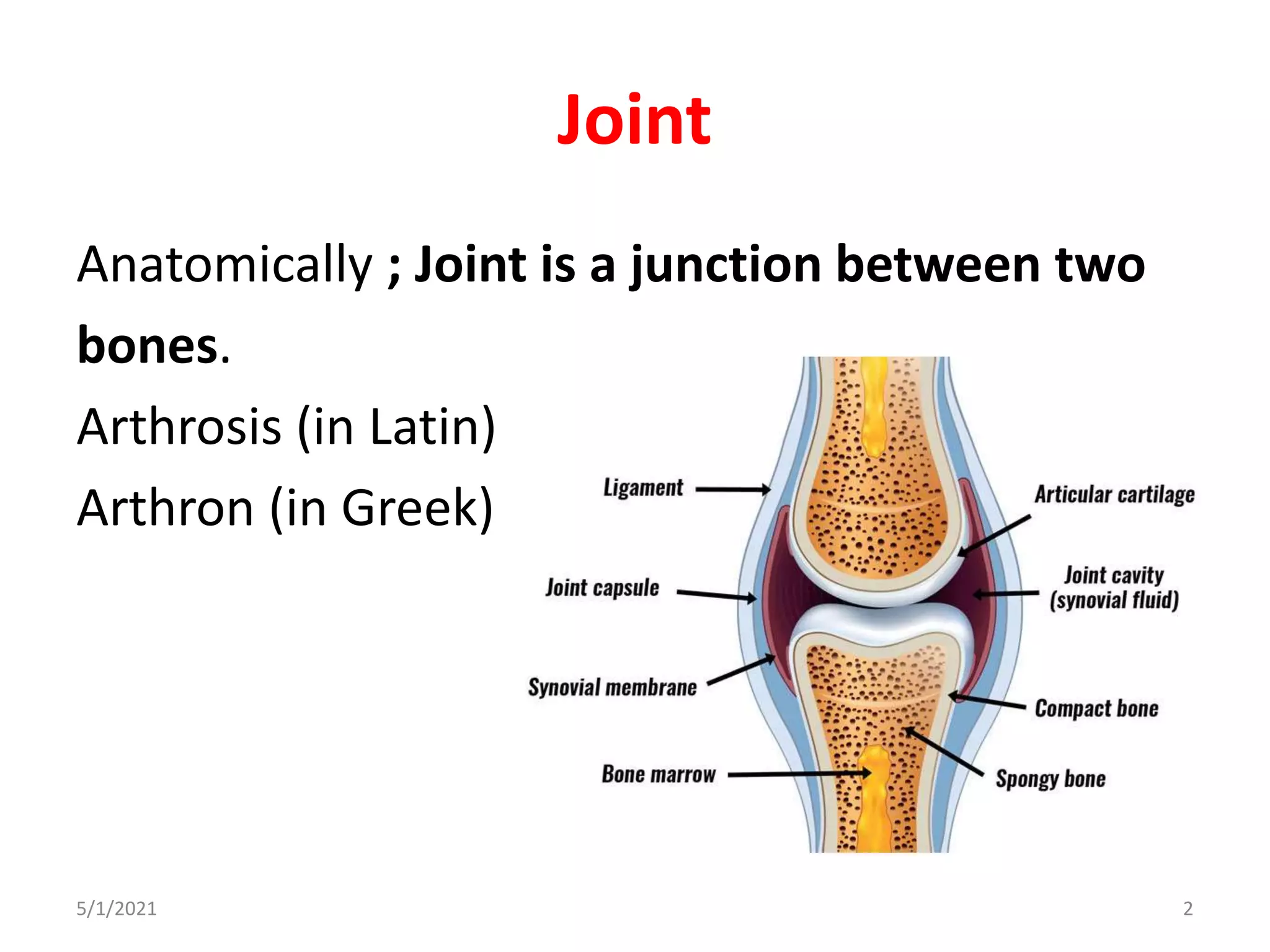
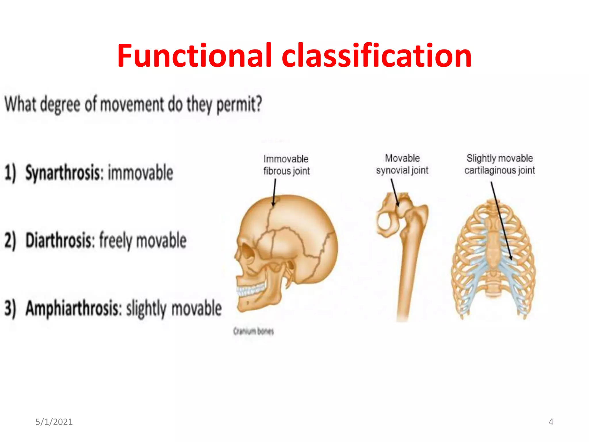
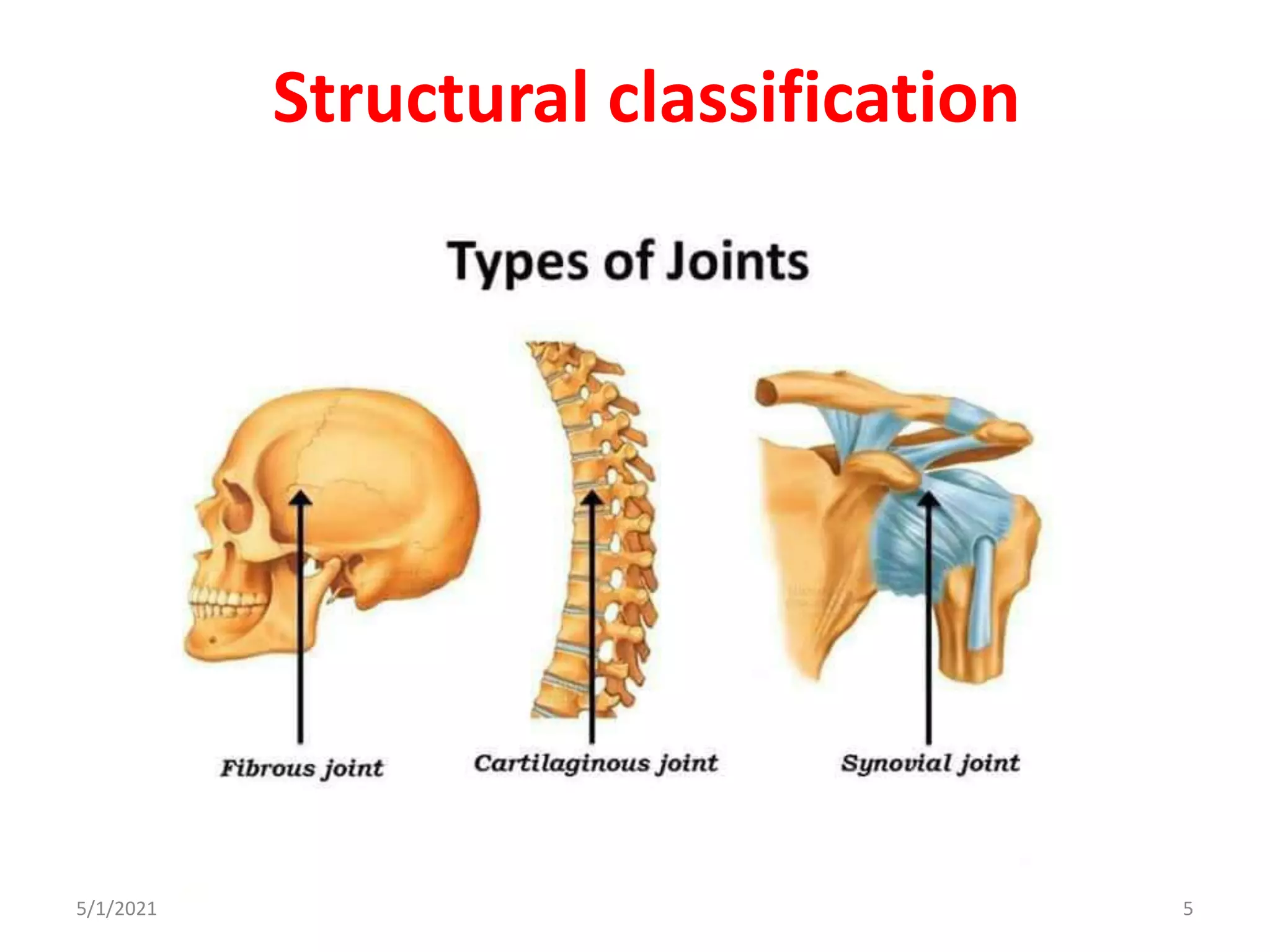
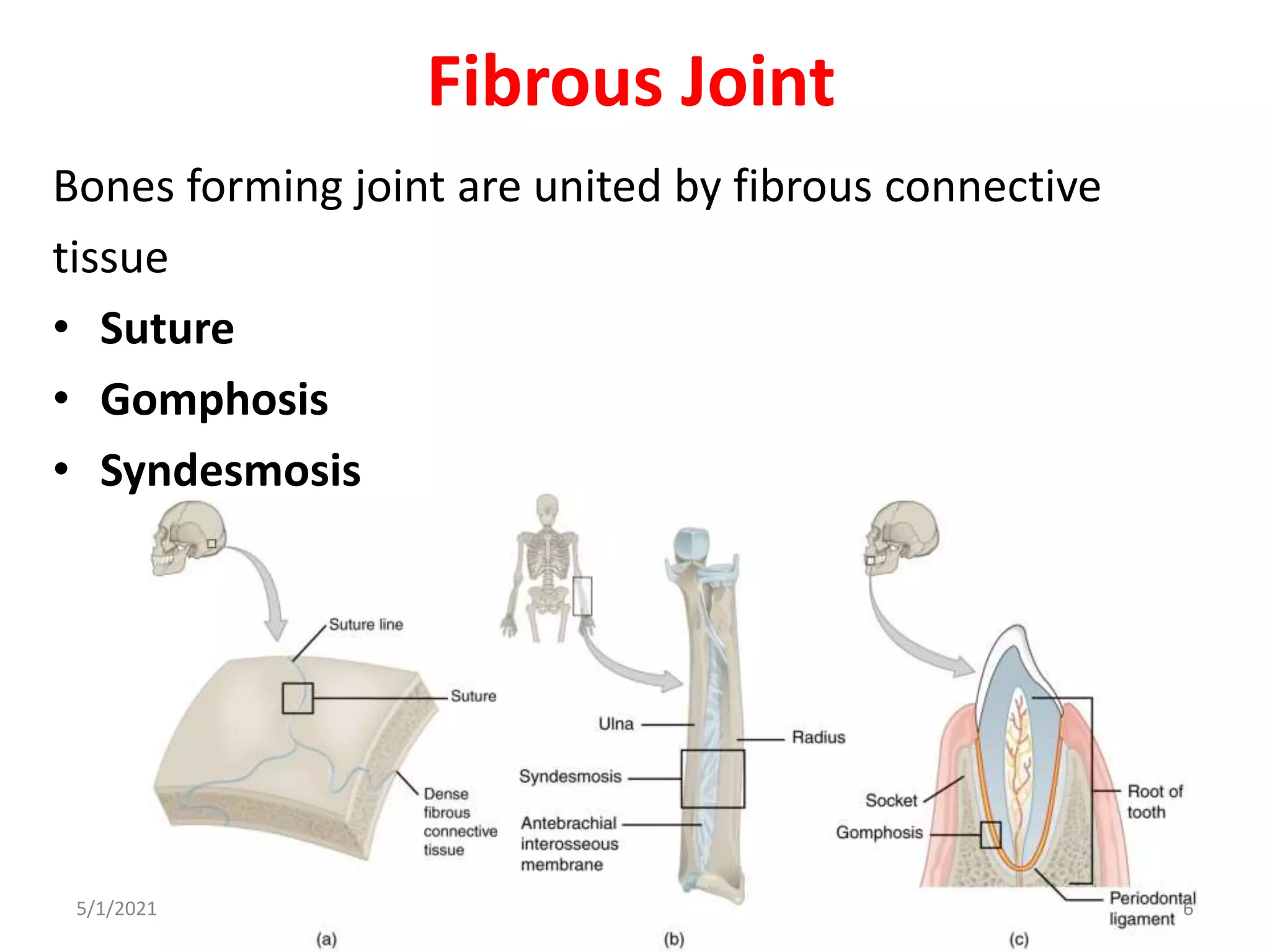
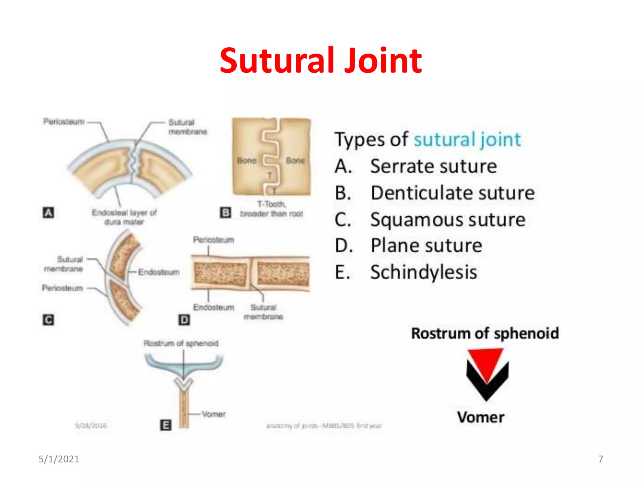
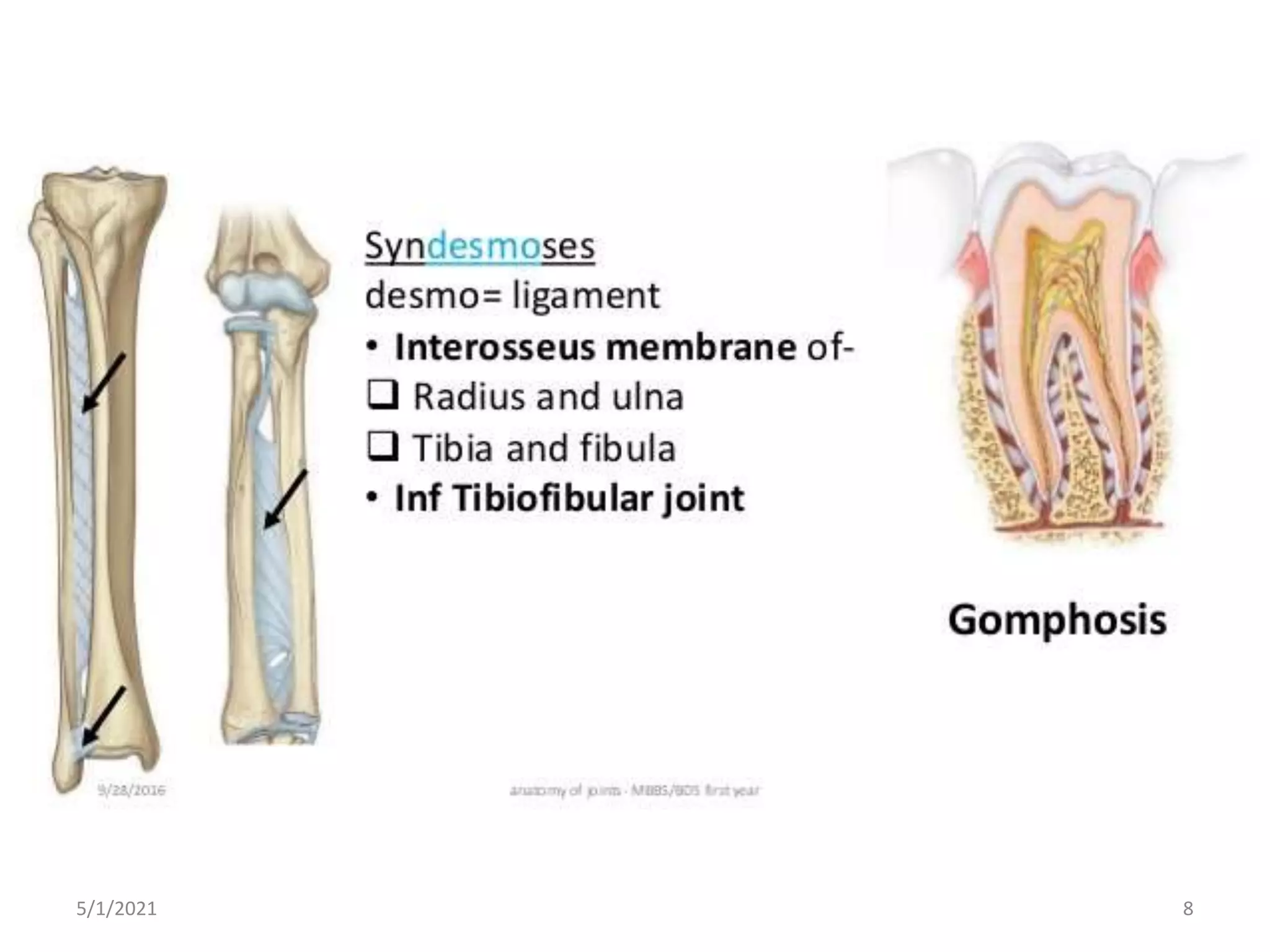
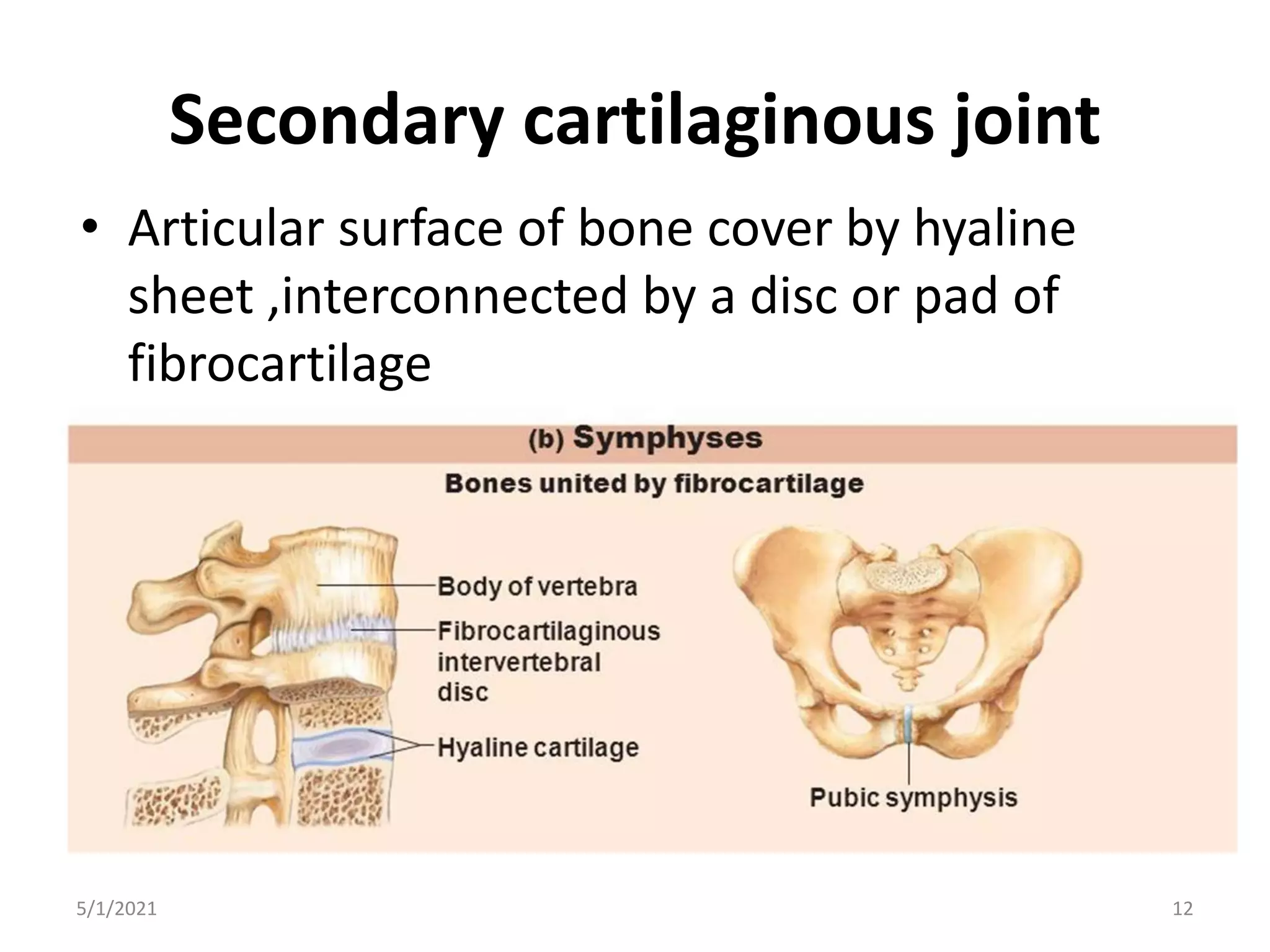
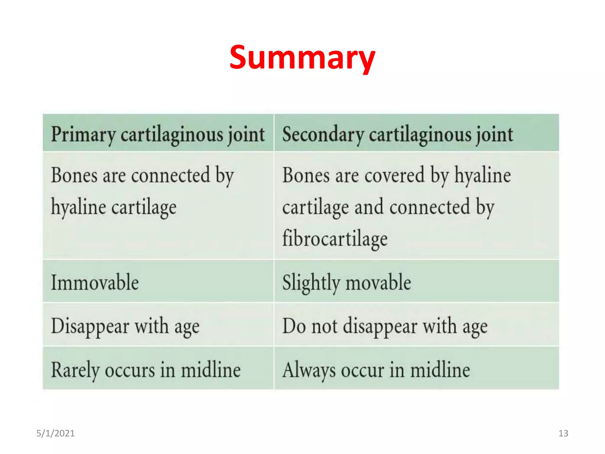
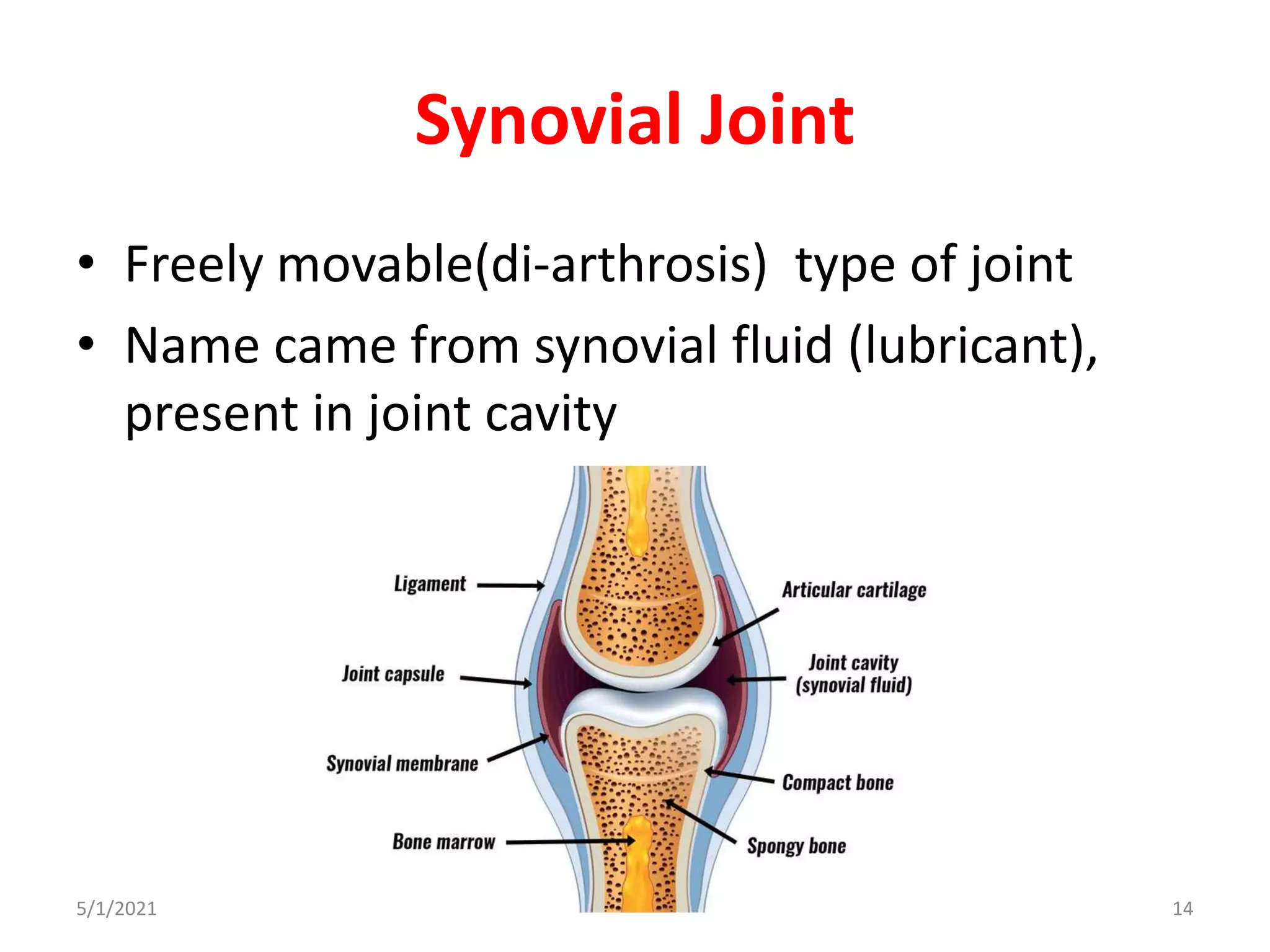
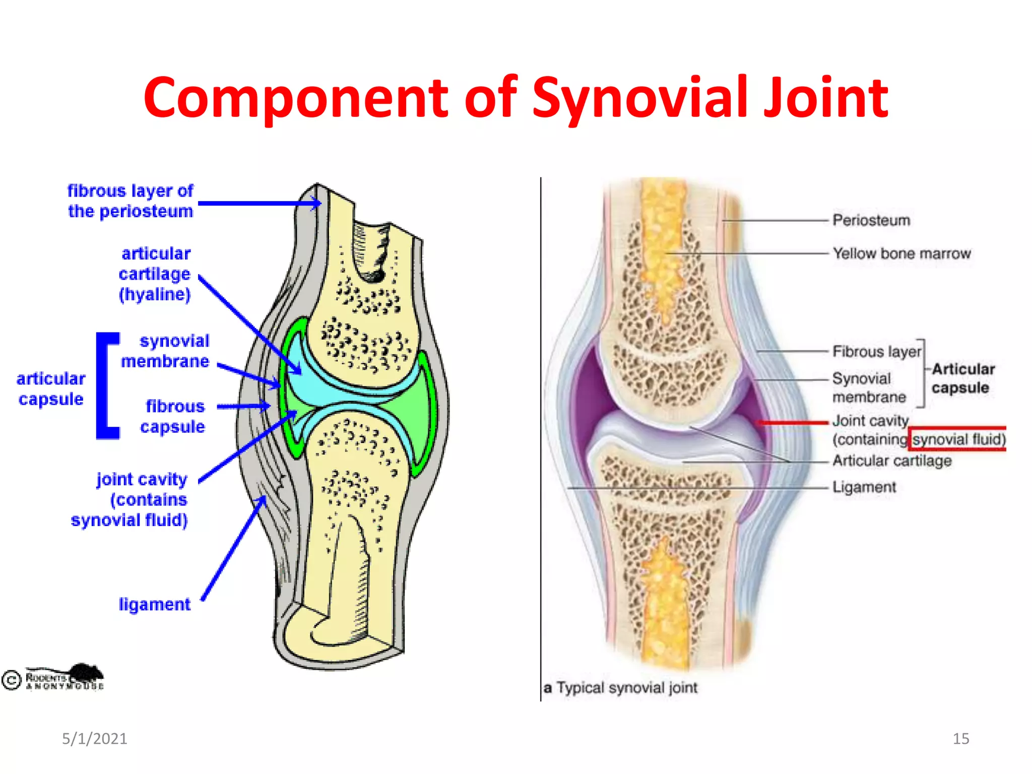
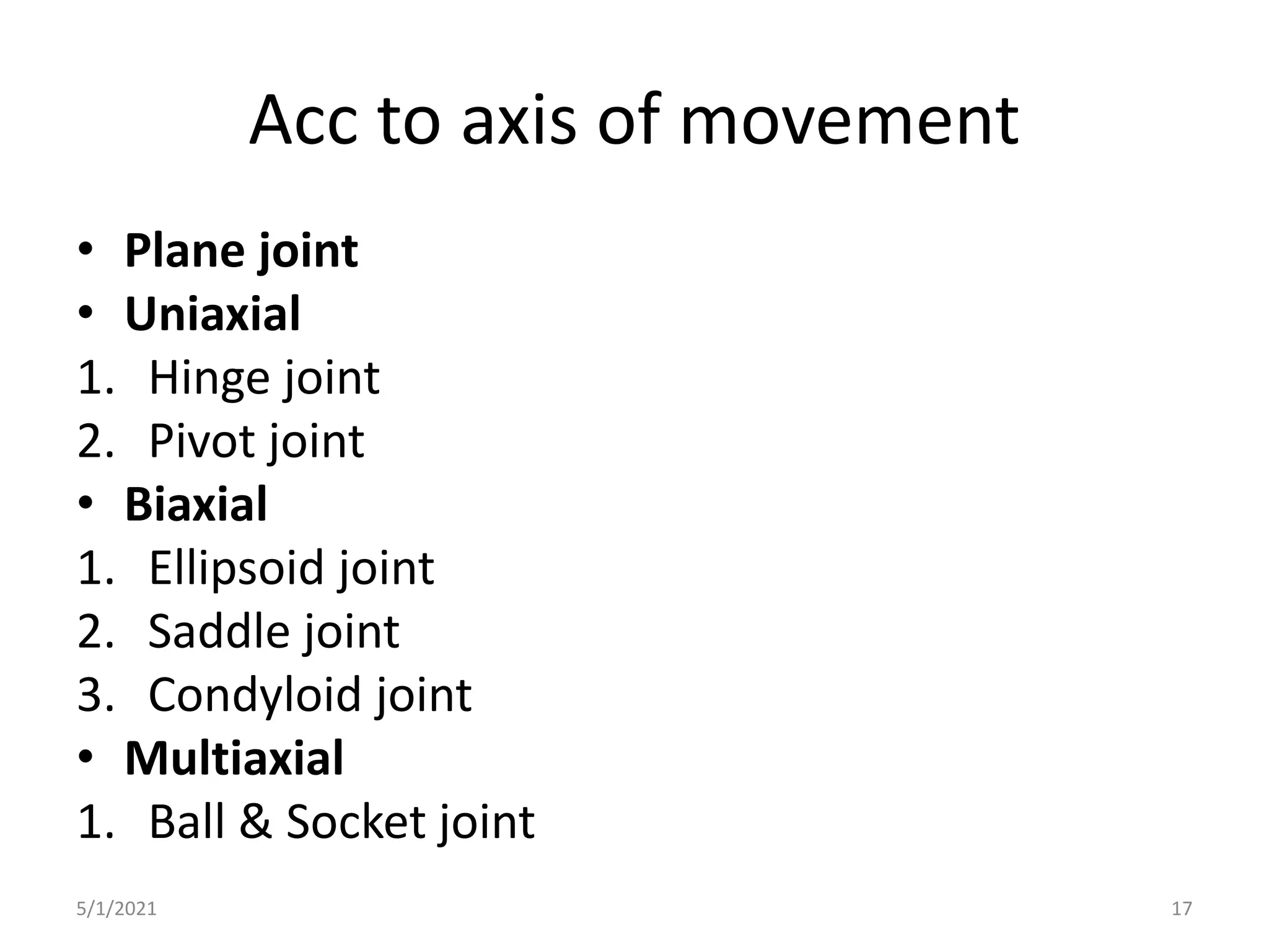
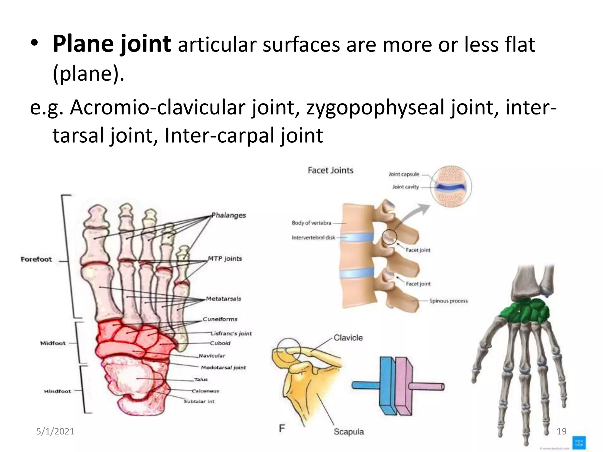
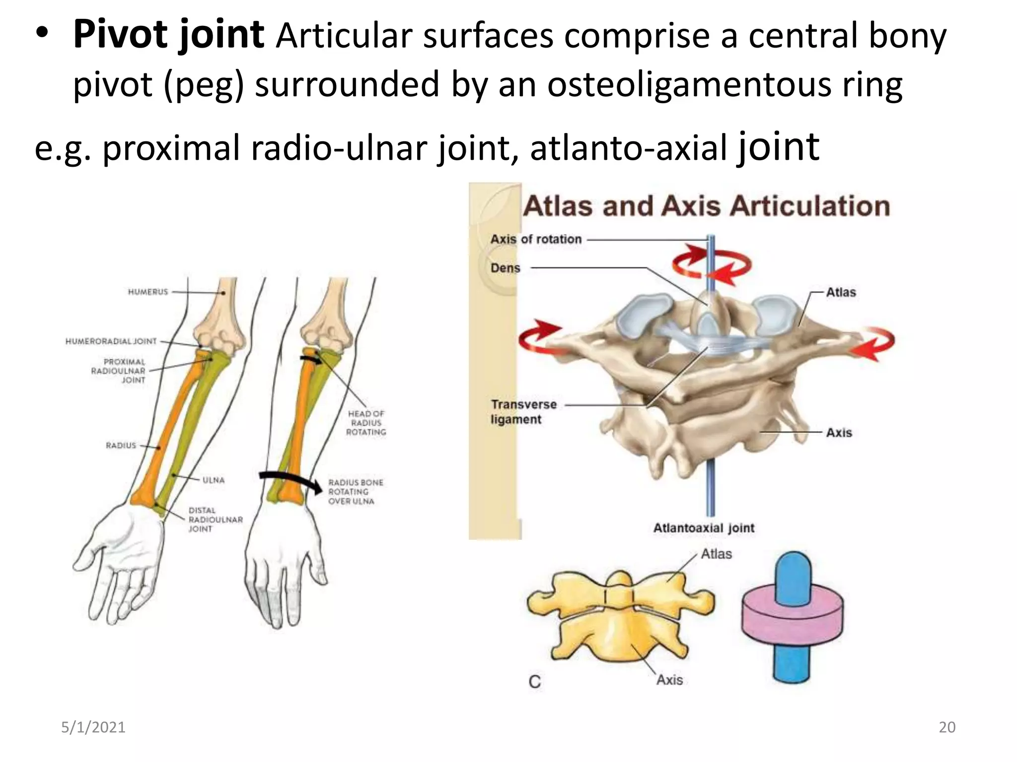
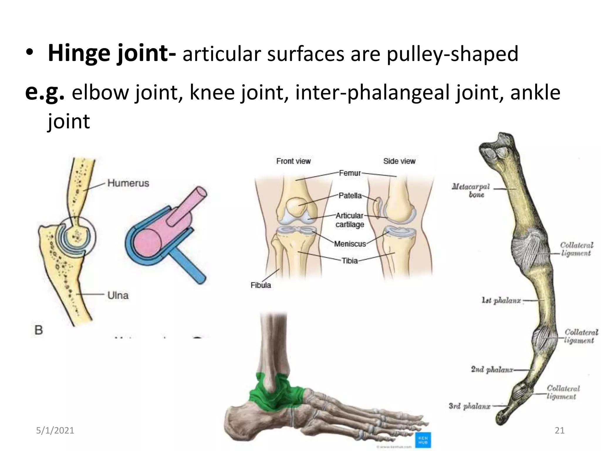
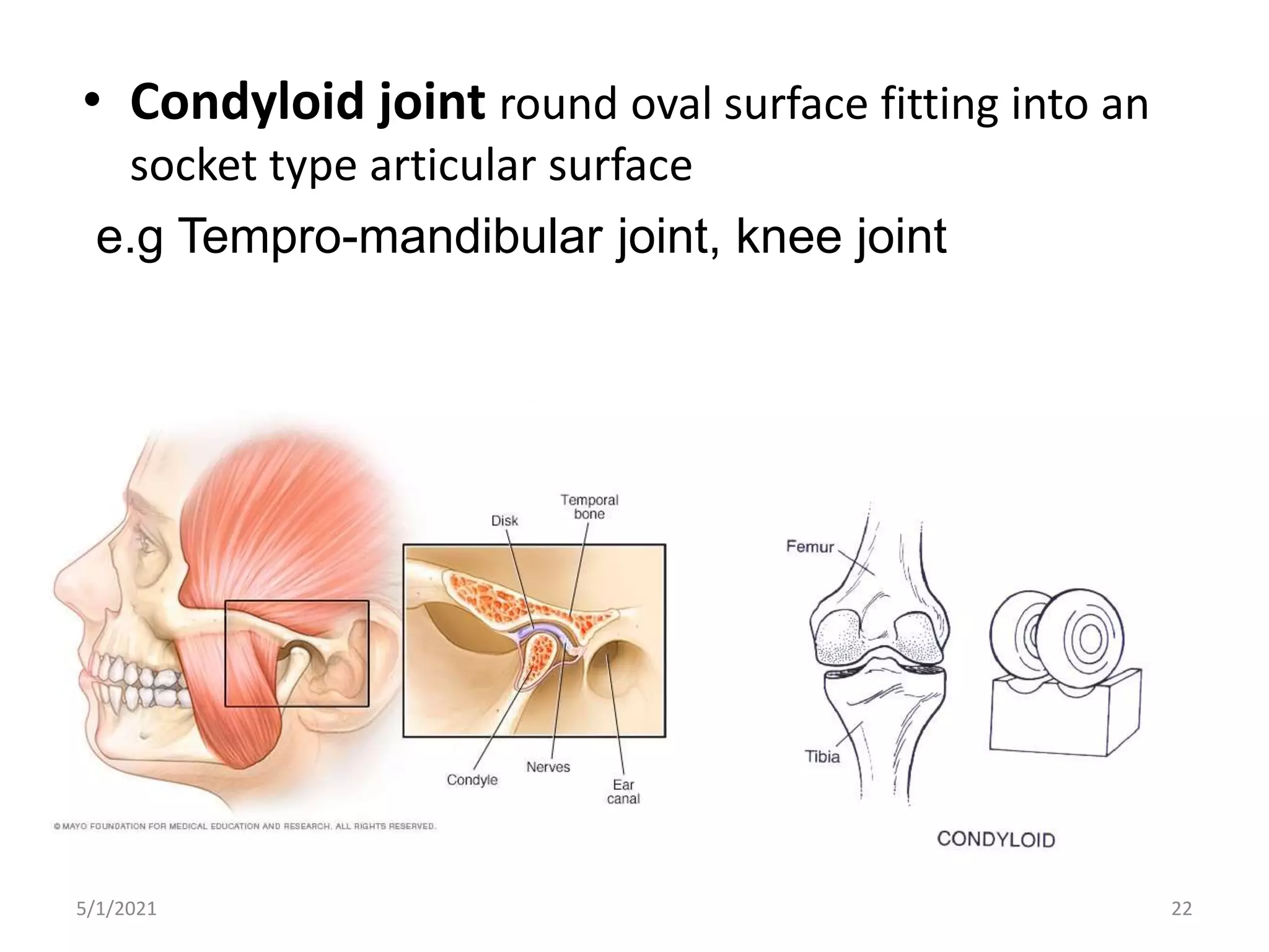
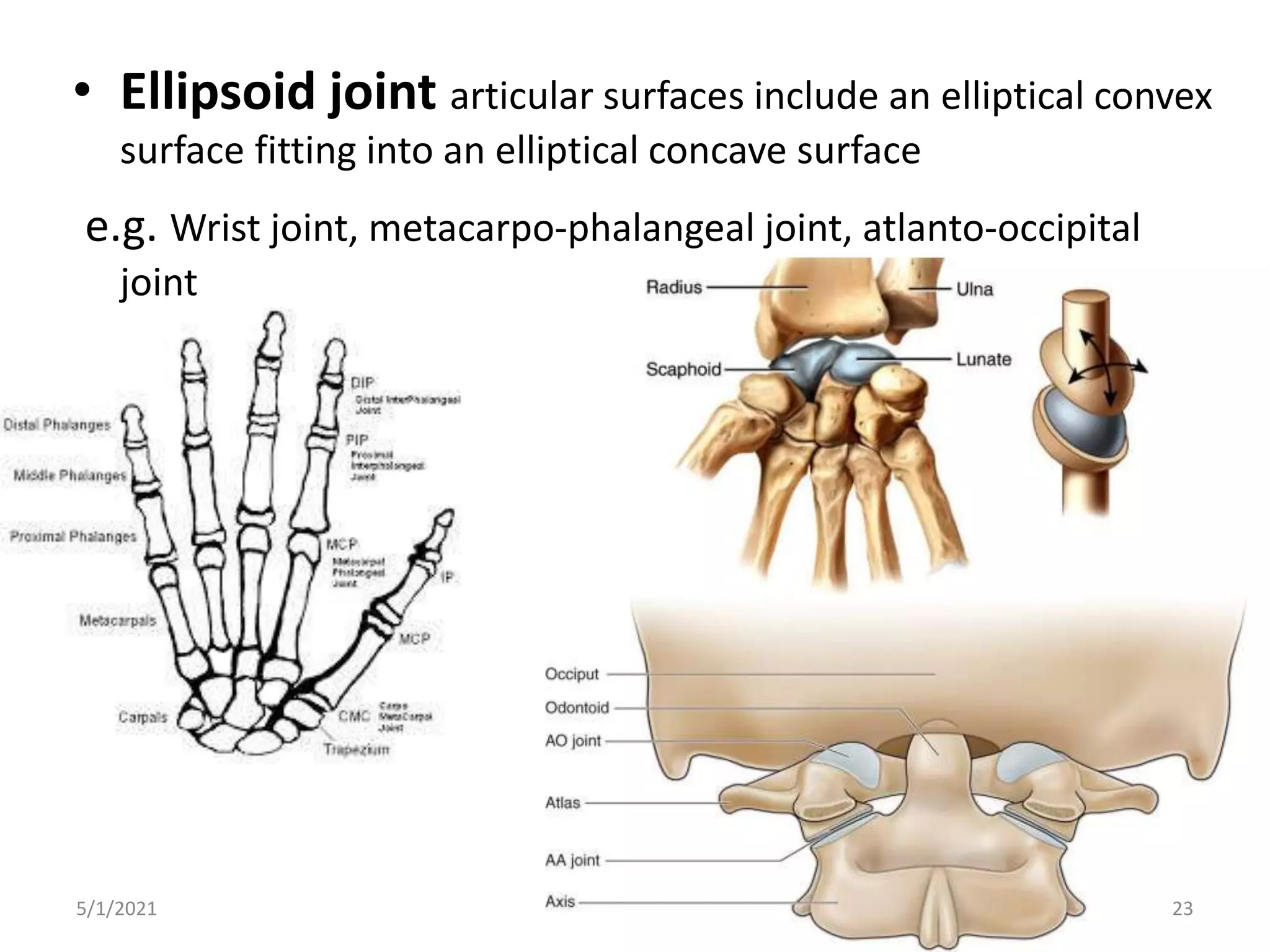
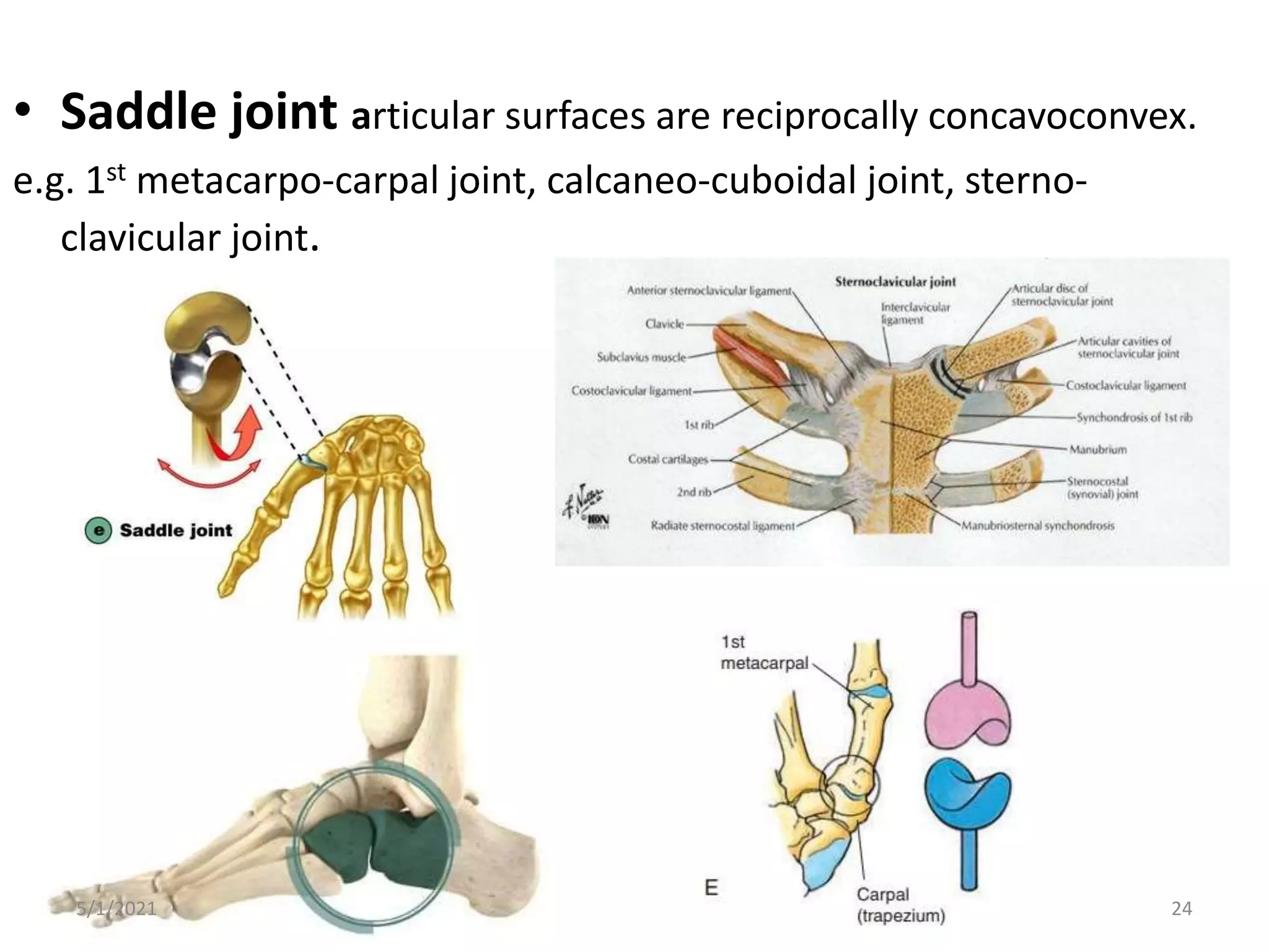
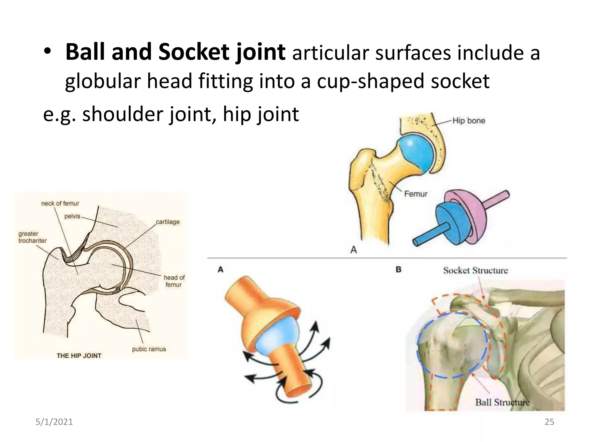
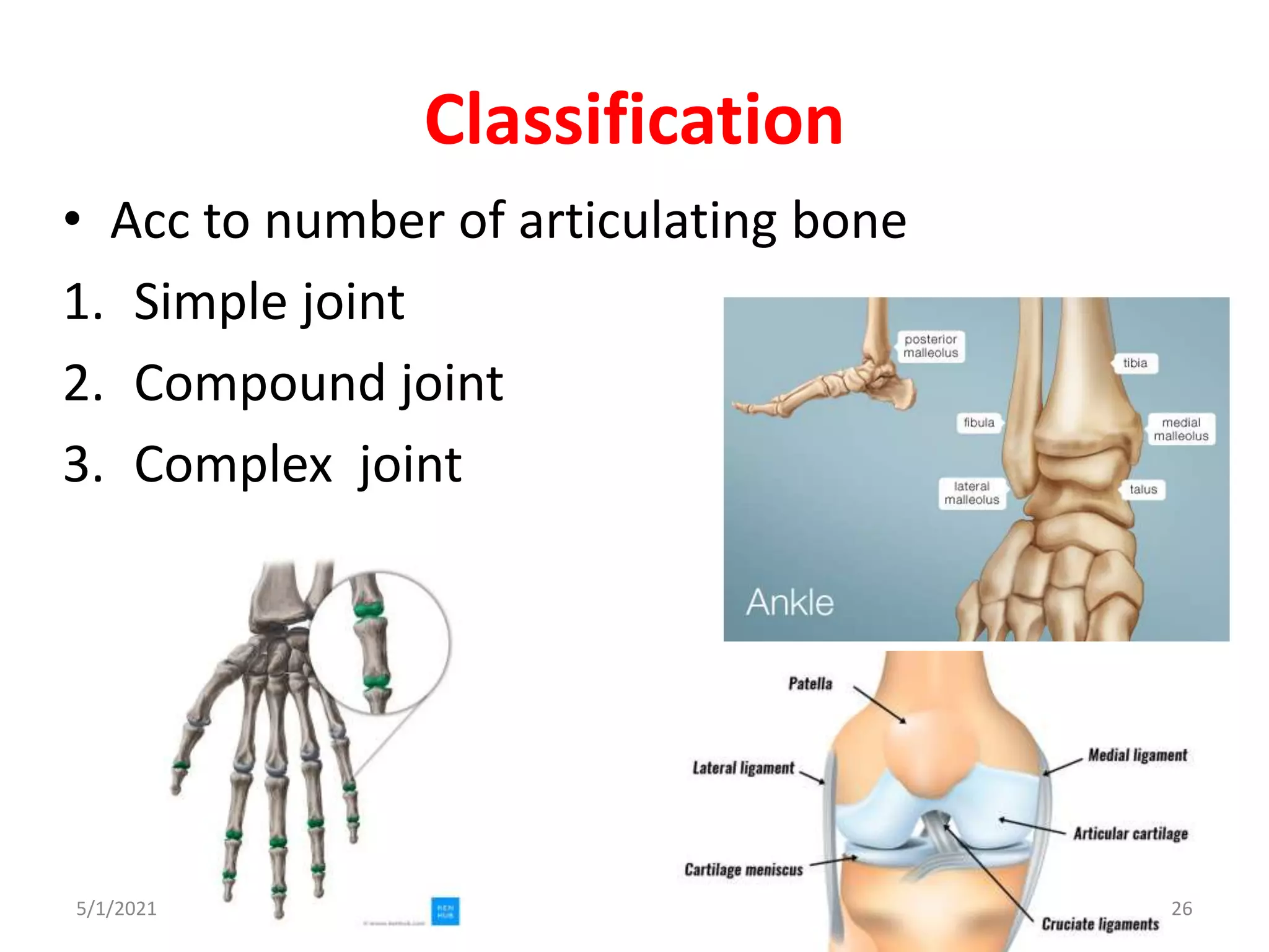
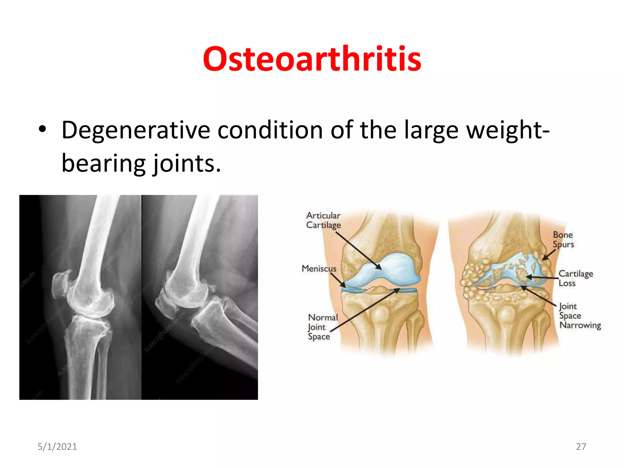
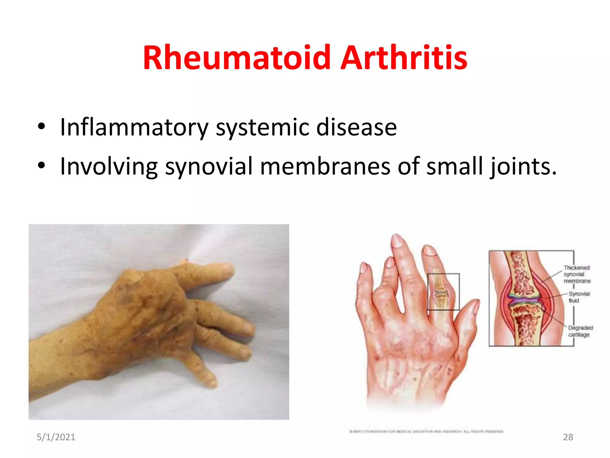
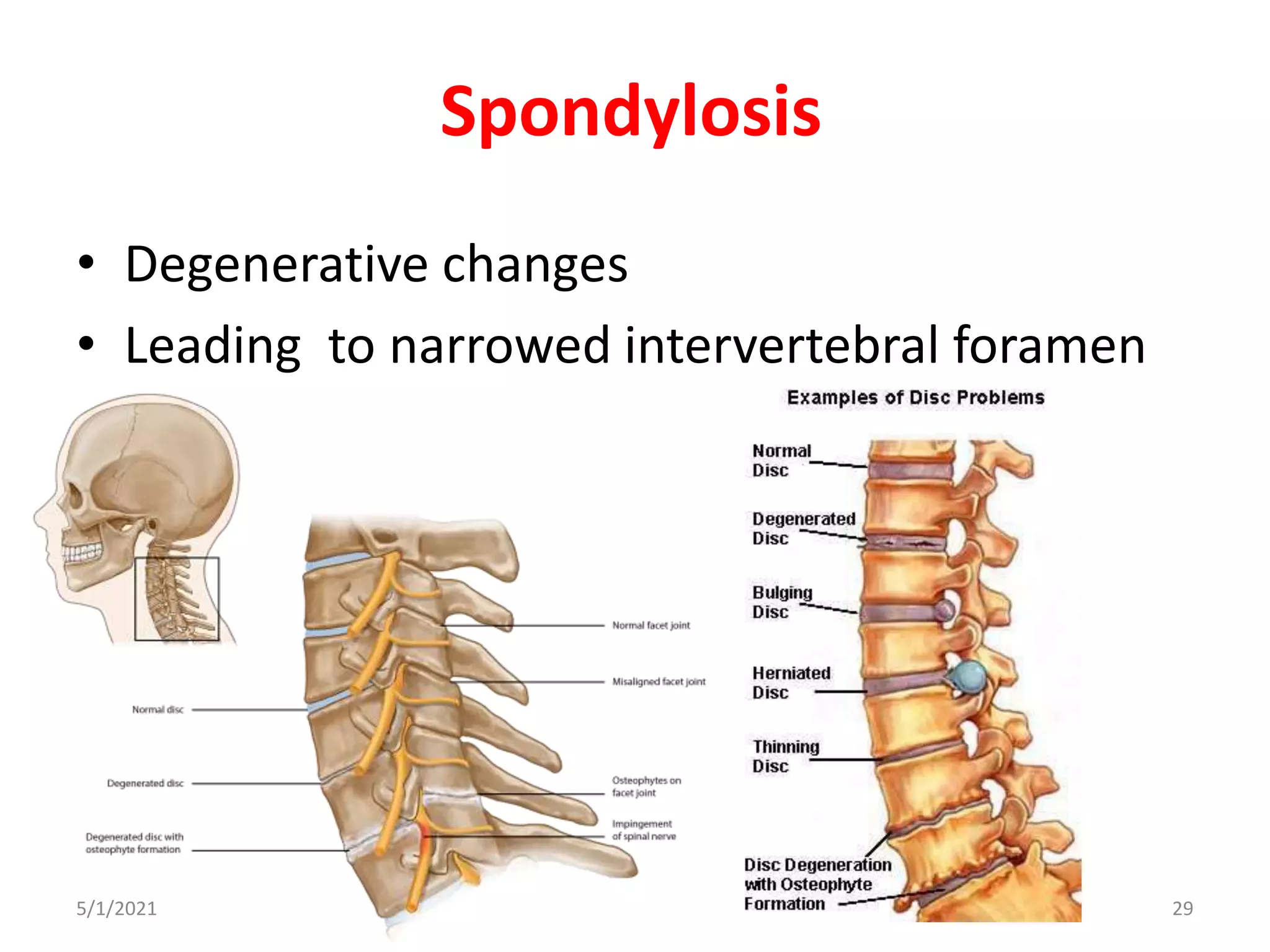
The document discusses the classification and types of joints in the body. It describes how joints can be classified structurally or functionally. Structural classification includes fibrous joints, cartilaginous joints, and synovial joints. Cartilaginous joints are further divided into primary and secondary joints. Synovial joints are classified based on their axis of movement into plane, uniaxial, biaxial, and multiaxial joints. Common examples of different joint types are provided. The presentation also mentions osteoarthritis, rheumatoid arthritis, and spondylosis as conditions affecting joints.
classification of joints. example of different types of joints. different types of joints on the basis of axis of movements. clinical aspects of joints. different between arthritis.
to download this presentation from this link.
https://mohmmed-ink.blogspot.com/2020/12/joints-of-upper-limb.html
anatomy of the upper limb joints. shoulder, elbow, wrist hand
Atlanto occipital and atlanto axial jointShubham Singh
Anatomy:
>Atlas is the topmost vertebra and chief peculiarity of atlas is that it has no body, it is ring like and consist of anterior and posterior arch and two lateral masses.
>Axis, the 2nd cervical vertebra has a concave under side and convex from side to side. The most distinctive characteristic of this bone is strong odontoid process, the dens.
TheJoint:
>Atlanto-occipital joint (articulation between the atlas and the occipital bone) consists of a pair of condyloid joints.
>The atlanto-occipital joints are synovial socket-type joints
Ligaments:
> Posterior atlanto-occipital membrane: extend from anterior arch of atlas to posterior margin of foramen magnum.
>Anterior atlanto-occipital membrane: extend from anterior arch of atlas to anterior margin of foramen magnum.
>The ligamentam flavam join laminae of adjacent vertebral arches.
>The interspinous ligaments expand to form the ligamentum nuchae which inserts along the posterior foramen magnum and external occipital condyle.
> The following four ligaments stabilize these joints:
1.Apical ligament: Connects the dens to the foramen magnum of the occipital bone.
2.Alar ligaments: Connect the dens to the lateral margins of the foramen magnum.
3.Cruciate ligament: Attaches the dens to the anterior arch of the atlas and the body of the axis to the foramen magnum of the occipital bone.
4.Tectorial membrane: Starts at the skull and becomes the posterior longitudinal ligament.
>Atlanto-axial articular capsules are thick and loose, and connect the margins of the lateral masses of the atlas with those of the posterior articular surfaces of the axis.
Muscles:
>Flexion is produced mainly by the action of longis capitis, rectus capitis anterior and sternocleidomastoid (anterior fibres)
>Extension by the rectus capitis posterior major and minor, the obliquus capitis superior, the semispinalis capitis, splenius capitis, longissimus capitis, sternocleidomastoid and upper fibres of the trapezius
>The recti lateralis are concerned in the lateral movement, assisted by the trapezius, splenius capitis, semispinalis capitis, and the sternocleidomastoid of the same side, all acting together.
Movements:
>Flexion and extension in the Sagittal axis, which give rise to the ordinary forward and backward nodding of the head.
>Lateral flexion to one or other side in the Frontal axis(titling of head
>Lateral AAJ Movement: It is a synovial joint which allows only gliding
>Medial AAJ Movement: This joint allows the rotation of the atlas the axis i.e round the dens.
Clinical anatomy:
> Headaches can arise from many different sources including dysfunctional muscles, tears in the ligaments, misalignment of the vertebral bodies, injury to cervical facets and degenerative discs.
>Excessive flexion could rupture the supraspinous ligament.
>Posterior atlanto-occipital membrane ossification cause migraine headaches due to compression of artery.
Branches/roots from L4-L5-S1 join and become superior gluteal nerve giving motor supply to abductor muscle of gluteus medius and gluteus minimus
Branches/roots from L5-S1-S2 join and form inferior gluteal nerve giving motor supply to gluteus maximus, this muscle has 2 function for extension and external rotation of the hip
Carpal Bone Anatomy Details PPT
Part-4 (UL Bone)
Carpal Bone names, attachments, clinical anatomy, General and specific points.
Carpal bones: 8
Like, share and comment.
Thank you
Ossification (Intracartilaginous and Intramembranous)Mohiuddin Masum
This presentation includes:
* Ossification definition
* Types of ossification
* Center of ossification
* Intramembranous ossification process
* Intracartilaginous ossification process
basic information about human body joints ( arthrology) .
topics cover under this ppt are- definition, classification and clinical anatomy of joints. this may be very helpful for the medical students for understand the basic concepts about joints.
A joint is an articulation between two bones in the body and are broadly classified by the tissue which connects the bones. The three main types of joints are: synovial, cartilaginous and fibrous.
JOINTS CLASSIFICATION AND DISORDERS.pptxMsSapnaSapna
A joint is the part of the body where two or more bones meet to allow movement. Generally speaking, the greater the range of movement, the higher the risk of injury because the strength of the joint is reduced. The six types of freely movable joint include ball and socket, saddle, hinge, condyloid, pivot and gliding.
Classification and Applied Aspects of JointsMathew Joseph
The structural classification divides joints into fibrous, cartilaginous, and synovial joints depending on the material composing the joint and the presence or absence of a cavity in the joint. The functional classification divides joints into three categories: synarthroses, amphiarthroses, and diarthroses
A joint is the site at which any two or more bones articulate or come together, meaning the ends or edges of the bones are held together by connective tissues.
It may allow flexibility and movement of the skeleton.
With the exception of the Hyoid bone, every bone in the body is connected to or form joints.
There are total 230 joints in the body.
Structural Classification of joints:
FIBROUS JOINT/ FIXED JOINT
CARTILAGENOUS JOINTS
SYNOVIAL JOINTS
The functional classification of joints is determined by the amount of mobility found between the adjacent bones.
Joints are thus functionally classified as a
Synarthrosis or immobile joint,
Amphiarthrosis or slightly moveable joint,
Diarthrosis, which is a freely moveable joint
The placenta is formed gradually during the first three months of pregnancy, while, after the fourth month, it grows parallel to the development of the uterus. Once completed, it resembles a spongy disc 20 cm in diameter and 3 cm thick.
The external human face develops between the 4th and 6th week of embryonic development. The development of the face is completed by the 6th week.
Between the 6th and 8th week, the palate begins to develop. Consequently, this causes a distinction between the nasal and oral cavities. This development is completed by the 12th week.
Development of Female Reproductive system.pptxRahul Sharma
the female reproductive system derives from four origins: mesoderm, primordial germ cells, coelomic epithelium, and mesenchyme. The uterus forms during Mullerian organogenesis accompanied by the development of the upper third of the vagina, the cervix, and both fallopian tubes.
Your cerebellum is part of your brain that helps coordinate and regulate a wide range of functions and processes in both your brain and body. While it's very small compared to your brain overall, it holds more than half of the neurons (cells that make up your nervous system) in your whole body.
An organelle is a subcellular structure that has one or more specific jobs to perform in the cell, much like an organ does in the body. Among the more important cell organelles are the nuclei, which store genetic information; mitochondria, which produce chemical energy; and ribosomes, which assemble proteins.
There are eight carpal bones in each wrist.
There are five metacarpal bones in each hand.
There are proximal, intermediate, and distal phalanges in each digit except for the thumb, which lacks an intermediate phalange.
What is the cardiovascular system? Your heart and many blood vessels in your body make up your cardiovascular system or circulatory system. Your heart uses the far-reaching, intricate network of blood vessels to deliver oxygen and other necessary things to your whole body.
The femoral triangle is a hollow region located in the supero-medial part of the anterior thigh. It appears most prominently with hip flexion, abduction and internal rotation. It is an easily accessible area through which multiple neurovascular structures pass through.
The brachial plexus is the network of nerves that sends signals from the spinal cord to the shoulder, arm and hand. A brachial plexus injury occurs when these nerves are stretched, compressed, or in the most serious cases, ripped apart or torn away from the spinal cord.
The four principal types of bones are long, short, flat and irregular. Bones that are longer than they are wide are called long bones. They consist of a long shaft with two bulky ends or extremities.
The ascending tracts carry sensory information from the body, like pain, for example, up the spinal cord to the brain. Descending tracts carry motor information, like instructions to move the arm, from the brain down the spinal cord to the body.
The anterior triangles refer to bilateral anatomic subdivisions of the neck comprising the anterior surface of the neck, deep to the superficial cervical fascia and platysma muscle. Laterally, the anterior triangle is bounded by the anterior border of the sternocleidomastoid muscle
The floor of the cranial cavity is divided into three distinct depressions. They are known as the anterior cranial fossa, middle cranial fossa and posterior cranial fossa. Each fossa accommodates a different part of the brain
Microtomy is a method for the preparation of thin sections for materials such as bones, minerals and teeth, and an alternative to electropolishing and ion milling. Microtome sections can be made thin enough to section a human hair across its breadth, with section thickness between 50 nm and 100 μm
What is Craniotomy?
What are the Indications for Craniotomy?
What are the Types of Craniotomy?
Equipment used in craniotomy?
What happen to the Bone flap?
What are the Tests Done Prior to Craniotomy?
What happens during surgery?
What are the risks?
References
Gross appearance of cerebellum
Structure of cerebellum
The functional division of the cerebellum
Afferent & efferent pathways
Clinical
MCQ’s
Clinical Vignettes
Seven cervical vertebrae
Identified by the presence of foramen in their transverse processes called foramen transversarium
3rd to 6th are typically have common features
1st, 2nd,and 7th are atypical
Ring-shaped and has no body and no spine
Consists of:
Right and left lateral masses
Short anterior arch and a long curved posterior arch
(c) Right and left transverse processes
2024.06.01 Introducing a competency framework for languag learning materials ...Sandy Millin
http://sandymillin.wordpress.com/iateflwebinar2024
Published classroom materials form the basis of syllabuses, drive teacher professional development, and have a potentially huge influence on learners, teachers and education systems. All teachers also create their own materials, whether a few sentences on a blackboard, a highly-structured fully-realised online course, or anything in between. Despite this, the knowledge and skills needed to create effective language learning materials are rarely part of teacher training, and are mostly learnt by trial and error.
Knowledge and skills frameworks, generally called competency frameworks, for ELT teachers, trainers and managers have existed for a few years now. However, until I created one for my MA dissertation, there wasn’t one drawing together what we need to know and do to be able to effectively produce language learning materials.
This webinar will introduce you to my framework, highlighting the key competencies I identified from my research. It will also show how anybody involved in language teaching (any language, not just English!), teacher training, managing schools or developing language learning materials can benefit from using the framework.
Biological screening of herbal drugs: Introduction and Need for
Phyto-Pharmacological Screening, New Strategies for evaluating
Natural Products, In vitro evaluation techniques for Antioxidants, Antimicrobial and Anticancer drugs. In vivo evaluation techniques
for Anti-inflammatory, Antiulcer, Anticancer, Wound healing, Antidiabetic, Hepatoprotective, Cardio protective, Diuretics and
Antifertility, Toxicity studies as per OECD guidelines
Francesca Gottschalk - How can education support child empowerment.pptxEduSkills OECD
Francesca Gottschalk from the OECD’s Centre for Educational Research and Innovation presents at the Ask an Expert Webinar: How can education support child empowerment?
Instructions for Submissions thorugh G- Classroom.pptxJheel Barad
This presentation provides a briefing on how to upload submissions and documents in Google Classroom. It was prepared as part of an orientation for new Sainik School in-service teacher trainees. As a training officer, my goal is to ensure that you are comfortable and proficient with this essential tool for managing assignments and fostering student engagement.
Unit 8 - Information and Communication Technology (Paper I).pdfThiyagu K
This slides describes the basic concepts of ICT, basics of Email, Emerging Technology and Digital Initiatives in Education. This presentations aligns with the UGC Paper I syllabus.
How to Make a Field invisible in Odoo 17Celine George
It is possible to hide or invisible some fields in odoo. Commonly using “invisible” attribute in the field definition to invisible the fields. This slide will show how to make a field invisible in odoo 17.
Honest Reviews of Tim Han LMA Course Program.pptxtimhan337
Personal development courses are widely available today, with each one promising life-changing outcomes. Tim Han’s Life Mastery Achievers (LMA) Course has drawn a lot of interest. In addition to offering my frank assessment of Success Insider’s LMA Course, this piece examines the course’s effects via a variety of Tim Han LMA course reviews and Success Insider comments.
9. Cartilaginous Joint
• Cartilaginous joint , where joint forming articular
surface are united by means of either hyaline
cartilage or fibrocartilage.
Two type,
1. Primary cartilaginous joint(Synchondroses )
2. Secondary cartilaginous joint(Symphyses)
5/1/2021 9
10. • Joints are united by means of –Hyaline
cartilage ,Fibro cartilage
5/1/2021 10
19. • Plane joint articular surfaces are more or less flat
(plane).
e.g. Acromio-clavicular joint, zygopophyseal joint, inter-
tarsal joint, Inter-carpal joint
5/1/2021 19
20. • Pivot joint Articular surfaces comprise a central bony
pivot (peg) surrounded by an osteoligamentous ring
e.g. proximal radio-ulnar joint, atlanto-axial joint
5/1/2021 20
21. • Hinge joint- articular surfaces are pulley-shaped
e.g. elbow joint, knee joint, inter-phalangeal joint, ankle
joint
5/1/2021 21
22. • Condyloid joint round oval surface fitting into an
socket type articular surface
e.g Tempro-mandibular joint, knee joint
5/1/2021 22
23. • Ellipsoid joint articular surfaces include an elliptical convex
surface fitting into an elliptical concave surface
e.g. Wrist joint, metacarpo-phalangeal joint, atlanto-occipital
joint
5/1/2021 23
24. • Saddle joint articular surfaces are reciprocally concavoconvex.
e.g. 1st metacarpo-carpal joint, calcaneo-cuboidal joint, sterno-
clavicular joint.
5/1/2021 24
25. • Ball and Socket joint articular surfaces include a
globular head fitting into a cup-shaped socket
e.g. shoulder joint, hip joint
5/1/2021 25
26. Classification
• Acc to number of articulating bone
1. Simple joint
2. Compound joint
3. Complex joint
5/1/2021 26
31. 1. A pt come with an X-ray of elbow , destroyed
epiphyseal plate can be seen ,what its lead to?
a) Abnormal growth
b) Shortening of limb
c) No Healing
d) A and b
A and b
32. 2. All of the following secondary cartilaginous
joints belongs to axial skeleton except?
a) Manubriosternal joint
b) Intervertebral discs
c) Symphysis pubis
d) Symphysis menti
a) Symphysis pubis
33. 3. Manubriosternal joint is an example of ?
a) Symphyses
b) Synchondrosis
c) Synovial
d) syndesmosis
Symphyses