Downloaded 360 times
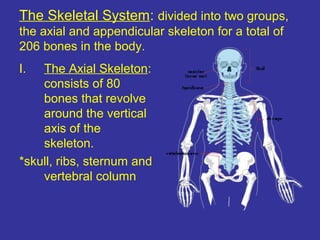
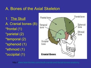
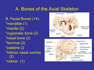
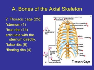
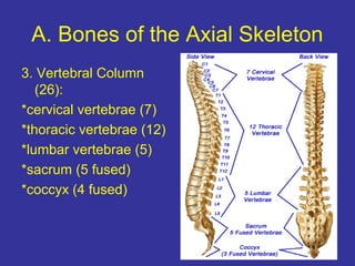
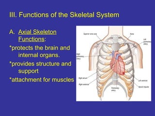
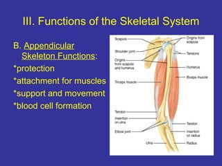
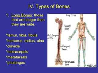
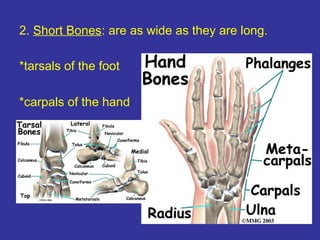


This document provides an overview of the skeletal and muscular systems. It begins by labeling the parts of a long bone, then describes the two groups that make up the skeletal system - the axial skeleton and appendicular skeleton. The axial skeleton includes the skull, ribs, sternum, and vertebral column. It then details the specific bones that make up these areas. The appendicular skeleton attaches to the axial skeleton and includes the pectoral girdle, upper limbs, pelvic girdle, and lower limbs. The document also outlines the functions of the skeletal system and describes the four types of bones. It defines anatomical terminology and discusses the roles of connective tissues like cartilage, ligaments, and tendons. Finally


