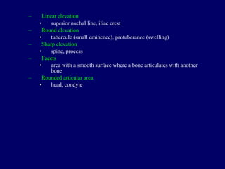
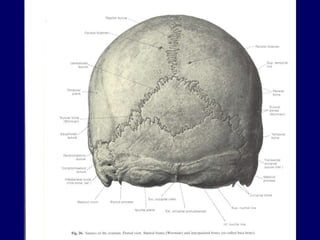
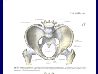
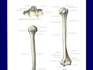
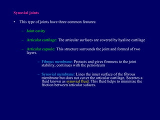
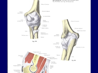
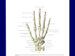
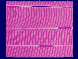
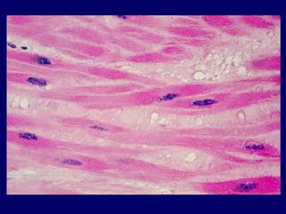
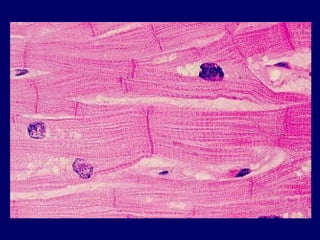
BONES O sseous tissue Skeletal System
The  study of bones is called osteology Rigid form of connective tissue forming the skeleton. Supporting framework of the body consists of over 200 bones. In average adult human skeleton has around 206 bones It comprises  around 14% of the total body weight
How many bones? A newborn baby has approximately  300  bones  An average an adult human has  206 bones  (these numbers can vary slightly).
Bones are living structures having a blood and nerve supply Periosteum (outer lining) Living bones have some  elasticity  (results from the organic matter) and  great rigidity  (results from microscopic tubes of inorganic calcium phosphate)
Function s  of the bones Protection for vital structures Support ;  forms a rigid  framework for the body Forms the mechanical basis  for movement Formation of blood cells (bone marrow) Storage of salts ;  calcium, phosphorus, iron, magnesium-thus  provide a mineral reservoir Bones are not entirely made of calcium, but a  mixture of chondroitin sulfate and hydroxyapatite  (70% of a bone) NEW:  Endocrine regulation by hormone Osteocalcin , (regulation of blood sugar and   fat deposition)
Types of Bones T here are t wo main types spongy  (cancellous)  and   compact bone
 
Bones forming the skeleton is classified as: Axial skeleton skull, vertebrae, ribs, and sternum Appendicular skeleton bones of the upper and lower limbs and the associated bones
Bones are also classified according to their shape Long bones  (humerus, bones of the fingers) have a shaft and two extremities Short bones  (foot and wrist bones) more or less cuboidal in shape Flat bones  (bones forming the calvaria, sternum, scapula) Irregular bones  (facial bones and the vertebrae) Pneumatic bones  (maxilla, frontal bone) contains air cells or sinuses Sesamoid bones round or oval nodules of bones that develop in certain tendons Accessory bones develops as a result of additional ossification center or lack of   fusion (mainly in foot) Heterotopic  bones develops as a result of trauma or constant strain (thigh muscles of horseback rider)
BONE MARKINGS Surfaces of the bones are not smooth. Bones display elevations,   depressions and holes. The surface features on the bones are given names to distinguish and define them.
Linear elevation superior nuchal line, iliac crest Round elevation tubercule (small eminence), protuberance (swelling) Sharp elevation spine, process Facets area with a smooth surface where a bone articulates with another bone Rounded articular area head, condyle


Depressions fossae (small depression), groove (sulcus, long narrow depressions) Foramen hole Canal a foramen having length Orifice opening Meatus a canal entering a structure
 
 
Anatomy of a Long Bone Diaphysis Shaft Composed of compact bone Epiphysis  Ends of the bone Composed mostly of spongy bone Figure 5.2a
Gross Anatomy of a Long Bone Epiphyseal line Thin line of bony tissue spanning the epiphysis that looks different from the rest.  Is the remnant of the epiphyseal plate. (growth) The  epiphyseal plate  (hyaline cartilage) is seen in young, growing bone. Figure 5.2a
Bone Growth Epiphyseal plates allow for growth of long bone during childhood New cartilage is continuously formed Older cartilage becomes ossified Cartilage is broken down Bone replaces cartilage
Clinical Note Avascular necrosis Bones are supplied by 1-3 nutrient arteries, if these rupture, or in some types of fracture there may be necrosis (death) of bone.
JOINTS
Arthrology is the study of joints . Joints are formed by the articulation between the articular surfaces of two or more bones. Articular system consists of joints and their associated bones and ligaments.
 
Classification of joints Structural classification Fibrous joints:  Joined by fibrous connective tissue Cartilaginous joints:   Joined by cartilage Synovial joints:  Joined by a capsule
Classification of joints Functional classification Synarthrosis  - permits little or no mobility. Most synarthrosis joints are fibrous joints (e.g. The Skull). Amphiarthrosis  - permits slight mobility. Most amphiarthrosis joints are cartilaginous joints (e.g. Vertebrae). Diarthrosis  - permits a variety of movements. All diarthrosis joints are synovial joints (e.g. Shoulder, Hip, Elbow, Knee etc)
Fibrous joints There is a fibrous connective tissue b et w een  the articular surfaces. Syndesmosis:  Articulating surfaces are connected firmly by a connective tissue e.g. distal tibio-fibular joint Suture:  Found between the skull bones Gomphosis:  Found between the dentures and the alveoles of the upper and lower jaw


Cartilaginous joints Cartilaginous hyaline tissue lies between the articular surfaces. These joints can make very limited amount of movement. Synchodros i s:  The cartilaginous tissue between the articular surfaces are lost within time and the joint loose the ability of movement e.g. sphenoid-occipital Symphys is :  There is a di s c made of fibrocartilaginous tissue between the articular surfaces. e.g. joints between the vertebrae (vertebral discs), symphysis of the pubis (pelvis). All symphysis are found in the midline.


Synovial joints This type of joints have three common features: Joint cavity Articular cartilage:  The articular surfaces are covered by hyaline cartilage Articular capsule:  This structure surrounds the joint and formed of two layers. Fibrous membrane:  Protects and gives firmness to the joint stability, continues with the periosteum Synovial membrane:  Lines the inner surface of the fibrous membrane but does not cover the articular cartilage. Secretes a fluid known as  synovial fluid . This fluid helps to minimize the friction between articular sufaces.
 
 
Synovial joints  (continued) Articular capsules  are usually strengthened by  articular ligaments These are from dense connective tissue and they connect the articulating bones to each other. Articular ligaments limit the undesired and/or excessive movements of the joints. Articular ligaments are classified as intrinsic and extrinsic ligaments. Intrinsic ligaments are the thickening of a portion of the articular capsule.
Synovial joints  (continued) In addition to the main features of the synovial joints there are some additional features that are common ly seen : Articular disc:  Help to hold the bones together. In some joints it is attached to only one of the bones. Labrum:  Some synovial joints have a fibrocartilaginous ring called labrum which deepens the articular surface for one of the bones. e.g. glenoid labrum
 
Synovial joints  (continued) Classification of the synovial joints Synovial joints are classified according to the shape of articulating surfaces and/or the type of movement they can make.
Synovial joints 1- Ball and socket joint 2- Condyloid joint  3- Saddle joint 4- Hinge joint 5- Pivot joint 6-  Plane
Synovial joints  (continued) Plane joints They permit gliding or sliding movements. Articular surfaces are almost flat. e.g. acromioclavicular joint, carpometacarpal joint
 
Synovial joints  (continued) Hinge joints (ginglymus, trochlear) This type of joints are also  uniaxial  and permits flex. and ext. around the transverse axis. Bones are joined with strong collateral ligaments. e.g. elbow and knee joints
 
Synovial joints  (continued) Condyloid joints These are  biaxial joints  that permit movement in in two axes (trans.-sagit.) e.g. radiocarpal joint, metacrpophalyngeal joints
 
Synovial joints  (continued) Saddle joints T he articular surfaces   resembl e  a saddle shape  and  are concave and convex  respectively . e.g. carpometacarpal joint of the thumb
 
Synovial joints  (continued) Ball and socket joints Multiaxial  (movable around all three axes) The spheroidal surface of a bone articulates with the socket shaped articular surface of another bone. e.g. shoulder joint
 
Synovial joints  (continued) Pivot joints Uniaxial joints that allow rotation. Rounded part of a bone rotates in a sleeve or ring like osteofibrous structure e.g.  prox-dist radioulnar joints
 
Nerve supply  of the joints According to the Hilton’s law the nerves innervating a joint are the branches of the nerves that innervate the muscles acting on that joint and the nerves innervating the sensory areas around that region. The nerves convey impulses regarding conscious proprioception (from the joint capsule) and pain (from the fibrous membrane).
Vessel  supply  of the joints There are a number of vessels that supply the blood circulation in a joint. These are the branches of surrounding vessels.
Features supplying the joint stability These features prevent the dislocation of the joint. The negative pressure within the joint cavity Joint capsule and the ligaments Muscles and their tendons around the joint Shapes of the articular surfaces
Clinical Note Degenerative Joint disease/ Osteoarthitis Over use of a joint wears the cartilage causing degenerative changes, common in hip, knee, vertebral column & hands. Increased friction causes pain. May require pain management, change in lifestyle, surgery or joint replacement.
MUSCLES
Myology, musculus (mus-mouse) Muscles move the skeleton, face and organs.
There are three types of muscles Striated muscle (skeletal muscles)- voluntarily controlled, though exceptions exist Non-striated muscle  (smooth muscle) -  involuntary Cardiac muscle
Striated muscle s are innervated by the  somatic nervous system Non-striated  and c ardiac muscle  are innervated by the  autonomic nervous system


Muscles are formed of fusiform type cells. Muscle cells are also referred as  muscle fibers .
Superficial fascia   and   deep fascia


Parts of a muscle Belly  (fleshy part) T endon   (in certain muscles termed as  aponeurosis )
Tendons attach to the bones and cartilage, skin or to the superficial fascia. Origin  and  insertion  (prox.-dist, fixed-moving)
Bursae Tendinous sheats Both are  soft tissue pads containing synovial fluid that prevents the friction of tendons from the surrounding tissue.
 
 
Muscle terminology Muscles are generally given names according to their shapes, location, number of their bellies, function, and size rhomboideus major palmaris longus biceps brachii tibialis posterior flexor carpi ulnaris teres major-teres minor
Contraction When a muscle contracts its length decreases by 1/3 or ½ Isotonic contraction   -   the length decreases Isometric contraction   -   the length does not decrease
Motor unit A motor neuron and  all  the muscle cells (fibers) inervated by this neuron
 
A single motor neuron may innervate thousands of muscle fibers but in certain muscles a motor neuron may innervate less number of muscles (e.g. eye mm. 3-4 fibers are innervated by a motor neuron). There are many motor units in a  single  muscle. Whole of the motor units does not necessarily contract during a muscle contraction.  Number of contracting units determines the force of contraction.
During a single joint movement a number of muscles contract. Prime mover Synergist muscles (support the movement) Antagonist muscles (oppose the movement) Fixator muscle (the muscle that prevents the undesired movements)
Innervation of the muscles Motor nerves  (somatic or autonomic) S ensory nerves ( for striated muscles;  muscle spindle-golgi tendon organs)

02 Bones, Joints, Muscles General Considerations

  • 1.
    BONES O sseoustissue Skeletal System
  • 2.
    The studyof bones is called osteology Rigid form of connective tissue forming the skeleton. Supporting framework of the body consists of over 200 bones. In average adult human skeleton has around 206 bones It comprises around 14% of the total body weight
  • 3.
    How many bones?A newborn baby has approximately 300 bones An average an adult human has 206 bones (these numbers can vary slightly).
  • 4.
    Bones are livingstructures having a blood and nerve supply Periosteum (outer lining) Living bones have some elasticity (results from the organic matter) and great rigidity (results from microscopic tubes of inorganic calcium phosphate)
  • 5.
    Function s of the bones Protection for vital structures Support ; forms a rigid framework for the body Forms the mechanical basis for movement Formation of blood cells (bone marrow) Storage of salts ; calcium, phosphorus, iron, magnesium-thus provide a mineral reservoir Bones are not entirely made of calcium, but a mixture of chondroitin sulfate and hydroxyapatite (70% of a bone) NEW: Endocrine regulation by hormone Osteocalcin , (regulation of blood sugar and fat deposition)
  • 6.
    Types of BonesT here are t wo main types spongy (cancellous) and compact bone
  • 7.
  • 8.
    Bones forming theskeleton is classified as: Axial skeleton skull, vertebrae, ribs, and sternum Appendicular skeleton bones of the upper and lower limbs and the associated bones
  • 9.
    Bones are alsoclassified according to their shape Long bones (humerus, bones of the fingers) have a shaft and two extremities Short bones (foot and wrist bones) more or less cuboidal in shape Flat bones (bones forming the calvaria, sternum, scapula) Irregular bones (facial bones and the vertebrae) Pneumatic bones (maxilla, frontal bone) contains air cells or sinuses Sesamoid bones round or oval nodules of bones that develop in certain tendons Accessory bones develops as a result of additional ossification center or lack of fusion (mainly in foot) Heterotopic bones develops as a result of trauma or constant strain (thigh muscles of horseback rider)
  • 10.
    BONE MARKINGS Surfacesof the bones are not smooth. Bones display elevations, depressions and holes. The surface features on the bones are given names to distinguish and define them.
  • 11.
    Linear elevation superiornuchal line, iliac crest Round elevation tubercule (small eminence), protuberance (swelling) Sharp elevation spine, process Facets area with a smooth surface where a bone articulates with another bone Rounded articular area head, condyle
  • 12.
  • 13.
  • 14.
  • 15.
    Depressions fossae (smalldepression), groove (sulcus, long narrow depressions) Foramen hole Canal a foramen having length Orifice opening Meatus a canal entering a structure
  • 16.
  • 17.
  • 18.
    Anatomy of aLong Bone Diaphysis Shaft Composed of compact bone Epiphysis Ends of the bone Composed mostly of spongy bone Figure 5.2a
  • 19.
    Gross Anatomy ofa Long Bone Epiphyseal line Thin line of bony tissue spanning the epiphysis that looks different from the rest. Is the remnant of the epiphyseal plate. (growth) The epiphyseal plate (hyaline cartilage) is seen in young, growing bone. Figure 5.2a
  • 20.
    Bone Growth Epiphysealplates allow for growth of long bone during childhood New cartilage is continuously formed Older cartilage becomes ossified Cartilage is broken down Bone replaces cartilage
  • 21.
    Clinical Note Avascularnecrosis Bones are supplied by 1-3 nutrient arteries, if these rupture, or in some types of fracture there may be necrosis (death) of bone.
  • 22.
  • 23.
    Arthrology is thestudy of joints . Joints are formed by the articulation between the articular surfaces of two or more bones. Articular system consists of joints and their associated bones and ligaments.
  • 24.
  • 25.
    Classification of jointsStructural classification Fibrous joints: Joined by fibrous connective tissue Cartilaginous joints: Joined by cartilage Synovial joints: Joined by a capsule
  • 26.
    Classification of jointsFunctional classification Synarthrosis - permits little or no mobility. Most synarthrosis joints are fibrous joints (e.g. The Skull). Amphiarthrosis - permits slight mobility. Most amphiarthrosis joints are cartilaginous joints (e.g. Vertebrae). Diarthrosis - permits a variety of movements. All diarthrosis joints are synovial joints (e.g. Shoulder, Hip, Elbow, Knee etc)
  • 27.
    Fibrous joints Thereis a fibrous connective tissue b et w een the articular surfaces. Syndesmosis: Articulating surfaces are connected firmly by a connective tissue e.g. distal tibio-fibular joint Suture: Found between the skull bones Gomphosis: Found between the dentures and the alveoles of the upper and lower jaw
  • 28.
  • 29.
  • 30.
  • 31.
    Cartilaginous joints Cartilaginoushyaline tissue lies between the articular surfaces. These joints can make very limited amount of movement. Synchodros i s: The cartilaginous tissue between the articular surfaces are lost within time and the joint loose the ability of movement e.g. sphenoid-occipital Symphys is : There is a di s c made of fibrocartilaginous tissue between the articular surfaces. e.g. joints between the vertebrae (vertebral discs), symphysis of the pubis (pelvis). All symphysis are found in the midline.
  • 32.
  • 33.
  • 34.
  • 35.
  • 36.
    Synovial joints Thistype of joints have three common features: Joint cavity Articular cartilage: The articular surfaces are covered by hyaline cartilage Articular capsule: This structure surrounds the joint and formed of two layers. Fibrous membrane: Protects and gives firmness to the joint stability, continues with the periosteum Synovial membrane: Lines the inner surface of the fibrous membrane but does not cover the articular cartilage. Secretes a fluid known as synovial fluid . This fluid helps to minimize the friction between articular sufaces.
  • 37.
  • 38.
  • 39.
    Synovial joints (continued) Articular capsules are usually strengthened by articular ligaments These are from dense connective tissue and they connect the articulating bones to each other. Articular ligaments limit the undesired and/or excessive movements of the joints. Articular ligaments are classified as intrinsic and extrinsic ligaments. Intrinsic ligaments are the thickening of a portion of the articular capsule.
  • 40.
    Synovial joints (continued) In addition to the main features of the synovial joints there are some additional features that are common ly seen : Articular disc: Help to hold the bones together. In some joints it is attached to only one of the bones. Labrum: Some synovial joints have a fibrocartilaginous ring called labrum which deepens the articular surface for one of the bones. e.g. glenoid labrum
  • 41.
  • 42.
    Synovial joints (continued) Classification of the synovial joints Synovial joints are classified according to the shape of articulating surfaces and/or the type of movement they can make.
  • 43.
    Synovial joints 1-Ball and socket joint 2- Condyloid joint 3- Saddle joint 4- Hinge joint 5- Pivot joint 6- Plane
  • 44.
    Synovial joints (continued) Plane joints They permit gliding or sliding movements. Articular surfaces are almost flat. e.g. acromioclavicular joint, carpometacarpal joint
  • 45.
  • 46.
    Synovial joints (continued) Hinge joints (ginglymus, trochlear) This type of joints are also uniaxial and permits flex. and ext. around the transverse axis. Bones are joined with strong collateral ligaments. e.g. elbow and knee joints
  • 47.
  • 48.
    Synovial joints (continued) Condyloid joints These are biaxial joints that permit movement in in two axes (trans.-sagit.) e.g. radiocarpal joint, metacrpophalyngeal joints
  • 49.
  • 50.
    Synovial joints (continued) Saddle joints T he articular surfaces resembl e a saddle shape and are concave and convex respectively . e.g. carpometacarpal joint of the thumb
  • 51.
  • 52.
    Synovial joints (continued) Ball and socket joints Multiaxial (movable around all three axes) The spheroidal surface of a bone articulates with the socket shaped articular surface of another bone. e.g. shoulder joint
  • 53.
  • 54.
    Synovial joints (continued) Pivot joints Uniaxial joints that allow rotation. Rounded part of a bone rotates in a sleeve or ring like osteofibrous structure e.g. prox-dist radioulnar joints
  • 55.
  • 56.
    Nerve supply of the joints According to the Hilton’s law the nerves innervating a joint are the branches of the nerves that innervate the muscles acting on that joint and the nerves innervating the sensory areas around that region. The nerves convey impulses regarding conscious proprioception (from the joint capsule) and pain (from the fibrous membrane).
  • 57.
    Vessel supply of the joints There are a number of vessels that supply the blood circulation in a joint. These are the branches of surrounding vessels.
  • 58.
    Features supplying thejoint stability These features prevent the dislocation of the joint. The negative pressure within the joint cavity Joint capsule and the ligaments Muscles and their tendons around the joint Shapes of the articular surfaces
  • 59.
    Clinical Note DegenerativeJoint disease/ Osteoarthitis Over use of a joint wears the cartilage causing degenerative changes, common in hip, knee, vertebral column & hands. Increased friction causes pain. May require pain management, change in lifestyle, surgery or joint replacement.
  • 60.
  • 61.
    Myology, musculus (mus-mouse)Muscles move the skeleton, face and organs.
  • 62.
    There are threetypes of muscles Striated muscle (skeletal muscles)- voluntarily controlled, though exceptions exist Non-striated muscle (smooth muscle) - involuntary Cardiac muscle
  • 63.
    Striated muscle sare innervated by the somatic nervous system Non-striated and c ardiac muscle are innervated by the autonomic nervous system
  • 64.
  • 65.
  • 66.
  • 67.
    Muscles are formedof fusiform type cells. Muscle cells are also referred as muscle fibers .
  • 68.
    Superficial fascia and deep fascia
  • 69.
  • 70.
  • 71.
  • 72.
    Parts of amuscle Belly (fleshy part) T endon (in certain muscles termed as aponeurosis )
  • 73.
    Tendons attach tothe bones and cartilage, skin or to the superficial fascia. Origin and insertion (prox.-dist, fixed-moving)
  • 74.
    Bursae Tendinous sheatsBoth are soft tissue pads containing synovial fluid that prevents the friction of tendons from the surrounding tissue.
  • 75.
  • 76.
  • 77.
    Muscle terminology Musclesare generally given names according to their shapes, location, number of their bellies, function, and size rhomboideus major palmaris longus biceps brachii tibialis posterior flexor carpi ulnaris teres major-teres minor
  • 78.
    Contraction When amuscle contracts its length decreases by 1/3 or ½ Isotonic contraction - the length decreases Isometric contraction - the length does not decrease
  • 79.
    Motor unit Amotor neuron and all the muscle cells (fibers) inervated by this neuron
  • 80.
  • 81.
    A single motorneuron may innervate thousands of muscle fibers but in certain muscles a motor neuron may innervate less number of muscles (e.g. eye mm. 3-4 fibers are innervated by a motor neuron). There are many motor units in a single muscle. Whole of the motor units does not necessarily contract during a muscle contraction. Number of contracting units determines the force of contraction.
  • 82.
    During a singlejoint movement a number of muscles contract. Prime mover Synergist muscles (support the movement) Antagonist muscles (oppose the movement) Fixator muscle (the muscle that prevents the undesired movements)
  • 83.
    Innervation of themuscles Motor nerves (somatic or autonomic) S ensory nerves ( for striated muscles; muscle spindle-golgi tendon organs)