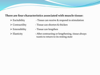
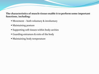
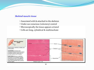
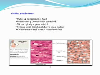
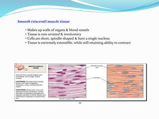
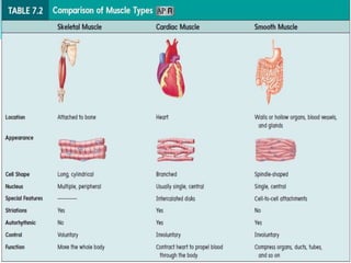
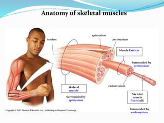
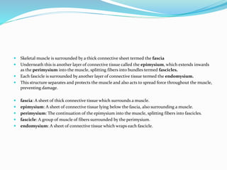
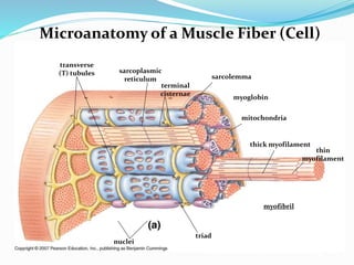
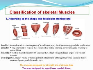
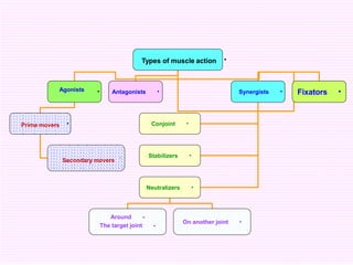
Dr Asma Lashari
University of Health Sciences
Lahore, Pakistan
There are four characteristics associated with muscle tissue:
 Excitability - Tissue can receive & respond to stimulation
 Contractility - Tissue can shorten & thicken
 Extensibility - Tissue can lengthen
 Elasticity - After contracting or lengthening, tissue always
wants to return to its resting state
The characteristics of muscle tissue enable it to perform some important
functions, including:
 Movement – both voluntary & involuntary
 Maintaining posture
 Supporting soft tissues within body cavities
 Guarding entrances & exits of the body
 Maintaining body temperature
Types of muscle tissue:
• Skeletal
• Cardiac
• Smooth (Visceral)
Skeletal muscle tissue
• Associated with & attached to the skeleton
• Under our conscious (voluntary) control
• Microscopically the tissue appears striated
• Cells are long, cylindrical & multinucleate
Cardiac muscle tissue
• Makes up myocardium of heart
• Unconsciously (involuntarily) controlled
• Microscopically appears striated
• Cells are short, branching & have a single nucleus
• Cells connect to each other at intercalated discs
Smooth (visceral) muscle tissue
• Makes up walls of organs & blood vessels
• Tissue is non-striated & involuntary
• Cells are short, spindle-shaped & have a single nucleus
• Tissue is extremely extensible, while still retaining ability to contract
Anatomy of skeletal muscles
Skeletal
muscle
fiber (cell)
Muscle Fascicle
Surrounded by
perimysium
Surrounded by
endomysium
endomysium
perimysium
Skeletal
muscle
Surrounded by
epimysium
epimysium
tendon
 Skeletal muscle is surrounded by a thick connective sheet termed the fascia
 Underneath this is another layer of connective tissue called the epimysium, which extends inwards
as the perimysium into the muscle, splitting fibers into bundles termed fascicles.
 Each fascicle is surrounded by another layer of connective tissue termed the endomysium.
 This structure separates and protects the muscle and also acts to spread force throughout the muscle,
preventing damage.
 fascia: A sheet of thick connective tissue which surrounds a muscle.
 epimysium: A sheet of connective tissue lying below the fascia, also surrounding a muscle.
 perimysium: The continuation of the epimysium into the muscle, splitting fibers into fascicles.
 fascicle: A group of muscle of fibers surrounded by the perimysium.
 endomysium: A sheet of connective tissue which wraps each fascicle.
Microanatomy of a Muscle Fiber (Cell)
sarcolemma
transverse
(T) tubules sarcoplasmic
reticulum
terminal
cisternae
myofibril
thin
myofilament
thick myofilament
triad
mitochondria
nuclei
myoglobin
Skeletal muscle movements at joints
Flexion/extension
Abduction/adduction
Rotation – left/right; internal(medial)/external(lateral)
pronation/supination
Elevation/depression
Protraction/retraction
Dorsiflexion/plantarflexion
Inversion/eversion
Parts of a skeletal muscle
Origin: it Is the proximal attachment which usually remains fixed during contraction
of muscles.
Fleshy part: a muscle may be fleshy partly or completely throughout its length.
Tendon: A tough band of fibrous tissue that usually connects a muscle with a bone.
Apponeurosis: A tough flat sheet of fibrous tissue that connects muscle with bones or
with the fascia of other muscles.
Insertion: it is the distal moveable end of skeletal muscle which moves during
contraction.
Naming of
skeletal
muscles
An Overview of the
Major Skeletal Muscles
Figure 7-11(a)
An Overview of the
Major Skeletal Muscles
Figure 7-11(b)
Classification of skeletal Muscles
1. According to the shape and fascicular architecture:
The muscles designed for strength are of pinnate type
The ones designed for speed have parallel fibers.
Parallel: A muscle with a common point of attachment, with fascicles running parallel to each other.
Circular: A ring like band of muscle that surrounds a bodily opening, constricting and relaxing to
control flow.
Pennate: A feather shaped muscle with fascicles that attach obliquely (at an angle) to a central
tendon.
Convergent: A muscle with a common point of attachment, although individual fascicles do not
necessarily run parallel to each other.
2) According to the myoglobin content:
• Red: contain more red fibers and they are responsible for movement,
which require slow action for a long time e.g antigravity muscles.
• White: contain more white fibers and they are responsible for movement,
which require rapid action for a short time.
3) According to the type of contractile activity:
a. Tonic muscles (stabilizers): it demonstrates continuous low level of
contractile activity which is required to maintain a given posture.
b. Phasic muscle (mobilizers): it demonstrates rapid (fast twitch) activity
which is required when changing from one position to another.
4) According to general limb appearance:
Contractors: those muscles pull the body into approximation of the fetal position
e.g. flexor adductors and medial rotators.
Expanders: those muscles which expand or open up the body
e.g. extensors abductors and lateral rotators.
5) According to the relative magnitude of their stabilizing and
rotatory components (muscle attachments):
Spurt: mainly rotator muscles which have their origin away from the joint and their
insertion near to the joint e.g. biceps muscle.
Shunt: mainly stabilizer muscles which have origin near the joint and their insertion
away from the joint e.g. brachio-radialis.
6) According to the orientation of the line of pull to the joint structure:
( e.g. flexors, extensors, abductors and adductors)
• The muscle located anterior to a joint may be extensor as in the case of the knee joint
or may be flexor as in the case of the elbow joint.
• The possible axes of motion are determined by the structure of the joint itself.
7) According to the number of joints over which the muscle
crosses:
• One joint muscle ( e.g. vastus mediales).
• Two joint muscle( e.g. rectus femoris).
• Multi-joint muscle ( e.g. finger flexors).
8) According to the type of muscle action or function (their interaction
in joint movement):
•
•
One action of the joint is not only the responsibility of one muscle but
it is the responsibility of different groups of muscles, which can be
classified as follows:
Agonists, antagonists, synergists, fixators.
antagonist: This type of muscle acts as opposing muscle to agonists, usually contracting as a means
of returning the limb to its original resting position.
agonist: These muscles are typically associates with the movement itself, and are sometimes
referred to as prime movers. They contract while another muscle relaxes.
synergist: This type of muscle acts around a movable joint to produce motion similar to or in
concert with agonist muscles.
Muscles classification according to development
 Mesodermal : All skeletal, smooth and cardiac muscles
 Ectodermal : Smooth muscle of iris, arrector pili muscle of skin
•Types of muscle action
•Agonists •Antagonists •Synergists •Fixators
•Prime movers
•
Secondary movers
•Conjoint
•Stabilizers
•Neutralizers
•
•
Around
The target joint
On another joint •
THANK YOU

The muscular system

  • 1.
    Dr Asma Lashari Universityof Health Sciences Lahore, Pakistan
  • 2.
    There are fourcharacteristics associated with muscle tissue:  Excitability - Tissue can receive & respond to stimulation  Contractility - Tissue can shorten & thicken  Extensibility - Tissue can lengthen  Elasticity - After contracting or lengthening, tissue always wants to return to its resting state
  • 3.
    The characteristics ofmuscle tissue enable it to perform some important functions, including:  Movement – both voluntary & involuntary  Maintaining posture  Supporting soft tissues within body cavities  Guarding entrances & exits of the body  Maintaining body temperature
  • 4.
    Types of muscletissue: • Skeletal • Cardiac • Smooth (Visceral)
  • 5.
    Skeletal muscle tissue •Associated with & attached to the skeleton • Under our conscious (voluntary) control • Microscopically the tissue appears striated • Cells are long, cylindrical & multinucleate
  • 6.
    Cardiac muscle tissue •Makes up myocardium of heart • Unconsciously (involuntarily) controlled • Microscopically appears striated • Cells are short, branching & have a single nucleus • Cells connect to each other at intercalated discs
  • 7.
    Smooth (visceral) muscletissue • Makes up walls of organs & blood vessels • Tissue is non-striated & involuntary • Cells are short, spindle-shaped & have a single nucleus • Tissue is extremely extensible, while still retaining ability to contract
  • 9.
    Anatomy of skeletalmuscles Skeletal muscle fiber (cell) Muscle Fascicle Surrounded by perimysium Surrounded by endomysium endomysium perimysium Skeletal muscle Surrounded by epimysium epimysium tendon
  • 10.
     Skeletal muscleis surrounded by a thick connective sheet termed the fascia  Underneath this is another layer of connective tissue called the epimysium, which extends inwards as the perimysium into the muscle, splitting fibers into bundles termed fascicles.  Each fascicle is surrounded by another layer of connective tissue termed the endomysium.  This structure separates and protects the muscle and also acts to spread force throughout the muscle, preventing damage.  fascia: A sheet of thick connective tissue which surrounds a muscle.  epimysium: A sheet of connective tissue lying below the fascia, also surrounding a muscle.  perimysium: The continuation of the epimysium into the muscle, splitting fibers into fascicles.  fascicle: A group of muscle of fibers surrounded by the perimysium.  endomysium: A sheet of connective tissue which wraps each fascicle.
  • 11.
    Microanatomy of aMuscle Fiber (Cell) sarcolemma transverse (T) tubules sarcoplasmic reticulum terminal cisternae myofibril thin myofilament thick myofilament triad mitochondria nuclei myoglobin
  • 12.
    Skeletal muscle movementsat joints Flexion/extension Abduction/adduction Rotation – left/right; internal(medial)/external(lateral) pronation/supination Elevation/depression Protraction/retraction Dorsiflexion/plantarflexion Inversion/eversion
  • 13.
    Parts of askeletal muscle Origin: it Is the proximal attachment which usually remains fixed during contraction of muscles. Fleshy part: a muscle may be fleshy partly or completely throughout its length. Tendon: A tough band of fibrous tissue that usually connects a muscle with a bone. Apponeurosis: A tough flat sheet of fibrous tissue that connects muscle with bones or with the fascia of other muscles. Insertion: it is the distal moveable end of skeletal muscle which moves during contraction.
  • 14.
  • 15.
    An Overview ofthe Major Skeletal Muscles Figure 7-11(a)
  • 16.
    An Overview ofthe Major Skeletal Muscles Figure 7-11(b)
  • 17.
    Classification of skeletalMuscles 1. According to the shape and fascicular architecture: The muscles designed for strength are of pinnate type The ones designed for speed have parallel fibers. Parallel: A muscle with a common point of attachment, with fascicles running parallel to each other. Circular: A ring like band of muscle that surrounds a bodily opening, constricting and relaxing to control flow. Pennate: A feather shaped muscle with fascicles that attach obliquely (at an angle) to a central tendon. Convergent: A muscle with a common point of attachment, although individual fascicles do not necessarily run parallel to each other.
  • 20.
    2) According tothe myoglobin content: • Red: contain more red fibers and they are responsible for movement, which require slow action for a long time e.g antigravity muscles. • White: contain more white fibers and they are responsible for movement, which require rapid action for a short time. 3) According to the type of contractile activity: a. Tonic muscles (stabilizers): it demonstrates continuous low level of contractile activity which is required to maintain a given posture. b. Phasic muscle (mobilizers): it demonstrates rapid (fast twitch) activity which is required when changing from one position to another.
  • 21.
    4) According togeneral limb appearance: Contractors: those muscles pull the body into approximation of the fetal position e.g. flexor adductors and medial rotators. Expanders: those muscles which expand or open up the body e.g. extensors abductors and lateral rotators. 5) According to the relative magnitude of their stabilizing and rotatory components (muscle attachments): Spurt: mainly rotator muscles which have their origin away from the joint and their insertion near to the joint e.g. biceps muscle. Shunt: mainly stabilizer muscles which have origin near the joint and their insertion away from the joint e.g. brachio-radialis.
  • 22.
    6) According tothe orientation of the line of pull to the joint structure: ( e.g. flexors, extensors, abductors and adductors) • The muscle located anterior to a joint may be extensor as in the case of the knee joint or may be flexor as in the case of the elbow joint. • The possible axes of motion are determined by the structure of the joint itself. 7) According to the number of joints over which the muscle crosses: • One joint muscle ( e.g. vastus mediales). • Two joint muscle( e.g. rectus femoris). • Multi-joint muscle ( e.g. finger flexors).
  • 23.
    8) According tothe type of muscle action or function (their interaction in joint movement): • • One action of the joint is not only the responsibility of one muscle but it is the responsibility of different groups of muscles, which can be classified as follows: Agonists, antagonists, synergists, fixators. antagonist: This type of muscle acts as opposing muscle to agonists, usually contracting as a means of returning the limb to its original resting position. agonist: These muscles are typically associates with the movement itself, and are sometimes referred to as prime movers. They contract while another muscle relaxes. synergist: This type of muscle acts around a movable joint to produce motion similar to or in concert with agonist muscles.
  • 24.
    Muscles classification accordingto development  Mesodermal : All skeletal, smooth and cardiac muscles  Ectodermal : Smooth muscle of iris, arrector pili muscle of skin
  • 25.
    •Types of muscleaction •Agonists •Antagonists •Synergists •Fixators •Prime movers • Secondary movers •Conjoint •Stabilizers •Neutralizers • • Around The target joint On another joint •
  • 26.