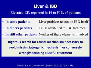
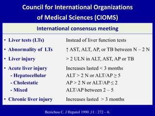
1) Liver tests are elevated in 10-50% of patients with inflammatory bowel disease (IBD). This can be due to the IBD itself, its treatment, or unrelated factors.
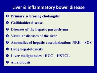
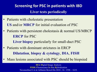
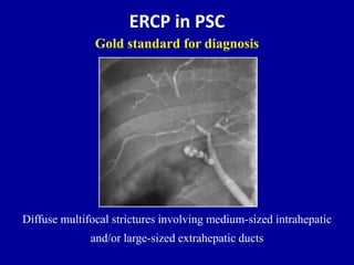
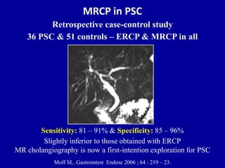
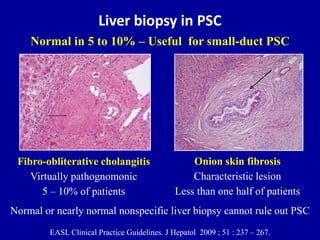
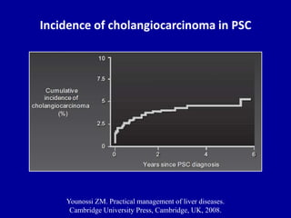
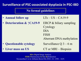
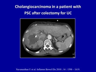
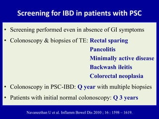
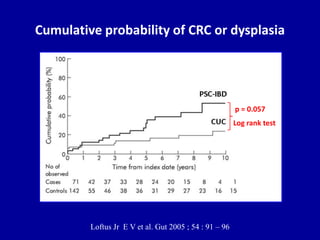
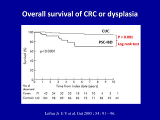
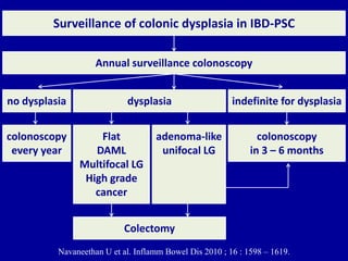
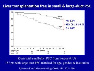
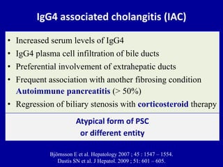
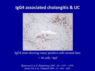
2) Primary sclerosing cholangitis (PSC) is a liver condition strongly associated with IBD. Screening and surveillance is important for PSC patients due to increased risk of cholangiocarcinoma.
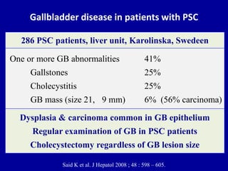
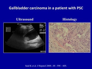
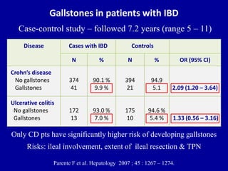
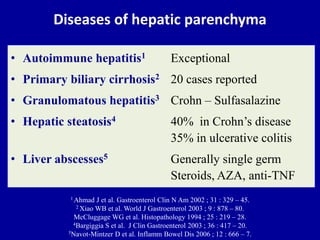
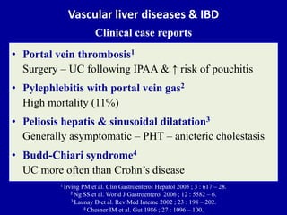
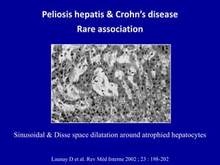
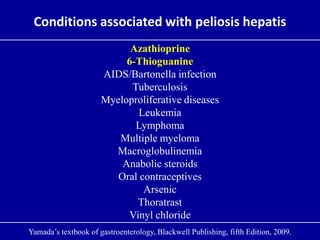
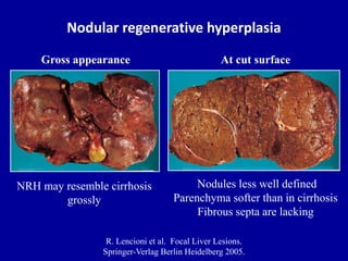
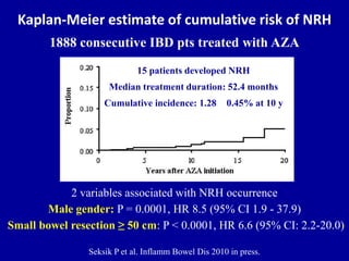
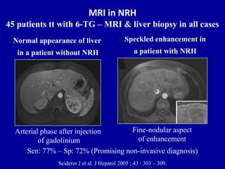
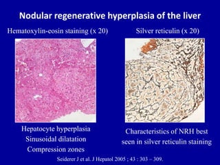
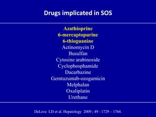
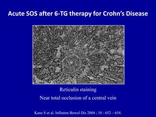
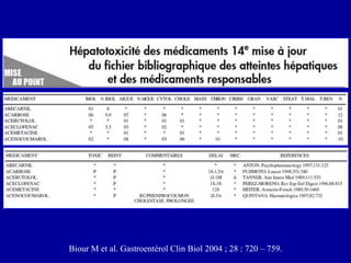
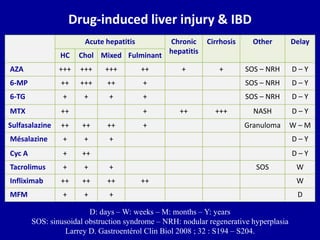
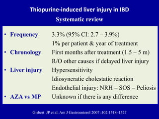
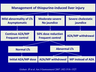
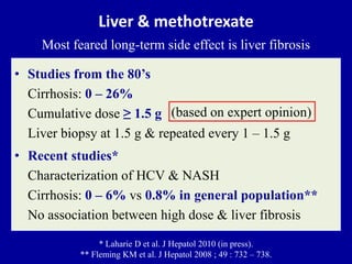
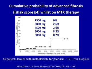
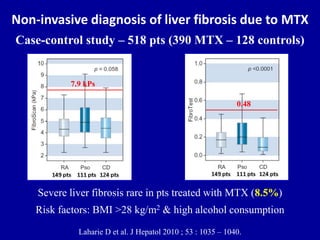
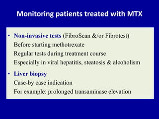
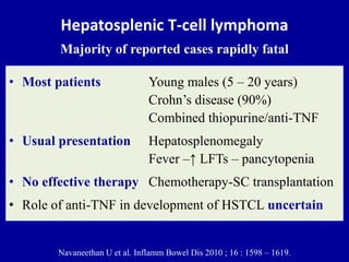
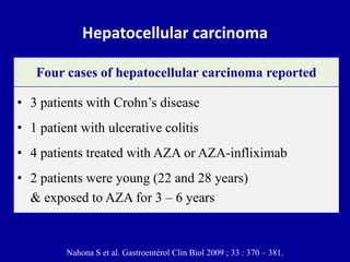
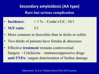
3) Several liver conditions have been reported in association with IBD or its treatments, including gallbladder disease, vascular diseases, nodular regenerative hyperplasia, and drug-induced hepatotoxicity from medications like azathioprine.
2. Liver & IBD
Elevated LTs reported in 10 to 50% of patients
Rigorous search for causal mechanism necessary to
avoid missing iatrogenic mechanism or conversely,
wrongly accusing a useful treatment
Nahona S et al. Gastroentérol Clin Biol 2009 ; 33 : 370 – 381.
• In some patients Liver problem related to IBD itself
• In others patients Cause attributed to IBD treatment
• In still other patients Neither of these elements involved
3. Liver & inflammatory bowel disease
Primary sclerosing cholangitis
Gallbladder disease
Diseases of the hepatic parenchyma
Vascular diseases of the liver
Anomalies of hepatic vascularization: NRH – SOS
Drug hepatotoxicity
Liver malignancies : HCC – HSTCL
Amyloidosis
5. Screening for PSC in patients with IBD
Liver tests periodically
• Patients with cholestatic presentation
US and/or MRCP for initial evaluation of PSC
• Patients with persistent cholestasis & normal US/MRCP
ERCP for PSC
Liver biopsy particularly for small-duct PSC
• Patients with dominant strictures in ERCP
Dilatation, biopsy & cytology, DIA, FISH
• Mass lesions associated with PSC should be biopsied
DIA: Digital Image Analysis
FISH: Fluorescence In Situ Hybridization
Navaneethan U et al. Inflamm Bowel Dis 2010 ; 16 : 1598 – 1619.
6. ERCP in PSC
Diffuse multifocal strictures involving medium-sized intrahepatic
and/or large-sized extrahepatic ducts
Gold standard for diagnosis
7. MRCP in PSC
Retrospective case-control study
36 PSC & 51 controls – ERCP & MRCP in all
Sensitivity: 81 – 91% & Specificity: 85 – 96%
Slightly inferior to those obtained with ERCP
MR cholangiography is now a first-intention exploration for PSC
Moff SL. Gastrointest Endosc 2006 ; 64 : 219 – 23.
8. Liver biopsy in PSC
Fibro-obliterative cholangitis
Virtually pathognomonic
5 – 10% of patients
Onion skin fibrosis
Characteristic lesion
Less than one half of patients
Normal in 5 to 10% – Useful for small-duct PSC
EASL Clinical Practice Guidelines. J Hepatol 2009 ; 51 : 237 – 267.
Normal or nearly normal nonspecific liver biopsy cannot rule out PSC
9. Incidence of cholangiocarcinoma in PSC
Younossi ZM. Practical management of liver diseases.
Cambridge University Press, Cambridge, UK, 2008.
10. Surveillance of PSC-associated dysplasia in PSC-IBD
DIA: Digital Image Analysis
FISH: Fluorescence In Situ Hybridization
Navaneethan U et al. Inflamm Bowel Dis 2010 ; 16 : 1598 – 1619.
No formal guidelines
• Annual follow up LTs – US – CA19-9
• Deterioration & ↑CA19-9 ERCP & biliary sampling
Cytology
DIA
FISH
Aberrant DNA methylation
• Questionable cytology Surveillance Q 3 – 6 m
• Liver mass on US CT or MRI – Biopsies
11. ERCP in PSC & cholangiocarcinoma
Dominant stricture of proximal CHD should be
considered suspicious for cholangiocarcinoma
12. Cholangiocarcinoma in a patient with
PSC after colectomy for UC
Navaneethan U et al. Inflamm Bowel Dis 2010 ; 16 : 1598 – 1619.
13. Screening for IBD in patients with PSC
• Screening performed even in absence of GI symptoms
• Colonoscopy & biopsies of TE: Rectal sparing
Pancolitis
Minimally active disease
Backwash ileitis
Colorectal neoplasia
• Colonoscopy in PSC-IBD: Q year with multiple biopsies
• Patients with initial normal colonoscopy: Q 3 years
Navaneethan U et al. Inflamm Bowel Dis 2010 ; 16 : 1598 – 1619.
14. Cumulative probability of CRC or dysplasia
p = 0.057
Log rank test
Loftus Jr E V et al. Gut 2005 ; 54 : 91 – 96
15. Overall survival of CRC or dysplasia
P = 0.001
Log rank test
Loftus Jr E V et al. Gut 2005 ; 54 : 91 – 96.
16. Navaneethan U et al. Inflamm Bowel Dis 2010 ; 16 : 1598 – 1619.
Surveillance of colonic dysplasia in IBD-PSC
Annual surveillance colonoscopy
Flat
DAML
Multifocal LG
High grade
cancer
Colectomy
dysplasia
colonoscopy
in 3 – 6 months
indefinite for dysplasiano dysplasia
colonoscopy
every year
adenoma-like
unifocal LG
17. Small-duct PSC
“formally pericholangitis”
• Diagnosis Biological cholestasis
Normal cholangiography
Histology compatible with PSC
• Frequency 10% in high-quality ERCP
• Evolution to PSC 25% after 10 years follow-up
• Prognosis Better than ‘‘classical’’ PSC
• Cholangiocarcinoma Never been described
Specific disease of BD with different natural history
18. Liver transplantation free in small & large-duct PSC
Björnsson E et al. Gastroenterology 2008 ; 134 : 975 – 980.
83 pts with small-duct PSC from Europe & US
157 pts with large-duct PSC matched for age, gender, & institution
HR: 3.04
95% CI: 1.82-5.06
P < .0001
19. IgG4 associated cholangitis (IAC)
• Increased serum levels of IgG4
• IgG4 plasma cell infiltration of bile ducts
• Preferential involvement of extrahepatic ducts
• Frequent association with another fibrosing condition
Autoimmune pancreatitis (> 50%)
• Regression of biliary stenosis with corticosteroid therapy
Björnsson E et al. Hepatology 2007 ; 45 : 1547 – 1554.
Dastis SN et al. J Hepatol. 2009 ; 51: 601 – 605.
Atypical form of PSC
or different entity
20. IgG4 associated cholangitis & UC
IgG4 stain showing many positive cells around duct
> 10 cells / hpf
Björnsson E et al. Hepatology 2007 ; 45 : 1547 – 1554.
Dastis SN et al. J Hepatol 2009 ; 51 : 601 – 605.
21. Gallbladder disease in patients with PSC
Said K et al. J Hepatol 2008 ; 48 : 598 – 605.
Dysplasia & carcinoma common in GB epithelium
Regular examination of GB in PSC patients
Cholecystectomy regardless of GB lesion size
One or more GB abnormalities 41%
Gallstones 25%
Cholecystitis 25%
GB mass (size 21, 9 mm) 6% (56% carcinoma)
286 PSC patients, liver unit, Karolinska, Swedeen
22. Gallbladder carcinoma in a patient with PSC
Said K et al. J Hepatol 2008 ; 48 : 598 – 605.
Ultrasound Histology
24. Ulcerative colitis
No gallstones
Gallstones
172
13
93.0 %
7.0 %
175
10
94.6 %
5.4 %
Gallstones in patients with IBD
Case-control study – followed 7.2 years (range 5 – 11)
Disease Cases with IBD Controls
N % N % OR (95% CI)
Crohn’s disease
No gallstones
Gallstones
374
41
90.1 %
9.9 %
394
21
94.9
5.1
Parente F et al. Hepatology 2007 ; 45 : 1267 – 1274.
Only CD pts have significantly higher risk of developing gallstones
Risks: ileal involvement, extent of ileal resection & TPN
2.09 (1.20 – 3.64)
1.33 (0.56 – 3.16)
25. Prophylactic cholecystectomy during
ileocolic resection in Crohn’ disease
• Patients 191 patients with Crohn's ileitis
Medical treatment – ileocolic resection
Matched control group for age & gender
• Retrospective Questionnaire – telephone interviews
• CholecystetomyCrohn ileitis 17/134
Control group 15/150
• Conclusion Prophylactic cholecystectomy not justified
Chew SS et al. Dis Colon Rectum 2003 ; 46 : 1484 – 8.
P = not significant
29. Vascular liver diseases & IBD
Clinical case reports
• Portal vein thrombosis1
Surgery – UC following IPAA & ↑ risk of pouchitis
• Pylephlebitis with portal vein gas2
High mortality (11%)
• Peliosis hepatis & sinusoidal dilatation3
Generally asymptomatic – PHT – anicteric cholestasis
• Budd-Chiari syndrome4
UC more often than Crohn’s disease
1 Irving PM et al. Clin Gastroenterol Hepatol 2005 ; 3 : 617 – 28.
2 Ng SS et al. World J Gastroenterol 2006 ; 12 : 5582 – 6.
3 Launay D et al. Rev Med Interne 2002 ; 23 : 198 – 202.
4 Chesner IM et al. Gut 1986 ; 27 : 1096 – 100.
30. PV gas & PV thrombosis
in fulminant Crohn’s colitis
PV gas & PV thrombosis
Gross ascites
Pneumoperitoneum
World J Gastroenterol 2006 ; 14 : 5582 – 5586.
31. Peliosis hepatis & Crohn’s disease
Rare association
Launay D et al. Rev Méd Interne 2002 ; 23 : 198-202
Sinusoidal & Disse space dilatation around atrophied hepatocytes
33. UC complicated by Budd-Chiari syndrome
Portal tract with rim of viable hepatocytes
Central veins occluded by fibrin thrombi
with surrounding haemorrhage & necrosis
(H & E x 68)
Chesner I M et al. Gut, 1986 , 27 , 1096 – 1100.
35. Nodular regenerative hyperplasia
R. Lencioni et al. Focal Liver Lesions.
Springer-Verlag Berlin Heidelberg 2005.
NRH may resemble cirrhosis
grossly
Gross appearance
Nodules less well defined
Parenchyma softer than in cirrhosis
Fibrous septa are lacking
At cut surface
36. Kaplan-Meier estimate of cumulative risk of NRH
1888 consecutive IBD pts treated with AZA
Seksik P et al. Inflamm Bowel Dis 2010 in press.
2 variables associated with NRH occurrence
Male gender: P = 0.0001, HR 8.5 (95% CI 1.9 - 37.9)
Small bowel resection ≥ 50 cm: P < 0.0001, HR 6.6 (95% CI: 2.2-20.0)
15 patients developed NRH
Median treatment duration: 52.4 months
Cumulative incidence: 1.28 0.45% at 10 y
37. MRI in NRH
45 patients tt with 6-TG – MRI & liver biopsy in all cases
Seiderer J et al. J Hepatol 2005 ; 43 : 303 – 309.
Speckled enhancement in
a patient with NRH
Fine-nodular aspect
of enhancement
Normal appearance of liver
in a patient without NRH
Arterial phase after injection
of gadolinium
Sen: 77% – Sp: 72% (Promising non-invasive diagnosis)
38. Nodular regenerative hyperplasia of the liver
Seiderer J et al. J Hepatol 2005 ; 43 : 303 – 309.
Hepatocyte hyperplasia
Sinusoidal dilatation
Compression zones
Hematoxylin-eosin staining (x 20)
Characteristics of NRH best
seen in silver reticulin staining
Silver reticulin (x 20)
39. Drugs implicated in SOS
Azathioprine
6-mercaptopurine
6-thioguanine
Actinomycin D
Busulfan
Cytosine arabinoside
Cyclophosphamide
Dacarbazine
Gemtuzumab-ozogamicin
Melphalan
Oxaliplatin
Urethane
DeLeve LD et al. Hepatology 2009 ; 49 : 1729 – 1764.
40. Acute SOS after 6-TG therapy for Crohn’s Disease
Reticulin staining
Near total occlusion of a central vein
Kane S et al. Inflamm Bowel Dis 2004 ; 10 : 652 – 654.
42. Council for International Organizations
of Medical Sciences (CIOMS)
Benichou C. J Hepatol 1990 ;11 : 272 – 6.
International consensus meeting
• Liver tests (LTs) Instead of liver function tests
• Abnormality of LTs ↑ AST, ALT, AP, or TB between N – 2 N
• Liver injury > 2 ULN in ALT, AST, AP or TB
• Acute liver injury Increases lasted < 3 months
- Hepatocellular ALT > 2 N or ALT/AP ≥ 5
- Cholestatic AP > 2 N or ALT/AP ≤ 2
- Mixed ALT/AP between 2 – 5
• Chronic liver injury Increases lasted > 3 months
43. Biour M et al. Gastroentérol Clin Biol 2004 ; 28 : 720 – 759.
44. Drug-induced liver injury & IBD
AZA +++ +++ +++ ++ + + SOS – NRH D – Y
6-MP ++ +++ ++ + SOS – NRH D – Y
6-TG + + + + SOS – NRH D – Y
MTX ++ + ++ +++ NASH D – Y
Sulfasalazine ++ ++ ++ + Granuloma W – M
Mésalazine + + + D – Y
Cyc A + ++ D – Y
Tacrolimus + + + SOS W
Infliximab ++ ++ ++ ++ W
MFM + + + D
D: days – W: weeks – M: months – Y: years
SOS: sinusoidal obstruction syndrome – NRH: nodular regenerative hyperplasia
Larrey D. Gastroentérol Clin Biol 2008 ; 32 : S194 – S204.
Acute hepatitis Chronic
hepatitis
Cirrhosis Other Delay
HC Chol Mixed Fulminant
45. Thiopurine-induced liver injury in IBD
Systematic review
• Frequency 3.3% (95% CI: 2.7 – 3.9%)
1% per patient & year of treatment
• Chronology First months after treatment (1.5 – 5 m)
R/O other causes if delayed liver injury
• Liver injury Hypersensitivity
Idiosyncratic cholestatic reaction
Endothelial injury: NRH – SOS – Peliosis
• AZA vs MP Unknown if there is any difference
Gisbert JP et al. Am J Gastroenterol 2007 ;102:1518–1527
46. Management of thiopurine-induced liver injury
Gisbert JP et al. Am J Gastroenterol 2007 ;102:1518–1527
Mild abnormality of LTs
Asymptomatic
Continue AZA/MP
Frequent control
Moderate-severe
No jaundice
50% dose reduction
Frequent control
AZA/MP withdrawal
Severe cholestatic
jaundice
Normal LTs
Initial AZA/MP dose
Abnormal LTs
AZA/MP withdrawal MP instead of AZA
47. Liver & methotrexate
Most feared long-term side effect is liver fibrosis
• Studies from the 80’s
Cirrhosis: 0 – 26%
Cumulative dose ≥ 1.5 g
Liver biopsy at 1.5 g & repeated every 1 – 1.5 g
• Recent studies*
Characterization of HCV & NASH
Cirrhosis: 0 – 6% vs 0.8% in general population**
No association between high dose & liver fibrosis
* Laharie D et al. J Hepatol 2010 (in press).
** Fleming KM et al. J Hepatol 2008 ; 49 : 732 – 738.
(based on expert opinion)
48. Cumulative probability of advanced fibrosis
(Ishak score ≥4) whilst on MTX therapy
Aithal GP et al. Aliment Pharmacol Ther 2004 ; 19 : 391 – 399.
1500 mg 0%
3000 mg 2.6%
4500 mg 2.6%
5000 mg 8.2%
6000 mg 8.2%
66 patients treated with methotrexate for psoriasis – 121 liver biopsies
49. Non-invasive diagnosis of liver fibrosis due to MTX
Case-control study – 518 pts (390 MTX – 128 controls)
Laharie D et al. J Hepatol 2010 ; 53 : 1035 – 1040.
Severe liver fibrosis rare in pts treated with MTX (8.5%)
Risk factors: BMI >28 kg/m2 & high alcohol consumption
7.9 kPs
0.48
50. Monitoring patients treated with MTX
• Non-invasive tests (FibroScan &/or Fibrotest)
Before starting methotrexate
Regular tests during treatment course
Especially in viral hepatitis, steatosis & alcoholism
• Liver biopsy
Case-by case indication
For example: prolonged transaminase elevation
52. Hepatosplenic T-cell lymphoma
Majority of reported cases rapidly fatal
• Most patients Young males (5 – 20 years)
Crohn’s disease (90%)
Combined thiopurine/anti-TNF
• Usual presentation Hepatosplenomegaly
Fever –↑ LFTs – pancytopenia
• No effective therapy Chemotherapy-SC transplantation
• Role of anti-TNF in development of HSTCL uncertain
Navaneethan U et al. Inflamm Bowel Dis 2010 ; 16 : 1598 – 1619.
53. Hepatocellular carcinoma
• 3 patients with Crohn’s disease
• 1 patient with ulcerative colitis
• 4 patients treated with AZA or AZA-infliximab
• 2 patients were young (22 and 28 years)
& exposed to AZA for 3 – 6 years
Four cases of hepatocellular carcinoma reported
Nahona S et al. Gastroentérol Clin Biol 2009 ; 33 : 370 – 381.
55. Secondary amyloidosis (AA type)
Rare but serious complication
• Incidence: < 1 % – Crohn’s/UC: 10/1
• M/F ratio: 3/1
• More common in ileocolitis than in ileitis or colitis
• Two-thirds of patients have fistulas & abscesses
• Effective treatment remains controversial:
Surgery – Colchicine – immunosuppressive drugs
anti-TNFα : targets deterioration of further damage
Oikonomou K et al. Inflamm Bowel Dis 2010 in press.
Patients with PSC and IBD may demonstrate a distinct pattern of clinical behavior compared with IBD patients without concomitant PSC, leading to the coined term of PSC-IBD as a separate clinical entity by the Mayo group
Prevalence of PSC in patients with IBD:1.5%–7.5% of patients with IBD will develop PSC during their disease course.DIA:Digital Image Analysis of cell samplesFISH:Fluorescence In Situ Hybridization of cell samples
MR cholangiography is now a first-intention exploration, ERCP being performed only for therapeutic purposes, or rarely when complementary diagnostic information is needed.GastrointestEndosc 2006;64:219-23.Background: We hypothesized that magnetic resonance cholangiography (MRC) may have less accuracy for the diagnosis and the assessment of the severity of primary sclerosingcholangitis (PSC) than endoscopic retrograde cholangiography (ERC).Objective: The aim of this study was to determine the diagnostic accuracy and interobserver agreement of both ERC and MRC in PSC.Design: A case-control study.Setting: University Hospital.Patients: ERCs and MRCs of 36 patients with PSC and 51 controls (normal/other biliary tract diseases) were read in an independent, blinded, and random fashion by 2 magnetic resonance radiologists and 2 interventional endoscopists by using a previously validated classification system. Readers had no access to clinical history, laboratory results, or patient mix.Results: Extrahepaticductal (EHD) and intrahepaticductal (IHD) visualization was excellent in 64% of 66% of MRCs and 86% of 74% of ERCs. Sensitivity and specificity for diagnosis of PSC for readers 1 to 4 were 91% and 85%, 88% and 90%, 81% and 96%, and 83% and 96%. respectively. Receiver operating curve values were excellent for all readers (all > 0.9). Interobserver agreement (k statistics) for the diagnosis of PSC (MRC, 0.83; ERC, 0.73) and for identifying the presence of IHD strictures (MRC, 0.64; ERC, 0.86) was good for both modalities, but only ERC (ERC, 0.55; MRC, 0.36) was good for the presence and the severity of EHD strictures. When assessment ofdisease severity was limited to the 36 patients with PSC, interobserver agreement was very poor for both MRC (0.23 and 0.07 for EHD and IHD, respectively) and ERC (0.24 and 0.34 for EHD and IHD, respectively).Limitations: The retrospective case–control study made it difficult to assess the impact of the diagnosis on patient management.Conclusions: ERC and MRC were comparable for diagnosing PSC, with very good interobserver agreement for the diagnosis of PSC and IHD strictures. Only ERC had good agreement for EHD strictures. Interobserver agreement was very poor for both MRC and ERC when disease severity of PSC was assessed.
The most common histological feature, the fibrous obliterating cholangitis, is absent in more than two thirds of the biopsy specimens due to the heterogeneous distribution of lesions within the liver. Periductal concentric (“onion-skin”) fibrosis is a classic histopathologic finding of PSC, but this observation is infrequent in PSC liver biopsy specimens and may also be observed in SSC.The histology report thus generally mentions lesions ‘‘compatible’’ with the diagnosis of bile duct disease: peribiliary portal inflammation, discrete atrophy of the bile ducts without periductal fibrosis, proliferative ductal reaction or ductopenia. The biopsy is found normal in 5-10% of cases. Consequently, when the context is suggestive, a normal or nearly normal nonspecific liver biopsy cannot rule out the diagnosis of primary sclerosingcholangitis.
A combination of cytology, FISH, and DIA was shown to have a sensitivity of 50%–64%, but with a specificity and a positive predictive value of 100%.
Prevalence of IBD during primary sclerosingcholangitis: Two thirds of cases.Ulcerative colitis is the main IBD involved.Prevalence of Crohn’s disease during PSC varies from 1 to 17%: always involves the colon.Ulcerative colitis is diagnosed before primary sclerosingcholangitis in about two thirds of patients but the inverse is also possible; the colitis may even begin after liver transplantation. There is no correlation between the severity of ulcerative colitis and the severity of primary sclerosingcholangitis; colectomy does not appear to modify the course of the primary sclerosingcholangitis.
PSC-IBD: a unique form of IBD associated with PSCGut 2005 ; 54 : 91 – 96.Background: Inflammatory bowel disease associated with primary sclerosingcholangitis (PSC-IBD) may have a high prevalence of rectal sparing, backwash ileitis, and colorectal neoplasia.Aims: To describe the clinical features and outcomes of PSC-IBD and compare these to a group of chronic ulcerative colitis (CUC) patients.Methods: The medical records of all patients with PSC-IBD evaluated at the Mayo Clinic Rochester between 1987 and 1992 were abstracted for information on endoscopic and histological features, colorectal neoplasia, surgery, and other clinical outcomes. Patients referred for colorectal neoplasia and those who did not undergo colonoscopy with biopsies were excluded. A control group of CUC patients matched for sex, duration of IBD at first clinic visit, and calendar year of first clinic visit was identified, and similar information was abstracted.Results: Seventy one PSC-IBD patients and 142 CUC patients without PSC were identified. Rectal sparing and backwash ileitis were more common in the PSC-IBD group (52% and 51%, respectively) than in controls (6% and 7%, respectively). Overall, colorectal neoplasia developed in 18 cases and 15 controls, including 11 cancers (seven cases and four controls). An increased risk of colorectal neoplasia or death was not detected in a matched analysis. Although the cumulative incidence of colorectal neoplasia was higher in cases (33%) than in controls (13%) at five years, this was of borderline statistical significance (p = 0.054, unmatched log rank test). Overall survival from first clinic visit was significantly worse among cases (79% v 97%) at five years (p,0.001, unmatched log rank test).Conclusion: PSC-IBD is frequently characterised by rectal sparing and backwash ileitis. Colorectal neoplasia develops in a substantial fraction and overall survival is worse. PSC-IBD may represent a distinct IBD phenotype.
Thus, in IBD patients presenting with elevation of alkaline phosphatase and a normal cholangiogram by ERCP/MRCP, and being excluded for other liver and biliary diseases, liver biopsy may be needed to rule out small-duct PSC.
Although an increased serum IgG4 level was observed in 9% of patients with PSCIAC appears to be a histologically and pathogenetically distinct entity with dramatic response to corticosteroid therapy in contrast to the progressive and refractory nature in PSC.IAC was described in 2 patients with concurrent IBD. The 2 patients were HLA-identical siblings and both had UC with coexisting IAC responsive to low-dose corticosteroid therapy.the question whether PSC, IAC, and AIP share a common pathogenesis or are variations of the same disease spectrum need to be further explored.IgG4 related systemic diseaseDiagnosis of IAC:Diagnosis is confirmed if biliary or other organ histology shows marked infiltration with IgG4 positive cells (> 10 cells/ hpf) or the stricture responds/resolves with steroid treatment.
Diagnosis of IAC is confirmed if biliary or other organ histology shows marked infiltration with IgG4 positive cells (10 cells/hpf) or the stricture responds/resolves with steroid treatment.
Gallbladder disease in patients with primary sclerosingcholangitisSaid K et al. J Hepatol 2008 ; 48 : 598 – 605.Background/Aims: Gallbladder abnormalities may be part of the spectrum in primary sclerosingcholangitis (PSC). The aim of the present study was to evaluate the occurrence and prognostic importance of gallbladder abnormalities in patients with PSC.Methods: Presence of gallbladder abnormalities was assessed in 286 patients with PSC treated at the Liver Unit, Karolinska University Hospital, Huddinge, between 1970 and 2005.Results: One or more gallbladder abnormalities were found in 41% of the patients. Gallstones were found in 25% and cholecystitis in 25%. Cholecystitis among patients with extrahepatic involvement of PSC (30% (65/214)) was significantly higher than among those with intrahepatic involvement (9% (6/70)) (P < 0.0001). A gallbladder mass lesion with a mean size of 21 (±9) mm (S.D.) was found in 18 (6%) patients, in 56% (10/18) of whom it constituted gallbladder carcinoma. In 9 patients without a gallbladder mass lesion, histological re-evaluation disclosed epithelial dysplasia of the gallbladder.Conclusions: Gallbladder disease is common in patients with PSC. Dysplasia and carcinoma are commonly found in gallbladder epithelium, suggesting that regular examination of the gallbladder in PSC patients could be of value for early detection of a gallbladder mass lesion. Cholecystectomy is recommended when such a lesion is detected, regardless of its size.
Chew SS, Ngo TQ, Douglas PR, Newstead GL, Selby W, Solomon MJ. Cholecystectomy in patients with Crohn’s ileitis. Dis Colon Rectum 2003;46:1484-8.Because of the higher prevalence of gallstone disease, prophylactic cholecystectomy could be warranted in patients undergoing ileal resection. An Australian team compared a group of patients with operated ilealCrohn’s disease (n = 89) with a group of patients with ilealCrohn’s disease treated medically (n = 45). Patients were contacted by phone; 20 had symptomatic gallstones (18/89 with ileal resection and 2/45 with medical treatment). In this work, the prevalence of cholecystectomy was the same in the two groups. Thus cholecystectomy would not appear to be warranted during ileal resection for Crohn’s disease.
Chew SS, Ngo TQ, Douglas PR, Newstead GL, Selby W, Solomon MJ. Cholecystectomy in patients with Crohn’s ileitis. Dis Colon Rectum 2003;46:1484-8.PURPOSE: Gallstone disease is reported to be higher in patients with Crohn's disease than in the general population. This study was designed to determine the prevalence of cholecystectomy in patients with Crohn's ileitis, attempt to identify any associated risk factors, and determine whether it is justified to perform prophylactic cholecystectomy during ileocolic resection.METHODS: A total of 191 patients with Crohn's ileitis who were treated medically or who had an ileocolic resection were retrospective reviewed. A questionnaire survey was performed. Telephone interviews were conducted for the nonrespondents. Further review of medical records was performed to determine the details of admissions for any gallstone disease and/or subsequent cholecystectomy. A.RESULTS: A total of 191 questionnaires were mailed, and the overall response rate was 70.2 percent (134/191) after telephone interview follow-up. There were 2 of 45 medical and 18 of 89 surgical patients with symptomatic cholelithiasis, i.e., 14.9 percent (20/134) of respondents. As a result, 2 patients (1.5 percent) required endoscopic sphincterotomy, 17 patients (12.7 percent) needed cholecystectomy, and 1 patient (0.7 percent) did not have any intervention. Only five patients had a cholecystectomy after their ileal resections. In the control group of 150 patients, 15 patients (14 females; mean age, 51.9 years; range, 34-78 years) had previous cholecystectomy. There was no significant difference with prevalence of cholecystectomy in Crohn's patients compared with controls (17/134 vs. 15/150; P = not significant). Furthermore, the number of ileal resections did not affect the cholecystectomy rate, but patients who had >30 cm of ileum resected were more likely to have cholecystectomy (P = 0.056).CONCLUSIONS: The prevalence of gallstone disease in Crohn's ileitis requiring cholecystectomy is similar to that of the general population with a female predominance. In addition, the number of patients requiring cholecystectomy after ileal resection was low. Thus, synchronous prophylactic cholecystectomy during ileocolic resection for Crohn's ileitis is not justified.
Bargiggia S, Maconi G, Elli M, Molteni P, Ardizzone S, Parente F, et al. Sonographic prevalence of liver steatosis and biliary tract stones in patients with inflammatory bowel disease: study of 511 subjects at a single center. J ClinGastroenterol 2003;36:417—20.An ultrasound study in 511 patients with IBD found hepatomegaly in 25.7% and moderate to severe hepatic steatosis (defined by the intensity gradient between the liver and the kidney) in 39.5% of patients with Crohn’s disease and 35% of those with ulcerative colitis.
RésuméIntroduction. – Les maladies inflammatoires chroniques de l’intestin se compliquent dans 10 à 30 % des cas d’anomalies hépatobiliaires. Parmi celles-ci, la stéatose hépatique et les lithiases biliaires sont les plus fréquentes dans la maladie de Crohn. À l’inverse, la péliose hépatique n’est qu’exceptionnellement décrite en association avec les maladies inflammatoires chroniques de l’intestin. Nousdécrivons un cas de péliose hépatique compliquant une maladie de Crohn jusqu’alors méconnue.Exégèse. – Une jeune femme âgée de 24 ans consultait pour une hépatomégalie et une altération de l’état général. L’interrogatoire retrouvait une diarrhée depuis trois mois et un épisode de rectorragie. Il existait une lésion aphtoïde de la langue. Le transit du grêle mettait en évidence un aspect typique d’iléite terminale évocateur de maladie de Crohn. Le foie était morphologiquement hétérogène. La ponction–biopsie hépatique permettait de poser le diagnostic de péliose hépatique pour laquelle les autres causes étaient écartées. Il s’agissait donc d’une péliose hépatique découverte devant une hépatomégalie et associée à une maladie de Crohn méconnue.Conclusion. – L’association entre une maladie de Crohn et une péliose hépatique n’a été qu‘exceptionnellement décrite. Il ne s’agit pas a priori d’un facteur de gravité des maladies inflammatoires chroniques de l’intestin. La signification physiopathologique de cette association est inconnue. La péliose hépatique est définie dans sa forme macrokystiquepar la présence de cavités multiples remplies d’hématies dans le parenchyme hépatique et souvent accompagnées par une dilatation des sinusoïdes. Dans les formes plus modérées, il existe une distension des sinusoïdes sans cavité mais avec un décollement endothélial ou des hématies. Causes de la péliose hépatique:Contraception orale - insuffisance cardiaque droite – tuberculose - infection par le virus de l’immunodéficience humaine- cancers – hémopathies - prise de vitamine A – azathioprine - dérivés C-17 α-alkyl de la testostérone.
Nodular regenerative hyperplasia is arare disorder defined by the presence of diffuse nodules of the hepatic parenchyma without annular fibrosis and may lead to severe portal hypertension.Diagnosis of NRH was suspected on acquired thrombocytopenia (i.e., platelet count under 150,000 per mm3), splenomegaly, or clinical signs of portal hypertension and proven by liver biopsy.
Incidence of NRH in IBD treatede with AZAInflamm Bowel Dis 2010;000:000–000Background: Nodular regenerative hyperplasia (NRH) is a rare hepatic disorder that may lead to severe portal hypertension. Cases of NRH have been reported in patients receiving thiopurines for inflammatory bowel disease (IBD). Since azathioprine (AZA) is used more and more frequently as a maintenance treatment in IBD, the risk of NRH must be known. The objective of this study was to evaluate the prevalence of NRH and its predictive factors in IBD patients treated with AZA.Materials and Methods: From the same tertiary referral center, 1888 consecutive IBD patients treated with AZA were studied. Clinical diagnosis of NRH was proven by liver biopsy in all cases except one. The cumulative risk of NRH was estimated with the Kaplan–Meier method. Factors associated with NRH were tested independently with the log-rank method and multivariate proportional hazards model with time-dependent covariates.Results: Fifteen patients developed NRH in a median treatment duration of 52.4 months (SE 1.6). The cumulative incidence of NRH was 1.28 6 0.45% at 10 years. Only two variables were independently associated with NRH occurrence: male gender (P = 0.0001, hazard ratio [HR] 8.5, 95% confidence interval [CI] 1.9– 37.9) and small bowel resection 50 cm (P < 0.0001, HR 6.6, 95% CI 2.2–20.0), either prior to or after AZA initiation. Conclusions: The risk of developing NRH during AZA treatment is low. This study suggests that male patients with small bowel resection 50 cm constitute the group with the higher risk of developing NRH while treated with AZA.
Background/Aims:Although 6-thioguanine (6-TG) has been suggested as an effective treatment option for patients with inflammatory bowel disease (IBD), the recent description of its hepatotoxicity has led to the recommendation not to consider this drug. We initiated a multicenter safety study in IBD-patients treated with 6-TG to investigate hepatic changes by liver biopsy and magnetic resonance imaging (MRI).Methods: Forty-five patients from three European centers treated with 6-TG (40–80 mg/d) at least for 8 weeks were enrolled. In all patients liver biopsy and MRI were performed. Slides and MR images were independently read by two pathologists and radiologists, respectively, and interpreted according to predefined criteria by consent.Results: In 8 patients nodular regenerative hyperplasia (NRH) was diagnosed by liver biopsy, in 8 additional patients NRH could not be excluded due to equivocal pathological findings. MRI demonstrated a sensitivity of 77% and a specificity of 72% in the detection of pathohistological findings consistent with and/or possibly related to NRH.Conclusions: Our study suggests that 6-TG therapy in IBD patients is associated with NRH of the liver. Based on a special MRI protocol, non-invasive diagnosis of NRH with promising sensitivity and specificity was demonstrated.
histological study of liver biopsy showing hyperplasticparenchymatous nodules without extensive fibrosis on silver reticulin staining, with compressed and atrophic internodular parenchyma.
Previously known as hepatic veno-occlusive disease.Partial or total non-thromboticfibrous obstruction of centrolobular veins.Manifestation: anictericcholestasis – ascites – PHT Few published cases in patients treated for 1- 2 years for UC or CD.
Acute Sinusoidal Obstruction Syndrome After 6-TG Therapy for Crohn’s DiseaseAbstract: 6-Thioguanine (6-TG), the active metabolite of 6-mercaptopurine and its prodrugazathioprine, are thought to be responsiblefor clinical efficacy in the treatment of active Crohn’s disease. Its use as a therapeutic agent for inflammatory bowel disease (IBD)has been limited to patients who are resistant to or intolerant of other antimetabolites. Short-term experience with this agent has not demonstrated an increased incidence of hematologic or hepatic toxicity; however long-term safety data are scarce. We herein report a patient who developed acute sinusoidal obstruction syndrome after 14 months of successful thioguanine treatment. This is the first report of such a complication in an adult treated with 6-TG for active Crohn’s disease.SOS: Partial or total non-thrombotic fibrous obstruction of CVManifestation: PHT – Ascites – AnictericcholestasisThere have been a few published cases of sinusoidal obstruction in patients treated for one or two years for ulcerative colitis or Crohn’s disease.
The term “liver tests” (LTs) should be used instead of “liver function tests.” “acute” liver injury was considered present when these increases had lasted less than 3 months“chronic” liver injury was used when these increases had lasted more than 3 monthsAcute drug-induced liver injury1- Drug-induced cholestatic injury: has been defined by an international consensus panel by an isolated elevation of serum alkaline phosphatase (AP) >2 ULN or an alanineaminotransferase (ALT)/AP ratio (both elevated above ULN) ≤ 2. 2- Drug-induced hepatocellular injury as the predominant form of DILI is defined by isolated ALT >2 ULN or an ALT/AP ratio (bothelevated above ULN) ≥ 5. 3- Mixed type injury is defined by an ALT/AP ratio of 2–5. Drug-induced cholestatic injury has a better prognosis than hepatocellular injury.For many drugs, the reported prevalence of DILI is between 1 in 10,000 and 1 in 100,000 patients, and about 30% of cases with DILI are cholestatic. However, these estimates are weakened by considerable underreporting of DILI. Both environmental and genetic factors may determine susceptibility. Genetically determined variations of hepatobiliary transporter and biotransformation enzyme expressionand function may be important risk factors for an individual’s susceptibility to cholestasis under conditions of xenobiotic stress by drugs.Cholestasis:By convention, cholestasis is considered chronic if it lasts >6 months.Isolated serum GGT elevation has little specificity for cholestasis, and may also result from enzyme induction in response to alcohol or drug intake.The cut-off levels of serum AP and GGT requiring diagnostic work-up are debated: AP levels higher than 1.5 times the upper limitof normal (ULN) and GGT levels >3 ULN have been proposed.
6-TG:During meetings arranged in Vienna and Prague in 2004, European experts applying 6-TG further on in IBD patientspresented data on the safety and efficacy of this drug. After thorough evaluation of its risk-benefit ratio, the group consented that 6-TG may still be considered as a rescue drug in stringently defined indications in IBD, albeit restricted to a clinical research setting.
The term “liver tests” (LTs) should be used instead of “liver function tests.” “Abnormality of LTs” was defined as an increase in aspartatetransaminase (AST), alaninetransaminase (ALT), alkaline phosphatase (AP), or total bilirubin between N (upper limit of the normal range) and 2 N. An isolated increase even over 2 N in AST, AP, or total bilirubin should be considered only a biochemical abnormality and not necessarilya sign of liver injury. “Liver injury” (or “hepatotoxicity”) should be used if there is an increase of over 2 N in ALT or conjugated bilirubin (or a combined increase in AST, AP, and total bilirubin provided one of them is above 2 N).
Le seuil de 1500 mg de méthotrexate en dose cumulée ne repose que sur un consensus d’experts.
Monitoring methotrexate-induced hepatic fibrosis in patients with psoriasisAithal GP et al. Aliment PharmacolTher 2004 ; 19 : 391 – 399.Background: Reports that up to 26% of subjects with psoriasis develop cirrhosis have led to a recommendation of serial liver biopsies after each cumulative dose of 1500 mg of methotrexate.Aim: To evaluate the progression of liver injury in patients with psoriasis and the impact of monitoring by liver biopsy on their management.Methods: One hundred and twenty-one liver biopsies from 66 subjects (aged 11–79 years) with psoriasis, receiving a median cumulative dose of 3206 mg of methotrexate over a period of 280.5 weeks, were evaluated.Results: The assessment of advanced fibrosis according to the Ishak system (≥ 4) correlated perfectly with that of the Scheuer system (≥ 3) and poorly with that of the Roenigk scale (≥ 3b) (r2 = 1.0 and 0.31, respectively). Two of 24 pre-treatment biopsies showed advanced fibrosis and both subjects were heavy drinkers. The cumulative probabilities of advanced fibrosis (Ishak≥ 4) were 0%, 2.6%, 2.6%, 8.2% and 8.2% at cumulative doses of 1500, 3000, 4500, 5000 and 6000 mg, respectively. None of the subjects developed cirrhosis during follow-up or discontinued therapy on the basis of liver biopsy findings.Conclusions: Advanced hepatic fibrosis with low-dose methotrexate therapy is much less frequent than previously reported. Pre-treatment or monitoring liver biopsies in accordance with the current guidelines have little impact on patient management.Ishak score system: 0 – 6 Scheuer scoring system: 0 – 4 Roenigk classification of methotrexate-associated liver damage: None – Mild – Moderate – Severe Le risque de fibrose hépatique sous méthotrexateestfaible.
Assessment of liver fibrosis with TE & FibroTest in pts treated with MTX for chronic inflammatory diseasesLaharie D et al. J Hepatol 2010 ; 53 : 1035 – 1040. Background & Aims: Although methotrexate (MTX) is used in the effective treatment of inflammatory disorders, its use is hampered by the risk of liver fibrosis. Non-invasive methods for the diagnosis of liver fibrosis, such as transient elastography (Fibro-Scan) and FibroTest could be useful for monitoring MTX–livertoxicity. The aim of this case–control study was to determine factors associated with liver fibrosis in a large cohort of patients requiring MTX.Methods: Consecutive adults with various benign inflammatory diseases were prospectively assessed using FibroScan and Fibro-Test when they were treated with MTX (cases) or before beginning treatment (controls).Results: Among 518 included patients, 44 patients (8.5%) had FibroScan and/or FibroTest results suggesting severe liver fibrosis. In a multivariate analysis, factors associated with abnormal markers of liver fibrosis were the body mass index >28 kg/m2 and high alcohol consumption. Neither long MTX duration nor cumulative doses were associated with elevated FibroScan or FibroTest results.Conclusions: Severe liver fibrosis is a rare event in patients treated with MTX and is probably unrelated to the total dose. Patients with other risk factors for liver disease should be closely monitored with non-invasive methods before and during MTX treatment.FibroScan >7.9 kPa suggesting severe liver fibrosis. > 12.5 kPa suggesting cirrhosisFibrotest > 0.48 suggesting severe liver fibrosis. > 0.74 suggesting cirrhosis
With wide use of biologics in IBD, there has been increasing reports of HSTCL in IBD patients.Usual presentation associated with HSTCL includes fever, fatigue, elevated LFTs, hepatosplenomegaly and pancytopenia. These symptoms may be seen in IBD patients and a high degree of suspicion, particularly in young patients on anti-TNF therapy, may be required todiagnose HSTCL. The FDA has issued a black box warning on the use of these drugs.The role of anti-TNF treatment in the development of HSTCL remains uncertain.Hepatosplenic T cell lymphoma in IBD – Leading article - Gut 2008;57:1639-1641 .The development of lymphoma in chronic inflammatory conditions has been a topic of great interest. Studies in inflammatory bowel disease (IBD) have demonstrated conflicting results, and it is often difficult to disassociate disease severity, disease duration and the risk attributable to drug exposure. Recently presented results from the very large French population-based CESAME study suggest a doubling of the risk of lymphoma in patients with IBD, with the majority of cases occurring in association with immunosuppressive therapy. Similarly, a meta-analysis of previous cohort studies concluded that the risk of lymphoma is increased fourfold in patients with IBD on thiopurine treatment (azathioprine and 6-mercaptopurine) compared with those not receiving such therapy. Such analyses cannot demonstrate causation, and it is possible that an increased lymphoma risk relates to more active underlying disease rather than thiopurine therapy, an interpretation supported by results from studies in IBD patients unexposed to such treatment. Whilst it is possible that thiopurine therapy carries a modestly increased relative risk of lymphoma, the absolute risk is still very small, and appears to be outweighed in the majority of patients by the benefits of better disease control. With the advent and success of antitumour necrosis factor (TNF) and other biological therapies, investigators have sought to define the place of concomitant immunomodulator therapy, and its effect on the risk–benefit equation. Early experience suggested that combination therapy was desirable to reduce the immunogenicity of infliximab, especially with episodic dosing. More recently, this approach has been challenged due to a lack of synergistic efficacy and the minimal reduction in immunogenicity when anti-TNF drugs are used …
Given the rarity, the asymptomatic nature in most presentations, and need for liver biopsy for diagnosis, the true incidence, natural history, and prognosis of hepatic amyloidosis in IBD remains unclear. Control of gut inflammation may reduce the severity of amyloiddeposition.