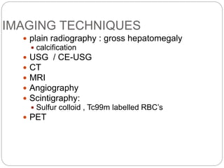
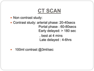
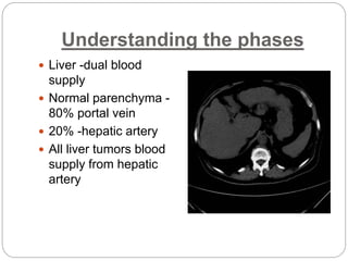
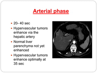
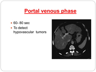
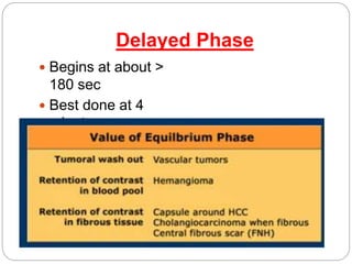
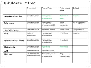
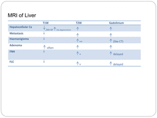
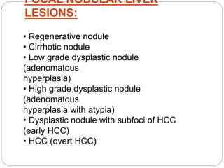
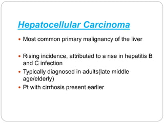
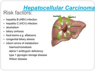
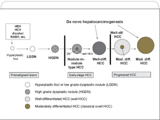
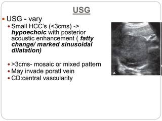
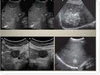
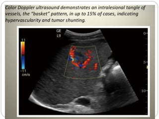
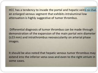
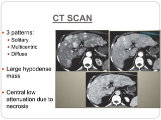
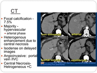
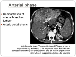
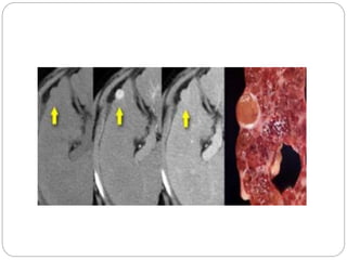
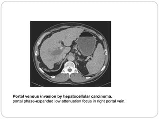
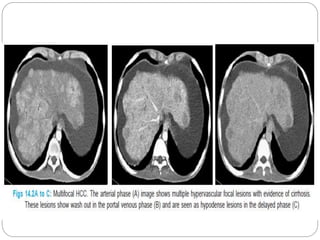
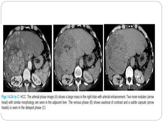
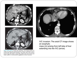
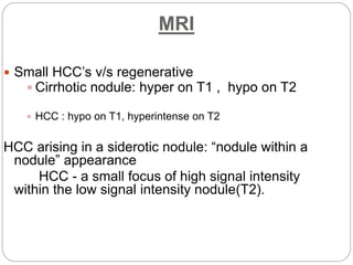
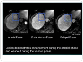
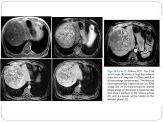
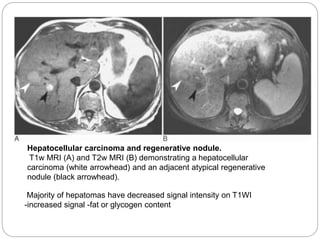
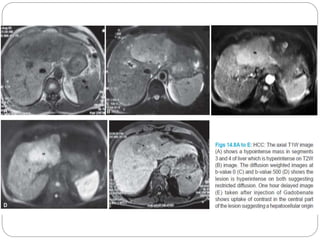
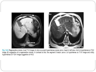
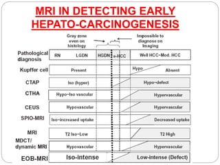
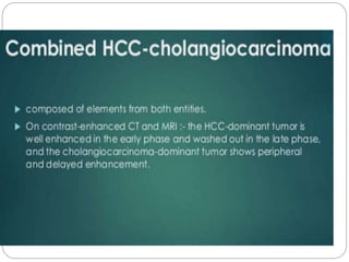
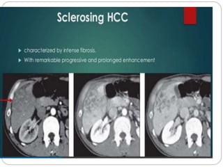
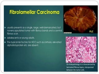
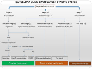
This document discusses imaging techniques for detecting and characterizing liver lesions. It focuses on multiphase CT and MRI protocols for hepatocellular carcinoma (HCC). CT involves non-contrast, arterial, portal, and delayed phase imaging. Arterial phase highlights hypervascular tumors fed by the hepatic artery. Portal phase detects hypovascular lesions. MRI features of HCC include hypointensity on T1-weighted imaging and hyperintensity on T2-weighted imaging. The Barcelona Clinic Liver Cancer staging system is also referenced.
Imaging assessment of malignant focal and diffuse liver lesions from Ultrasound to Mri with overview of interventional modalities and diagnostic snippets,
Hepatocellular carcinoma—role of interventional radiologist Dr. Muhammad Bin ...Dr. Muhammad Bin Zulfiqar
In these presentation we will discuss the merits, demrits and outcomes of various interventional radiology modalities for the treatment of hepatocellular carcinoma
Triphasic CT (TPCT) Scan of the liver is essential in view of the dual blood supply of the liver. TPCT allows characterisaiton of all liver lesions and close to pathological correlaiton by non invasive imaging alone. Additionally providing segmental vascular analysis as a surgicical guide.
Purpose of this presentation is to educate non radiologist about basic CT anatomy of abdominal viscera and basic interpretation of very common diseases
Macroeconomics- Movie Location
This will be used as part of your Personal Professional Portfolio once graded.
Objective:
Prepare a presentation or a paper using research, basic comparative analysis, data organization and application of economic information. You will make an informed assessment of an economic climate outside of the United States to accomplish an entertainment industry objective.
Welcome to TechSoup New Member Orientation and Q&A (May 2024).pdfTechSoup
In this webinar you will learn how your organization can access TechSoup's wide variety of product discount and donation programs. From hardware to software, we'll give you a tour of the tools available to help your nonprofit with productivity, collaboration, financial management, donor tracking, security, and more.
Acetabularia Information For Class 9 .docxvaibhavrinwa19
Acetabularia acetabulum is a single-celled green alga that in its vegetative state is morphologically differentiated into a basal rhizoid and an axially elongated stalk, which bears whorls of branching hairs. The single diploid nucleus resides in the rhizoid.
Honest Reviews of Tim Han LMA Course Program.pptxtimhan337
Personal development courses are widely available today, with each one promising life-changing outcomes. Tim Han’s Life Mastery Achievers (LMA) Course has drawn a lot of interest. In addition to offering my frank assessment of Success Insider’s LMA Course, this piece examines the course’s effects via a variety of Tim Han LMA course reviews and Success Insider comments.
Introduction to AI for Nonprofits with Tapp NetworkTechSoup
Dive into the world of AI! Experts Jon Hill and Tareq Monaur will guide you through AI's role in enhancing nonprofit websites and basic marketing strategies, making it easy to understand and apply.
Read| The latest issue of The Challenger is here! We are thrilled to announce that our school paper has qualified for the NATIONAL SCHOOLS PRESS CONFERENCE (NSPC) 2024. Thank you for your unwavering support and trust. Dive into the stories that made us stand out!
9. Pre contrast Arterial Phase Portal venous
phase
Delayed
Hepatocelluar Ca Low attenuation Homogenous
enhancement
Washout of
lesion
Isodense
Adenoma Low attenuation Homogenous
enhancement 85%
Iso or
hypodense
Iso or hypodense
Haemangioma Low attenuation Peripheral puddles Partial Fill in Complete fill in
FNH Iso/Low
attenuation
Homogenous
enhancement
Hypodense Isodense
Hypervascular Mets Low attenuation Homogenous
enhancement
Hypodense
Metastasis Low attenuation Hypodense Hypodense
Cyst Low attenuation No enhancement
Abscess Low attenuation may
have irregular margins
Transient regional
enhancement
Ring
enhancement
Multiphasic CT of Liver
10. T1W T2W Gadolinium
Hepatocellular Ca
,iso or (fat degeneration)
Metastasis
Haemanigioma
++ (like CT)
Adenoma
often
FNH
+ delayed
FLC
+ delayed
MRI of Liver
11. FOCAL FAT SPARING
Diagnostic confusion
with tumors
Common sites
Periportal region of the
medial segment of left
lobe (segment IV)
Either side of falciform
ligament
Cranial aspect of GB
fossa
Characteristic features:
Geographic appearance
Lack of mass effect
Vessels through the
lesion
12. FOCAL NODULAR LIVER
LESIONS:
• Regenerative nodule
• Cirrhotic nodule
• Low grade dysplastic nodule
(adenomatous
hyperplasia)
• High grade dysplastic nodule
(adenomatous
hyperplasia with atypia)
• Dysplastic nodule with subfoci of HCC
(early HCC)
• HCC (overt HCC)
13. Hepatocellular Carcinoma
Most common primary malignancy of the liver
Rising incidence, attributed to a rise in hepatitis B
and C infection
Typically diagnosed in adults(late middle
age/elderly)
Pt with cirrhosis present earlier
14. Risk factors:
hepatitis B (HBV) infection
hepatitis C (HCV) infection
alcoholism
biliary cirrhosis
food toxins e.g. aflatoxins
congenital biliary atresia
inborn errors of metabolism
haemochromatosis
alpha-1 antitrypsin deficiency
type 1 glycogen storage disease
Wilson disease
Hepatocellular Carcinoma
15.
16. USG
USG - vary
Small HCC’s (<3cms) ->
hypoechoic with posterior
acoustic enhancement ( fatty
change/ marked sinusoidal
dilatation)
>3cms- mosaic or mixed pattern
May invade poratl vein
CD:central vascularity
17.
18.
19.
20.
21.
22. CT SCAN
3 patterns:
Solitary
Multicentric
Diffuse
Large hypodense
mass
Central low
attenuation due to
necrosis
23. CT
Focal calcification -
7.5%
Majority -
hypervascular
arterial phase
Heterogenous
enhancement due to
central necrosis
Isodense on delayed
images
Angioinvasive: portal
vein /IVC
Central Necrosis-
Hetrogeneous +C
24. Arterial phase
Demonstration of
arterial branches
tumour
Arterio portal shunts
Arterio-portal shunt: The arterial phase CT image shows a
large enhancing lesion (m) in the segments 3 and 4 of liver with
contrast in the left hepatic artery (arrow) and left branch of portal vein
(arrow head) suggesting arterio-portal shunting
25.
26. Portal venous invasion by hepatocellular carcinoma.
portal phase-expanded low attenuation focus in right portal vein.
27.
28.
29. IVC invasion: The axial CT image shows
an exophytic
mass (m) arising from left lobe of liver
extending into the IVC (arrow)
30. MRI
Small HCC’s v/s regenerative
Cirrhotic nodule: hyper on T1 , hypo on T2
HCC : hypo on T1, hyperintense on T2
HCC arising in a siderotic nodule: “nodule within a
nodule” appearance
HCC - a small focus of high signal intensity
within the low signal intensity nodule(T2).
31.
32.
33. Hepatocellular carcinoma and regenerative nodule.
T1w MRI (A) and T2w MRI (B) demonstrating a hepatocellular
carcinoma (white arrowhead) and an adjacent atypical regenerative
nodule (black arrowhead).
Majority of hepatomas have decreased signal intensity on T1WI
-increased signal -fat or glycogen content