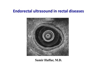
SlideShare a Scribd company logo
Samir Haffar, M.D.
Endorectal ultrasound in rectal diseases
"It is necessary to see before reflecting,
to seize appearances before probing the causes;
and our ideas on any external object are vague
if they are not for us so many images.”
Xavier Bichat (1771 - 1802)
French anatomist and physiologist
Father of modern histology and
descriptive anatomy
(1) Normal ultrasound-anatomy of the rectum
(2) Endorectal ultrasound (ERUS) in rectal diseases
Rectal tumors
Submucosal lesions
Endorectal ultrasound for anorectal lesions
(1) Normal ultrasound-anatomy of the rectum
Santoro JA & Di Falco G. Endosonographic anatomy of the normal anal canal.
In: Santoro JA & Di Falco G (eds), Benign anorectal diseases. Springer-Verlag, Italia, 2006.
Coronal anatomy of the anal canal
Patient preparation
• Patients given routine enema two hours before examination
• Sedation not necessary
• Examination in left lateral decubitus, in knee-chest position
• Digital rectal exam before insertion of the probe into rectum:
Identify lesion size, location, & mobility of the tumor
Kim MJ. Ultrasonography 2015; 34:19-31.
Santoro GA. Recents advances in colorectal polyps. Genoa, 11 April 2014.
Good visualization depends on
Maintain probe in center of lumen
Distension of water-filled balloon
Good contact with rectal wall
Five layers of the rectal wall
2 - 3 mm thick
Normal blood vessels in the rectal wall
Axial ERUS image
Continuity of hypoechoic vessels more than cross-sectional diameter
is criterion used to distinguish vessels from hypoechoic lymph nodes
Blood vessels appear to branch or extend longitudinally
Santoro GA & Di Falco G. Stage uN1: Lymph node metastases.
In: Santoro GA & Di Falco G (eds), Atlas of endoanal & endorectal US. Springer, Italia, 2004.
Axial ERUS image Axial ERUS image
Santoro JA & Di Falco G. Endosonographic anatomy of the normal anal canal.
In: Santoro JA & Di Falco G (eds), Benign anorectal diseases. Springer-Verlag, Italia, 2006.
Normal peri-rectal US anatomy in males
Seminal vesicles Prostate
Uterus
Axial ERUS image
Vagina1
Ovary
Uterus
Bladder
Axial ERUS image
Uterus, ovary & bladder2
(1) Santoro JA & Di Falco G. Endosonographic anatomy of the normal anal canal.
In: Santoro JA & Di Falco G (eds), Benign anorectal diseases. Springer-Verlag, Italia, 2006.
(2) Roseau G. World J Gastrointest Endosc 2014; 6(11): 525-533.
Normal peri-rectal US anatomy in females
Iliac vessels
Long tubular anechoic structure near recto-sigmoid junction
Iliac region is important for nodal staging
Cooper ST & Sanders MK. Radial endoscopic ultaround.
In: Shami VM & Kahaleh M (eds), Endoscpic ultrasound. Springer, New York, 2010.
(2) Endorectal ultrasound in rectal diseases
Rectal tumors
• Tumor depth T staging
• Nodal metastasis N staging
• Circumferential resection margin Prostate & seminal vesicules
Vagina & uterus
Mesorectal fascia (MRF)
Loco-regional staging of rectal
adenoma/adenocarcinoma
Make every effort to image all 5 layers at all points of tumor because
tumor infiltration can differ significantly along the body of the tumor
uT staging in rectal cancer
uTx Primary tumor cannot be assessed
uT0 Noninvasive lesion confined to mucosa
uT1 Tumor confined to mucosa & submucosa
uT2 Tumor penetrates into but not through MP
uT3 Tumor extends into perirectal fat
uT4 Tumor involves adjacent structure
Berton F et al. AJR 2008; 190:1495–1504.
uT0 rectal lesion
Lesion expands inner hypoechoic (mucosa)
surrounded by uniform middle hyperechoic layer
Axial ERUS image
Santoro GA & Di Falco G. Endoluminal ultrasound in preoperative staging of rectal carcinoma.
In: Santoro GA et al (eds), Atlas of endoanal & endorectal ultrasonography. Springer, Italia, 2004.
uT1 rectal lesion
Diaphragmatic representation of T1
rectal tumor invading submucosa
Diaphragmatic representation
Tumor extended to hyperechoic
submucosa layer surrounded by
uniform hypoechoic muscularis layer
Axial ERUS image
Submucosal invasion of rectal cancer
Kudo differentiate 3 types of early invasive cancers
• SM-1 tumor: invading superior third of submucosa
• SM-2 tumor: invading superficial two thirds of submucosa
• SM-3 tumor: invading deep third of submucosa
Kudo S. Endoscopy 1993; 25: 455-461.
Increased rate of lymph node metastasis with increased
submucosal invasion
Kudo's classification
uT2 rectal lesion
Diaphragmatic representation of T2
rectal tumor invading muscolaris propria
Invasion of muscularis propria
based on its thickness
Intact perirectal fat interface
Diaphragmatic representation Axial ERUS image
Santoro GA & Di Falco G. Endoluminal ultrasound in preoperative staging of rectal carcinoma.
In: Santoro GA et al (eds), Atlas of endoanal & endorectal ultrasonography. Springer, Italia, 2004.
uT3 rectal lesion
T3 rectal tumor infiltrating
into peri-rectal fat
Diaphragmatic representation
Perirectal fat invasion based on
irregularities of outer hyperechoic layer
Axial ERUS image
Santoro GA & Di Falco G. Endoluminal ultrasound in preoperative staging of rectal carcinoma.
In: Santoro GA et al (eds), Atlas of endoanal & endorectal ultrasonography. Springer, Italia, 2004.
uT4 rectal lesion / Invasion of the prostate
Axial ERUS image2
Tumor invading into the
prostate anteriorly (arrow)
(1) Santoro GA & al. Endoluminal ultrasound in preoperative staging of rectal carcinoma.
In: Santoro GA et al (eds), Atlas of endoanal & endorectal ultrasonography. Springer, Italia, 2004.
(2) Chern H et al. Clinical Staging. In: Czito BG et al, Rectal cancer. Springer, New York, 2010.
Diaphragmatic representation1
Tumor involves adjacent structure
uT4 rectal lesion / Invasion of seminal vesicule
Intact seminal vesicula
Fatty tissue plane between tumor (Tm)
& right seminal vesicule (S)
Axial ERUS image Axial ERUS image
Same patient at another plane
Invasion of left seminal vesicule
Tm: tumor – S: seminal vesicule
Engin G. J Ultrasound Med 2006; 25:57–73.
Residual strictured lumen represented by small amount of echogenic air
Wall thickened & hypoechoic with no residual normal wall layers
Invasion of perirectal fat (T3 lesion)
Berton F et al. AJR 2008; 190:1495–1504.
ERUS in stenotic rectal lesion
less accuracy for T staging of stenotic tumors
Axial ERUS image
T staging of rectal cancer by ERUS
Meta-analysis and systematic review
ERUS should be strongly considered for T staging of rectal tumors
3630 references, 42 studies, N = 5039 pts
• T1: sensitivity 87.8% specificity 98.3%
• T2: sensitivity 80.5% specificity 95.6% - least accurately assessed
• T3: sensitivity 96.4% specificity 90.6%
• T4: sensitivity 95.4% specificity 98.3%
Puli Sr et al. Ann Surg Oncol 2009;16:254-6.
Can ERUS predict early rectal cancers that can
be resected endoscopically?
Meta-analysis & systematic review
• Data collection: Medline and PubMed
• 11 studies (N= 1791)
• TRUS for T0: 97.3% sensitivity 96.3% specificity
ERUS helps physicians accurately stage T0 rectal
cancers & offer endoscopic treatment to these patients
Puli SR et al. Dig Dis Sci 2010;55:1221-9.
N staging in rectal cancer (N)
• Nx: Regional lymph nodes cannot be assessed
• N0: No regional lymph nodes
• N1: Metastasis in 1 to 3 pericolic or perirectal LNs
• N2: Metastasis in 4 or more pericolic or perirectal LNs
uN+: lymph node metastasis
• Node diameter > 5 mm
• Hypoechoic or echogenicity similar to the primary tumor
• Lack of echogenic central area
• Circular rather than oval
• Discrete borders
• Adjacent or proximal to the tumor
Lymph node metastasis
Several rounded hypoechoic lymph nodes in mesorectal fascia (arrows)
Intact seminal vesicules anteriorly
Gollub MJ et al. Radiol Clin N Am 2007; 45: 85–118.
ERUS accuracy for nodal invasion in rectal cancer
Meta-analysis and systematic review
• 3610 references, 35 studies, N= 2732 pts
• N+: sensitivity 73.2% specificity 75.8%
• Criteria are needed to improve the diagnostic accuracy
Puli Sr et al. Ann Surg Oncol 2009;16:1255-65.
Circumferential resection margin in rectal cancer
• Seminal vesicles, prostate, and vagina
EUS can identify circumferential resection margin
• Mesorectal fascia (MRF)
MRI has better performance for assessment of MRF
One study reported strong correlation between 3D-ERUS & MRI2
(1) Kim MJ. Ultrasonography 2015;34:19-31.
(2) Phang PT et al. Dis Colon Rectum 2012;55:59-64.
The most powerful predictor for local recurrence is the shortest
distance from the tumor to the mesorectal fascia
T3* poses a higher risk for recurrence than T3△
Kim MJ. J Korean Med Assoc 2009; 52(5): 509 – 517.
Mesorectal fascia & rectal cancer
T2-weighted axial MRI1
MRF in healthy
subject (white arrows)
T3 rectal cancer (black arrow)
invading mesorectal fat
Intact MRF (white arrows)
T2-weighted MRI1
T3 rectal cancer (arrow
MRF involvement
(arrowheads)
T2-weighted MRI2
(1) Kim MJ. J Korean Med Assoc 2009; 52(5): 509 – 517.
(2) Heo SH et al. World J Gastroenterol 2014; 20(15): 4244-4255.
Mesorectal fascia (MRF)
Misinterpretation of ERUS in rectal cancer
• Close proximity to anal verge
• Improper balloon inflation
• Artifact from air or stool
• Refraction artifact
• Post-biopsy or surgical change
• Hemorrhage
• Pedunculated tumor
Marone P et al. World J Gastrointest Endosc 2015; 7(7): 688-701.
Overstaging in rectal cancer
Hemorrhage in biopsy area
Axial ERUS after endoscopic biopsy
T1 overstaging adenocarcinoma
Mass confined with adventitial layer (arrows) at 6-o’clock
Overstaging of T1 tumor caused by hemorrhage in biopsy area
Engin G. J Ultrasound Med 2006; 25:57–73.
Thickening of submucosal layer at 9-o’clock
Appearance of T1 rectal tumor confined to submucosa (arrows)
Normal examination when repeated after negative endoscopy results
False-positive finding due to a sharp bend in the rectum
Engin G. J Ultrasound Med 2006; 25:57–73.
Axial ERUS image
False positive diagnosis of rectal tumor
MRI ERUS
Availability Radiology department Office
Patient contraindications Metal implants, claustrophobia None
Anatomic location Good Excellent
Tissue resolution Excellent Good
Anatomic coverage Wide Narrow
Operator dependency High Very high
Early cancer T1 vs. T2 Poor Good
T1/T2 vs. T3 Good Good
T4 Excellent Only ant tumors
Mesorectal nodes Moderate Moderate
Internal Iliac/sup rectal nodes Good Poor
Relationship to mesorectal fascia Excellent Poor
Infiltration of levator muscle Good Moderate
Infiltration of anal sphincter Moderate Good
Kim MJ. Ultrasonography 2015;34:19-31.
MRI & ERUS in rectal tumors
ERUS and MRI play a complementary role in
the loco-regional staging of rectal tumors
ERUS
uT0 uT1-T2 N0 uT3-T4 &/or N+
Surgery MRI
Chemoradiation
Endoscopic resection
Therapeutic strategy in rectal cancer
sub-mucosa
scar tissue
ERUS following endoscopic excision
ERUS two months after endoscopic excision
Focal widening of hypoechoic layer (muscolaris mucosa)
Disruption of hyperechoic layer (submucosa)
Changes consistent with post-fulguration scar tissue
Santoro GA & Di Falco G. Postsurgical evaluation.
In: Santoro GA et al (eds), Atlas of endoanal & endorectal ultrasonography. Springer, Italia, 2004.
site of excision
submucosa
Hypoechoic lesion in muscolaris propria of 1.5 cm adjacent to prostate
Intact peri-rectal fat interface (T2)
No residual tumor was identified at histopathologic analysis
Rectal wall scarring misinterpreted as T2 lesion
Santoro GA & Di Falco G. Postsurgical evaluation.
In: Santoro GA et al (eds), Atlas of endoanal & endorectal ultrasonography. Springer, Italia, 2004.
ERUS three months after endoscopic excision
ERUS following endoscopic excision
ERUS after neo-adjuvant therapy (NAT)
Low accuracy in restaging rectal cancer
Difficulty to differentiate inflammation & fibrosis from
actual residual cancer
Marone P et al. World J Gastrointest Endosc 2015; 7(7): 688-701.
Effect of radiation on rectal wall
The rectal wall is thickened, more hypoechoic, and hypervascularized
Different layers less clearly visualized
Santoro GA & Di Falco G. Staging following preoperative chemoradiotherapy for rectal cancer.
In: Santoro GA et al (eds), Atlas of endoanal & endorectal ultrasonography. Springer, Italia, 2004.
perirectal fat
blood vessel
submucosa
ERUS following neoadjuvant chemoradiation
perirectal fat
MP
sub-mucosa
perirectal
fat
residual
tumor ?
Irregular peri-rectal fat interface
3 hypoechoic LNs (arrows)
EUS staging: uT3N1
Probable tumor in left anterolateral wall
Regular peri-rectal fat interface
EUS restaging: uT2N0
Pathologic staging: pT0N0
ERUS before chemoradiation ERUS following chemoradiation
Santoro GA & Di Falco G. Staging following preoperative chemoradiotherapy for rectal cancer.
In: Santoro GA et al (eds), Atlas of endoanal & endorectal ultrasonography. Springer, Italia, 2004.
ERUS following surgical treatment
• ERUS has high sensitivity but low specificity in local recurrences
Inability to differentiate postoperative changes & benign lesion
from recurrence
• EUS-FNA increases specificity but there are no clear data that
it influence patients survival after surgery for rectal cancer
Marone P et al. World J Gastrointest Endosc 2015; 7(7): 688-701.
ERUS of a stapled anastomosis
Clips
Suturing clips visible as bright spots with dorsal shadow
Typical structure of rectal wall interrupted by the anastomosis
presenting as ring with hyperechoic thick inner layer
followed by small echo poor layer
surrounded by small white interface layer (perirectal fat interface)
Santoro GA & Di Falco G. Postsurgical evaluation.
In: Santoro GA et al (eds), Atlas of endoanal & endorectal ultrasonography. Springer, Italia, 2004.
Recurrent posterior extraluminal rectal carcinoma at site of resection
appearing like 4 x 4 cm mixed echogenicity rounded mass
Santoro GA & Di Falco G. Local recurrences.
In: Santoro GA et al (eds), Atlas of endoanal & endorectal ultrasonography. Springer, Italia, 2004.
ERUS following surgical resection
• 3D-US Better T & N staging compared to 2D-ERUS & CT scan
Correct visualization of mesorectal fascia (MRF)
• CDUS Quantifying tumor angiogenesis with prognostic information
• CEUS Limited information on its role in rectal cancer
Quantifying tumour angiogenesis (anti-angiogenic therapy)
Differentiate benig/malignant LN (correct use of EUS-FNA)
• Elastography May differentiate benign from malignant lesions
NAT: neoadjuvent therapy - CDUS: color Doppler US - CEUS: contrast enhanced ultrasound
Marone P et al. World J Gastrointest Endosc 2015; 7(7): 688-701.
Future perspectives of ERUS in rectal cancer
Probe creates parallel axial images packaged to create a 3D volume
Shobeiri SA. 2D/3D endovaginal & endoanal instrumentation and techniques.
In Shobeiri SA (Ed), Practical pelvic floor ultrasonography, Springer, New York, 2014.
3-D endorectal ultrasound
Miro AGF et al. SA. Preoperative staging of rectal cancer: role of endorectal ultrasound.
In Giulio A. Santoro (Eds), Rectal cancer - A multidisciplinary approach to management,
Intech, 2011.
uN+ on 3-D endorectal ultrasound
3-D ERUS in loco-regional staging of rectal cancer
Recently published data show excellent results for T & N staging
• T staging: T1 tumor 97%
T2 tumor 94%
T3 tumor 96%
T4 tumor 98.5%
• N staging: diagnostic accuracy 87%
Compensate for some limitations imposed by ERUS
Kolev NY et al. Int Surg 2014; 99: 106-111.
Typical hypervascularity
Color demarcates tumor from
normal rectal wall on left side
Technique helpful for tumor staging
Hypoechoic tumor
Destruction of submucosa & involvement
of muscularis propria on right side
Berton F et al. AJR 2008; 190:1495–1504.
Color Doppler in rectal cancer
58-year-old man with T2 rectal cancer
Axial ERUS image Color Doppler ERUS image
Contrast-enhanced US
B mode image
T3 rectal lesion
Contrast harmonic image
before contrast injection
Hyperenhanced areas alternating
with avascular (necrotic) areas
Contrast harmonic image
15s after contrast injectio
Cârțână ET et al. World J Gastroenterol 2016; 22(5): 1756-1766.
Principle of elastography
may allow differentiation of benign from malignant lesions
• Adding color overlay coding for different elasticity values
Red color: soft tissue
Blue color: hard tissue
• Tissue strain: calculated by integrated software application
Strain ratio reports elasticity of selected normal tissue to target lesion
Strain ratio cut-off value of 1.25 differentiate adenoma/adenocarcinoma
Cârțână ET et al. World J Gastroenterol 2016; 22(5): 1756-1766.
Normal appearance of anal canal in elastography
IAS consisted of softer areas than the EAS, appears in red color
EAS consisted of harder elements than the IAS appears in blue
Albuquerque A. World J Gastrointest Endosc 2015; 7(6): 575-581.
Elastography in rectal adenoma
Rectal tumor from 2 to 7 o’clock position
Difficult to determine adenoma from early adenocarcinoma
Mucosal layer not clearly distinguished from submocosal layer
Real-time Elastogram
Tumor appears softer (more red)
than same-depth reference tissue
US image with strain ratio
Strain ratio = 0.54 (indicate adenoma)
Confirmed on resection specimen
Waage JER et al. Colorectal Dis 2014; 17, 124–132.
Elastography in rectal adenocarcinoma
Adenocarcinoma situated from 11 to 3 o’clock
Elastogram
Tumor appears harder (more blue)
than same depth reference tissue
US image with strain ratio
Strain ratio = 5.56
Indicative of adenocarcinoma
Waage JER et al. Colorectal Dis 2014; 17, 124–132.
ERUS in loco-regional staging of rectal cancer
Summary
• Significant learning curve
• Highly operator dependent
• Accurate in determining depth of invasion (T staging)
• Moderately accurate in assessment of LN involvement (N staging)
• Inaccurate in assessment of mesorectal fascia (MRF)
• Excellent results of 3D-ERUS for T and N staging
Rectal lymphoma
64-year-old man with known lymphoma & GI involvement
Axial ERUS image of posterior rectal wall
Intact mucosa & submucosal layers which excludes adenocarcinoma
Extensive hypoechoic tumor involving deep layers of rectal wall
Diffuse extension into perirectal fat evident as many hypoechoic bands
Berton F et al. AJR 2008; 190:1495–1504.
Submucosal lesions
Rectal submucosal lesions
limited reports on use
• Broad spectrum of intramural lesions and extrinsic compressions
• Differential diagnosis based on originating wall layer & echostructure
• Cytological & histological confirmation by performing EUS-FNA
Cârțână ET et al. World J Gastroenterol 2016; 22(5): 1756-1766.
Rectal submucosal lesions
• Intramural lesions Gastrointestinal stromal tumor (GIST)
"broad spectrum" Leiomyoma
Neuroendocrine tumor (NET)
Lipoma
Lymphangioma
Granular cell tumor
Varices
Pneumatosis coli
Linitis plastica
Duplication cyst
Endometriosis
• Extramural compressions mimicking intramural lesions
De Parades V et al. Acta Endoscopica 2006; 36(1): 87-100.
Rectal gastrointestinal stromal tumor (GIST)
59-year-old woman – asymptomatic tumor at routine examination
Axial ERUS image
Solid well-defined round mass arising from muscularis propria
Tumor is growing with submucosal pattern
Mucosal surface bulges into fluid-filled lumen
Berton F et al. AJR 2008; 190:1495–1504.
Rectal neuroendocrine tumor (NET)
81 year-old male patient underwent colonoscopy for
intermittent hematochezia
Sessile lesion in the distal
rectum measuring 15 mm
Endoscopic image
Hypoechoic lesion from the second layer
Biopsies: well differentiated NET
Axial ERUS image
www.grupuge.com.pt
Polyp mass with 0.9 cm central ulceration
Otherwise normal surface mucosa
Endoscopic image
Hyperechoic round lesion
at the submucosal layer
Axial ERUS
Lim CS et al. Endoscopy 2012; 44: E306–E307.
Rectal lipoma
53-year-old woman with a 3- month history of
intermittent hematochezia
Rectal varices
Endoscopic image
Color flow images of rectal varices
and inflowing vessel
Color Doppler ERUS
Sato T et al. Diagnosis and endoscopic treatments of rectal varices.
In: Da Rocha JR (ed), Endoscopic procedures in colon and rectum. InTech, 2011.
Pneumatosis coli
Multiple smooth submucosal lesions in
sigmoid about 20 cm from insertion site
Endoscopic image Axial EUS image
Air interface in the submucosa
with acoustic shadowing
http://www.healio.com/gastroenterology/news/blogs/
Linitis plastica of the rectum
secondary to another close or distant cancer but can also be primitive
Rectal linitis plastica by extension from vesical carcinoma
Circumferential hypoechogenic & heterogeneous thickness of rectal wall
Complete disorganization of the usual five layer structure
Deep biopsies are often negative
Similar appearance in radiation proctitis, IBD, & SURS
De Parades V et al. Acta Endoscopica 2006; 36(1): 87-100.
Subacute radiation proctitis
Nodular ulcerated mass (large arrow)
Telangiectasias typical of radiation
proctitis (small arrow)
Endoscopic image
Full wall thickness without mass
Thick mucosa (large white arrow)
Thick perirectal fat (small white arrow)
Prostate anterior to rectum (black arrow)
Axial ERUS
Williams G & Brian MY. J Ultrasound Med 2010; 29:1495–1498.
ERUS in inflammatory bowel disease
can differentiate active UC from active Crohn's disease
Total wall thickness
Thickened mucosal layer (bracket)
Patient with active UC
Total wall thickness
Thickened submucosal layer (bracket)
Patient with active Crohn’s disease
Ellrichmann M et al. Aliment Pharmacol Ther 2014; 39: 823–833.
Solitary rectal ulcer syndrome (SRUS)
24-year-old man – blood & mucus in stool for 6 months
Loss of first interface layer & muscularis mucosa
Thickening of submucosa & muscularis propria
Axial ERUS at 9 cm from the anal verge
Sharma M & Somasundaram A. Gastroenterology 2011;141:e7–e8.
Rectal duplication
Axial ERUS image
Rounded anechoic cystic lesion in the retrorectal region
Hypoechoic layer in cystic wall strongly suggestive
but atypical forms are frequent
Transformation is a rare but concerning complication
Case courtesy of Dr Maulik S Patel, Radiopaedia.org, rID: 25874
Epidermoid retrorectal (presacral cyst)
Axial ERUS image
Anechoic circular image in posterior middle & lower rectum
Epidermoid retrorectal/presacral cyst
dos Reis Lima DM et al. J Coloproctol (rio j) 2015;35(1):53–58.
Infiltrating endometriosis
• Endometriosis of digestive tract
Rectum & sigmoid are most frequent locations: 90% of cases
Hypoechoic thickening of MP from superficial to deep part
Sub-mucosae infiltration in advanced disease
Sensitivity 87-100% and specificity 66-100% in different series
• Other pelvic endometriosis locations
Ovary - torus uterinus - bladder - uterosacral ligament (USL)
ERUS can localize USL only when it is infiltrated by endometriosis
Roseau G. World J Gastrointest Endosc 2014; 16; 6(11): 525-533.
Classification of intestinal endometriosis
Rossini & Ribeiro
Classification of depth of intestinal infiltration
ueT1 Extra intestinal lesion
ueT2 Infiltrate serosa
ueT3 Infiltrates serosa & MP
ueT4 From serosa to submucosa
ueT5 From serosa to mucosa
MP: muscularis propria
Rossini LGB et al. Endoscopic Ultrasound 2012; 1(1): 23-35.
Classification of intestinal endometriosis
ERUS - Linear probe
ueT1
ueT2
ueT3
ueT5
ueT4
Rossini LGB et al. Endoscopic Ultrasound 2012; 1(1): 23-35.
Endometriosis
Heterogeneous lesion in the rectovaginal septum
Vieira AM et al. Rev Esp Enferm Dig (Madrid) 2010; 102(5): 308-313.
Axial ERUS image
Pelvic endometriosis locations
Infiltrated torus uterinus
Infiltrated utero-sacral ligament
Bilateral ovarian endometriomas
Bladder nodule
Roseau G. World J Gastrointest Endosc 2014; 16; 6(11): 525-533.
Accuracy of imaging modalities in endometriosis
• ERUS Accurate for recto-vaginal septum & recto-
sigmoid walls
• TVUS Accurate for endometriomas
• MRI Accurate for torus, uterosacral ligament &
small bladder lesion
TVUS: transvaginal ultrasound
Gauche Cazalis C et al. Gynecol Obstet Fertil 2012; 40: 634-641.
Thank You

More Related Content

What's hot

Ca Rectum Imaging
Ca Rectum ImagingCa Rectum Imaging
Ca Rectum Imaging
Dr. Manoj Krishnan Sarojam
 
Transanal total mesorectal excision
Transanal total mesorectal excisionTransanal total mesorectal excision
Transanal total mesorectal excision
Abhishek Thakur
 
Ablation of HCC
Ablation of HCCAblation of HCC
Ablation of HCC
PAIRS WEB
 
Doppler ultrasound of the portal system - Pathological findings
Doppler ultrasound of the portal system - Pathological findingsDoppler ultrasound of the portal system - Pathological findings
Doppler ultrasound of the portal system - Pathological findings
Samir Haffar
 
Hepatocellular carcinoma—role of interventional radiologist Dr. Muhammad Bin ...
Hepatocellular carcinoma—role of interventional radiologist Dr. Muhammad Bin ...Hepatocellular carcinoma—role of interventional radiologist Dr. Muhammad Bin ...
Hepatocellular carcinoma—role of interventional radiologist Dr. Muhammad Bin ...
Dr. Muhammad Bin Zulfiqar
 
Soft tissue sarcoma
Soft tissue sarcomaSoft tissue sarcoma
Soft tissue sarcoma
Kiran Ramakrishna
 
Total Neoadjuvant therapy in locally advanced carcinoma Rectum
Total Neoadjuvant therapy in locally advanced carcinoma RectumTotal Neoadjuvant therapy in locally advanced carcinoma Rectum
Total Neoadjuvant therapy in locally advanced carcinoma Rectum
Cancer surgery By Royapettah Oncology Group
 
Ultrasound of the abdominal wall hernias
Ultrasound of the abdominal wall herniasUltrasound of the abdominal wall hernias
Ultrasound of the abdominal wall herniasSamir Haffar
 
Ultrasound of the gallbladder
Ultrasound of the gallbladderUltrasound of the gallbladder
Ultrasound of the gallbladder
Samir Haffar
 
Surgery of Rectal Cancer : Potentials and Limitations - Dimitris P. Korkolis
Surgery of Rectal Cancer : Potentials and Limitations - Dimitris P. KorkolisSurgery of Rectal Cancer : Potentials and Limitations - Dimitris P. Korkolis
Surgery of Rectal Cancer : Potentials and Limitations - Dimitris P. Korkolis
Dimitris P. Korkolis
 
Breast imaging.pptx
Breast imaging.pptxBreast imaging.pptx
Breast imaging.pptx
Pradeep Pande
 
Pelvis ct pathology
Pelvis ct pathologyPelvis ct pathology
Pelvis ct pathology
Sarah Shara
 
Doppler ultrasound of portal vein thrombosis
Doppler ultrasound of portal vein thrombosisDoppler ultrasound of portal vein thrombosis
Doppler ultrasound of portal vein thrombosisSamir Haffar
 
Cystic liver lesions - An ultrasound perspective
Cystic liver lesions - An ultrasound perspectiveCystic liver lesions - An ultrasound perspective
Cystic liver lesions - An ultrasound perspectiveSamir Haffar
 
Breast ultrasound
Breast ultrasoundBreast ultrasound
Breast ultrasound
airwave12
 
Doppler ultrasound of the portal system - Normal findings
Doppler ultrasound of the portal system - Normal findingsDoppler ultrasound of the portal system - Normal findings
Doppler ultrasound of the portal system - Normal findingsSamir Haffar
 
MANAGEMENT OF METASTASIS RENAL CELL CARCINOMA
MANAGEMENT OF METASTASIS RENAL CELL CARCINOMAMANAGEMENT OF METASTASIS RENAL CELL CARCINOMA
MANAGEMENT OF METASTASIS RENAL CELL CARCINOMA
GovtRoyapettahHospit
 
Staging and Diagnostic approach of rectal cancer
 Staging and Diagnostic approach  of rectal cancer Staging and Diagnostic approach  of rectal cancer
Staging and Diagnostic approach of rectal cancer
Dr.Bhavin Vadodariya
 
Imaging of urinary bladder carcinoma
Imaging of urinary bladder carcinomaImaging of urinary bladder carcinoma
Imaging of urinary bladder carcinoma
Milan Silwal
 
surgical manag of colorectal liver mets
surgical manag of colorectal liver metssurgical manag of colorectal liver mets
surgical manag of colorectal liver mets
Dr Dharma ram Poonia
 

What's hot (20)

Ca Rectum Imaging
Ca Rectum ImagingCa Rectum Imaging
Ca Rectum Imaging
 
Transanal total mesorectal excision
Transanal total mesorectal excisionTransanal total mesorectal excision
Transanal total mesorectal excision
 
Ablation of HCC
Ablation of HCCAblation of HCC
Ablation of HCC
 
Doppler ultrasound of the portal system - Pathological findings
Doppler ultrasound of the portal system - Pathological findingsDoppler ultrasound of the portal system - Pathological findings
Doppler ultrasound of the portal system - Pathological findings
 
Hepatocellular carcinoma—role of interventional radiologist Dr. Muhammad Bin ...
Hepatocellular carcinoma—role of interventional radiologist Dr. Muhammad Bin ...Hepatocellular carcinoma—role of interventional radiologist Dr. Muhammad Bin ...
Hepatocellular carcinoma—role of interventional radiologist Dr. Muhammad Bin ...
 
Soft tissue sarcoma
Soft tissue sarcomaSoft tissue sarcoma
Soft tissue sarcoma
 
Total Neoadjuvant therapy in locally advanced carcinoma Rectum
Total Neoadjuvant therapy in locally advanced carcinoma RectumTotal Neoadjuvant therapy in locally advanced carcinoma Rectum
Total Neoadjuvant therapy in locally advanced carcinoma Rectum
 
Ultrasound of the abdominal wall hernias
Ultrasound of the abdominal wall herniasUltrasound of the abdominal wall hernias
Ultrasound of the abdominal wall hernias
 
Ultrasound of the gallbladder
Ultrasound of the gallbladderUltrasound of the gallbladder
Ultrasound of the gallbladder
 
Surgery of Rectal Cancer : Potentials and Limitations - Dimitris P. Korkolis
Surgery of Rectal Cancer : Potentials and Limitations - Dimitris P. KorkolisSurgery of Rectal Cancer : Potentials and Limitations - Dimitris P. Korkolis
Surgery of Rectal Cancer : Potentials and Limitations - Dimitris P. Korkolis
 
Breast imaging.pptx
Breast imaging.pptxBreast imaging.pptx
Breast imaging.pptx
 
Pelvis ct pathology
Pelvis ct pathologyPelvis ct pathology
Pelvis ct pathology
 
Doppler ultrasound of portal vein thrombosis
Doppler ultrasound of portal vein thrombosisDoppler ultrasound of portal vein thrombosis
Doppler ultrasound of portal vein thrombosis
 
Cystic liver lesions - An ultrasound perspective
Cystic liver lesions - An ultrasound perspectiveCystic liver lesions - An ultrasound perspective
Cystic liver lesions - An ultrasound perspective
 
Breast ultrasound
Breast ultrasoundBreast ultrasound
Breast ultrasound
 
Doppler ultrasound of the portal system - Normal findings
Doppler ultrasound of the portal system - Normal findingsDoppler ultrasound of the portal system - Normal findings
Doppler ultrasound of the portal system - Normal findings
 
MANAGEMENT OF METASTASIS RENAL CELL CARCINOMA
MANAGEMENT OF METASTASIS RENAL CELL CARCINOMAMANAGEMENT OF METASTASIS RENAL CELL CARCINOMA
MANAGEMENT OF METASTASIS RENAL CELL CARCINOMA
 
Staging and Diagnostic approach of rectal cancer
 Staging and Diagnostic approach  of rectal cancer Staging and Diagnostic approach  of rectal cancer
Staging and Diagnostic approach of rectal cancer
 
Imaging of urinary bladder carcinoma
Imaging of urinary bladder carcinomaImaging of urinary bladder carcinoma
Imaging of urinary bladder carcinoma
 
surgical manag of colorectal liver mets
surgical manag of colorectal liver metssurgical manag of colorectal liver mets
surgical manag of colorectal liver mets
 

Viewers also liked

Renal artery aneurysm
Renal artery aneurysmRenal artery aneurysm
Renal artery aneurysmSamir Haffar
 
Doppler ultrasound of normal venous flow
Doppler ultrasound of normal venous flowDoppler ultrasound of normal venous flow
Doppler ultrasound of normal venous flowSamir Haffar
 
Normal & abnormal swallows in chicago classification version 3.0
Normal & abnormal swallows in chicago classification version 3.0Normal & abnormal swallows in chicago classification version 3.0
Normal & abnormal swallows in chicago classification version 3.0
Samir Haffar
 
TB or not TB: a diagnostic challenge
TB or not TB: a diagnostic challengeTB or not TB: a diagnostic challenge
TB or not TB: a diagnostic challengeSamir Haffar
 
Doppler ultrasound of the kidneys
Doppler ultrasound of the kidneysDoppler ultrasound of the kidneys
Doppler ultrasound of the kidneys
Samir Haffar
 
Arterio-Portal Fistula Syndrome (APFS)
Arterio-Portal Fistula Syndrome (APFS)Arterio-Portal Fistula Syndrome (APFS)
Arterio-Portal Fistula Syndrome (APFS)Samir Haffar
 
Esophageal motility disorders in Chicago classification v3.0
Esophageal motility disorders in Chicago classification v3.0Esophageal motility disorders in Chicago classification v3.0
Esophageal motility disorders in Chicago classification v3.0
Samir Haffar
 
Combined esophageal pH-Multiple Intraluminal Impedence
Combined esophageal pH-Multiple Intraluminal ImpedenceCombined esophageal pH-Multiple Intraluminal Impedence
Combined esophageal pH-Multiple Intraluminal ImpedenceSamir Haffar
 
Carcinoma rectum
Carcinoma   rectumCarcinoma   rectum
Carcinoma rectum
barun kumar
 
Classification of esophageal motility disorders
Classification of esophageal motility disordersClassification of esophageal motility disorders
Classification of esophageal motility disordersSamir Haffar
 
Artifacts in esophageal high resolution manometry
Artifacts in esophageal high resolution manometryArtifacts in esophageal high resolution manometry
Artifacts in esophageal high resolution manometry
Samir Haffar
 
Doppler ultrasound of carotid arteries
Doppler ultrasound of carotid arteriesDoppler ultrasound of carotid arteries
Doppler ultrasound of carotid arteriesSamir Haffar
 
Doppler ultrasound of lower limb arteries
Doppler ultrasound of lower limb arteriesDoppler ultrasound of lower limb arteries
Doppler ultrasound of lower limb arteriesSamir Haffar
 
LinkedIn SlideShare: Knowledge, Well-Presented
LinkedIn SlideShare: Knowledge, Well-PresentedLinkedIn SlideShare: Knowledge, Well-Presented
LinkedIn SlideShare: Knowledge, Well-Presented
SlideShare
 
Radial Margin Positivity as a Poor Prognostic Factor for Colon Cancer
Radial Margin Positivity as a Poor Prognostic Factor for Colon CancerRadial Margin Positivity as a Poor Prognostic Factor for Colon Cancer
Radial Margin Positivity as a Poor Prognostic Factor for Colon Cancer
Ramzi Amri
 
Cancer de mama
Cancer de mamaCancer de mama
Cancer de mama
Lilian Lemus
 
How to search the medical litterature on the net?
How to search the medical litterature on the net?How to search the medical litterature on the net?
How to search the medical litterature on the net?Samir Haffar
 
Mixed cryoglobulinemia & HEV infection
Mixed cryoglobulinemia & HEV infectionMixed cryoglobulinemia & HEV infection
Mixed cryoglobulinemia & HEV infection
Samir Haffar
 

Viewers also liked (20)

Renal artery aneurysm
Renal artery aneurysmRenal artery aneurysm
Renal artery aneurysm
 
Doppler ultrasound of normal venous flow
Doppler ultrasound of normal venous flowDoppler ultrasound of normal venous flow
Doppler ultrasound of normal venous flow
 
Normal & abnormal swallows in chicago classification version 3.0
Normal & abnormal swallows in chicago classification version 3.0Normal & abnormal swallows in chicago classification version 3.0
Normal & abnormal swallows in chicago classification version 3.0
 
TB or not TB: a diagnostic challenge
TB or not TB: a diagnostic challengeTB or not TB: a diagnostic challenge
TB or not TB: a diagnostic challenge
 
Doppler ultrasound of the kidneys
Doppler ultrasound of the kidneysDoppler ultrasound of the kidneys
Doppler ultrasound of the kidneys
 
Arterio-Portal Fistula Syndrome (APFS)
Arterio-Portal Fistula Syndrome (APFS)Arterio-Portal Fistula Syndrome (APFS)
Arterio-Portal Fistula Syndrome (APFS)
 
Esophageal motility disorders in Chicago classification v3.0
Esophageal motility disorders in Chicago classification v3.0Esophageal motility disorders in Chicago classification v3.0
Esophageal motility disorders in Chicago classification v3.0
 
Combined esophageal pH-Multiple Intraluminal Impedence
Combined esophageal pH-Multiple Intraluminal ImpedenceCombined esophageal pH-Multiple Intraluminal Impedence
Combined esophageal pH-Multiple Intraluminal Impedence
 
Hemorrhoides
HemorrhoidesHemorrhoides
Hemorrhoides
 
Carcinoma rectum
Carcinoma   rectumCarcinoma   rectum
Carcinoma rectum
 
Classification of esophageal motility disorders
Classification of esophageal motility disordersClassification of esophageal motility disorders
Classification of esophageal motility disorders
 
Artifacts in esophageal high resolution manometry
Artifacts in esophageal high resolution manometryArtifacts in esophageal high resolution manometry
Artifacts in esophageal high resolution manometry
 
Doppler ultrasound of carotid arteries
Doppler ultrasound of carotid arteriesDoppler ultrasound of carotid arteries
Doppler ultrasound of carotid arteries
 
Doppler ultrasound of lower limb arteries
Doppler ultrasound of lower limb arteriesDoppler ultrasound of lower limb arteries
Doppler ultrasound of lower limb arteries
 
LinkedIn SlideShare: Knowledge, Well-Presented
LinkedIn SlideShare: Knowledge, Well-PresentedLinkedIn SlideShare: Knowledge, Well-Presented
LinkedIn SlideShare: Knowledge, Well-Presented
 
Radial Margin Positivity as a Poor Prognostic Factor for Colon Cancer
Radial Margin Positivity as a Poor Prognostic Factor for Colon CancerRadial Margin Positivity as a Poor Prognostic Factor for Colon Cancer
Radial Margin Positivity as a Poor Prognostic Factor for Colon Cancer
 
Cervicitis
CervicitisCervicitis
Cervicitis
 
Cancer de mama
Cancer de mamaCancer de mama
Cancer de mama
 
How to search the medical litterature on the net?
How to search the medical litterature on the net?How to search the medical litterature on the net?
How to search the medical litterature on the net?
 
Mixed cryoglobulinemia & HEV infection
Mixed cryoglobulinemia & HEV infectionMixed cryoglobulinemia & HEV infection
Mixed cryoglobulinemia & HEV infection
 

Similar to Endorectal ultrasound in rectal diseases

Thyroid incidentaloma
Thyroid incidentalomaThyroid incidentaloma
Thyroid incidentaloma
florence keli
 
Dermatoglyphics of prostate cancer patients
Dermatoglyphics of prostate cancer patientsDermatoglyphics of prostate cancer patients
Dermatoglyphics of prostate cancer patientsCA. Sanjay Ruia
 
Metastatic Tumors of the Spinal Column: Diagnosis and Treatment
Metastatic Tumors of the Spinal Column: Diagnosis and TreatmentMetastatic Tumors of the Spinal Column: Diagnosis and Treatment
Metastatic Tumors of the Spinal Column: Diagnosis and Treatment
George Sapkas
 
Presentation1, radiological imaging of anal carcinoma.
Presentation1, radiological imaging of anal carcinoma.Presentation1, radiological imaging of anal carcinoma.
Presentation1, radiological imaging of anal carcinoma.
Abdellah Nazeer
 
Overview of thyroid imaging
Overview of thyroid imagingOverview of thyroid imaging
Overview of thyroid imaging
Durre Sabih
 
Approach to ovarian masses (NEW)
Approach to ovarian masses (NEW)Approach to ovarian masses (NEW)
Approach to ovarian masses (NEW)
Ameen Rageh
 
Lecture metastatic breast carcinoma to the spine (final version)
Lecture metastatic breast carcinoma to the spine (final version)Lecture metastatic breast carcinoma to the spine (final version)
Lecture metastatic breast carcinoma to the spine (final version)
Spiro Antoniades
 
Thyroid cancer imaging
Thyroid cancer imagingThyroid cancer imaging
Thyroid cancer imaging
Stalinsurgeon Joseph Antonymuthu
 
Thyroid cancer imaging
Thyroid cancer imagingThyroid cancer imaging
Thyroid cancer imaging
Stalinsurgeon Joseph Antonymuthu
 
Organ Preservation Surgery For Laryngeal Cancer
Organ Preservation Surgery For Laryngeal CancerOrgan Preservation Surgery For Laryngeal Cancer
Organ Preservation Surgery For Laryngeal Cancerfondas vakalis
 
Managememt of Carcinoma Nasopharynx
Managememt  of Carcinoma NasopharynxManagememt  of Carcinoma Nasopharynx
Managememt of Carcinoma Nasopharynx
Isha Jaiswal
 
rectal cancer staging;Imaging for rectal cancer staging -Endoscopic ultrasoun...
rectal cancer staging;Imaging for rectal cancer staging -Endoscopic ultrasoun...rectal cancer staging;Imaging for rectal cancer staging -Endoscopic ultrasoun...
rectal cancer staging;Imaging for rectal cancer staging -Endoscopic ultrasoun...
jim kuok
 
Prostate Biopsy--New Emerging Trends, Dr. Muhammad Bin Zulfiqar
Prostate Biopsy--New Emerging Trends, Dr. Muhammad Bin Zulfiqar Prostate Biopsy--New Emerging Trends, Dr. Muhammad Bin Zulfiqar
Prostate Biopsy--New Emerging Trends, Dr. Muhammad Bin Zulfiqar
Dr. Muhammad Bin Zulfiqar
 
Rectal MRI .pptx
Rectal MRI .pptxRectal MRI .pptx
Rectal MRI .pptx
rojelio101
 
Ca oropharynx
Ca oropharynxCa oropharynx
Ca oropharynx
DrAyush Garg
 
Surgical management of early laryngeal cancer dr.bhavin
Surgical management of early laryngeal cancer  dr.bhavinSurgical management of early laryngeal cancer  dr.bhavin
Surgical management of early laryngeal cancer dr.bhavin
Dr.Bhavin Vadodariya
 
Metastatic Tumors of the Spinal Column
Metastatic Tumors of the Spinal Column Metastatic Tumors of the Spinal Column
Metastatic Tumors of the Spinal Column
George Sapkas
 
cancer staging.pptx
cancer staging.pptxcancer staging.pptx
cancer staging.pptx
Mona Quenawy
 

Similar to Endorectal ultrasound in rectal diseases (20)

Thyroid incidentaloma
Thyroid incidentalomaThyroid incidentaloma
Thyroid incidentaloma
 
Dermatoglyphics of prostate cancer patients
Dermatoglyphics of prostate cancer patientsDermatoglyphics of prostate cancer patients
Dermatoglyphics of prostate cancer patients
 
Metastatic Tumors of the Spinal Column: Diagnosis and Treatment
Metastatic Tumors of the Spinal Column: Diagnosis and TreatmentMetastatic Tumors of the Spinal Column: Diagnosis and Treatment
Metastatic Tumors of the Spinal Column: Diagnosis and Treatment
 
Presentation1, radiological imaging of anal carcinoma.
Presentation1, radiological imaging of anal carcinoma.Presentation1, radiological imaging of anal carcinoma.
Presentation1, radiological imaging of anal carcinoma.
 
1 sk jain
1 sk jain1 sk jain
1 sk jain
 
Overview of thyroid imaging
Overview of thyroid imagingOverview of thyroid imaging
Overview of thyroid imaging
 
Approach to ovarian masses (NEW)
Approach to ovarian masses (NEW)Approach to ovarian masses (NEW)
Approach to ovarian masses (NEW)
 
Lecture metastatic breast carcinoma to the spine (final version)
Lecture metastatic breast carcinoma to the spine (final version)Lecture metastatic breast carcinoma to the spine (final version)
Lecture metastatic breast carcinoma to the spine (final version)
 
Thyroid cancer imaging
Thyroid cancer imagingThyroid cancer imaging
Thyroid cancer imaging
 
Thyroid cancer imaging
Thyroid cancer imagingThyroid cancer imaging
Thyroid cancer imaging
 
Organ Preservation Surgery For Laryngeal Cancer
Organ Preservation Surgery For Laryngeal CancerOrgan Preservation Surgery For Laryngeal Cancer
Organ Preservation Surgery For Laryngeal Cancer
 
Managememt of Carcinoma Nasopharynx
Managememt  of Carcinoma NasopharynxManagememt  of Carcinoma Nasopharynx
Managememt of Carcinoma Nasopharynx
 
rectal cancer staging;Imaging for rectal cancer staging -Endoscopic ultrasoun...
rectal cancer staging;Imaging for rectal cancer staging -Endoscopic ultrasoun...rectal cancer staging;Imaging for rectal cancer staging -Endoscopic ultrasoun...
rectal cancer staging;Imaging for rectal cancer staging -Endoscopic ultrasoun...
 
Prostate Biopsy--New Emerging Trends, Dr. Muhammad Bin Zulfiqar
Prostate Biopsy--New Emerging Trends, Dr. Muhammad Bin Zulfiqar Prostate Biopsy--New Emerging Trends, Dr. Muhammad Bin Zulfiqar
Prostate Biopsy--New Emerging Trends, Dr. Muhammad Bin Zulfiqar
 
Rectal MRI .pptx
Rectal MRI .pptxRectal MRI .pptx
Rectal MRI .pptx
 
Ca oropharynx
Ca oropharynxCa oropharynx
Ca oropharynx
 
Surgical management of early laryngeal cancer dr.bhavin
Surgical management of early laryngeal cancer  dr.bhavinSurgical management of early laryngeal cancer  dr.bhavin
Surgical management of early laryngeal cancer dr.bhavin
 
Metastatic Tumors of the Spinal Column
Metastatic Tumors of the Spinal Column Metastatic Tumors of the Spinal Column
Metastatic Tumors of the Spinal Column
 
cancer staging.pptx
cancer staging.pptxcancer staging.pptx
cancer staging.pptx
 
Oite 2010 disease
Oite 2010 diseaseOite 2010 disease
Oite 2010 disease
 

More from Samir Haffar

Diagnosis of sliding hiatal hernia
Diagnosis of sliding hiatal herniaDiagnosis of sliding hiatal hernia
Diagnosis of sliding hiatal hernia
Samir Haffar
 
Ultrasound of thyroid nodules
Ultrasound of thyroid nodulesUltrasound of thyroid nodules
Ultrasound of thyroid nodules
Samir Haffar
 
Ultrasound of carpal tunnel syndrome
Ultrasound of carpal tunnel syndromeUltrasound of carpal tunnel syndrome
Ultrasound of carpal tunnel syndrome
Samir Haffar
 
Assessment of liver fibrosis by us elastography
Assessment of liver fibrosis by us elastographyAssessment of liver fibrosis by us elastography
Assessment of liver fibrosis by us elastography
Samir Haffar
 
Doppler ultrasound of visceral arteries
Doppler ultrasound of visceral arteriesDoppler ultrasound of visceral arteries
Doppler ultrasound of visceral arteries
Samir Haffar
 
Ultrasound of groin & anterior abdominal wall hernias
Ultrasound of groin & anterior abdominal wall herniasUltrasound of groin & anterior abdominal wall hernias
Ultrasound of groin & anterior abdominal wall hernias
Samir Haffar
 
Extended focus assessment with sonography for trauma
Extended focus assessment with sonography for traumaExtended focus assessment with sonography for trauma
Extended focus assessment with sonography for trauma
Samir Haffar
 
Acute appendicitis - Ultrasound first
Acute appendicitis  - Ultrasound firstAcute appendicitis  - Ultrasound first
Acute appendicitis - Ultrasound first
Samir Haffar
 
Carotid intima-media thickness
Carotid intima-media thicknessCarotid intima-media thickness
Carotid intima-media thickness
Samir Haffar
 
Esophageal pH monitoring in pediatrics
Esophageal pH monitoring in pediatricsEsophageal pH monitoring in pediatrics
Esophageal pH monitoring in pediatrics
Samir Haffar
 
JNET classification of colo rectal polyps
JNET classification of colo rectal polypsJNET classification of colo rectal polyps
JNET classification of colo rectal polyps
Samir Haffar
 
Types of clinical studies
Types of clinical studiesTypes of clinical studies
Types of clinical studies
Samir Haffar
 
MCQs in evidence based practice
MCQs in evidence based practiceMCQs in evidence based practice
MCQs in evidence based practice
Samir Haffar
 
Understanding scientific peer review
Understanding scientific peer reviewUnderstanding scientific peer review
Understanding scientific peer review
Samir Haffar
 
Indications, examination protocol & results of conventional anorectal manometry
Indications, examination protocol & results of conventional anorectal manometryIndications, examination protocol & results of conventional anorectal manometry
Indications, examination protocol & results of conventional anorectal manometry
Samir Haffar
 
Ultrasound & doppler ultrasound in liver transplantation
Ultrasound & doppler ultrasound in liver transplantationUltrasound & doppler ultrasound in liver transplantation
Ultrasound & doppler ultrasound in liver transplantation
Samir Haffar
 
Types of graphs used in medicine
Types of graphs used in medicineTypes of graphs used in medicine
Types of graphs used in medicine
Samir Haffar
 
Vaccine of hepatitis E virus
Vaccine of hepatitis E virusVaccine of hepatitis E virus
Vaccine of hepatitis E virusSamir Haffar
 
Side effects of Peg-Interferon & Ribavirin in treatment of chronic hepatitis C
Side effects of Peg-Interferon & Ribavirin in treatment of chronic hepatitis CSide effects of Peg-Interferon & Ribavirin in treatment of chronic hepatitis C
Side effects of Peg-Interferon & Ribavirin in treatment of chronic hepatitis CSamir Haffar
 

More from Samir Haffar (19)

Diagnosis of sliding hiatal hernia
Diagnosis of sliding hiatal herniaDiagnosis of sliding hiatal hernia
Diagnosis of sliding hiatal hernia
 
Ultrasound of thyroid nodules
Ultrasound of thyroid nodulesUltrasound of thyroid nodules
Ultrasound of thyroid nodules
 
Ultrasound of carpal tunnel syndrome
Ultrasound of carpal tunnel syndromeUltrasound of carpal tunnel syndrome
Ultrasound of carpal tunnel syndrome
 
Assessment of liver fibrosis by us elastography
Assessment of liver fibrosis by us elastographyAssessment of liver fibrosis by us elastography
Assessment of liver fibrosis by us elastography
 
Doppler ultrasound of visceral arteries
Doppler ultrasound of visceral arteriesDoppler ultrasound of visceral arteries
Doppler ultrasound of visceral arteries
 
Ultrasound of groin & anterior abdominal wall hernias
Ultrasound of groin & anterior abdominal wall herniasUltrasound of groin & anterior abdominal wall hernias
Ultrasound of groin & anterior abdominal wall hernias
 
Extended focus assessment with sonography for trauma
Extended focus assessment with sonography for traumaExtended focus assessment with sonography for trauma
Extended focus assessment with sonography for trauma
 
Acute appendicitis - Ultrasound first
Acute appendicitis  - Ultrasound firstAcute appendicitis  - Ultrasound first
Acute appendicitis - Ultrasound first
 
Carotid intima-media thickness
Carotid intima-media thicknessCarotid intima-media thickness
Carotid intima-media thickness
 
Esophageal pH monitoring in pediatrics
Esophageal pH monitoring in pediatricsEsophageal pH monitoring in pediatrics
Esophageal pH monitoring in pediatrics
 
JNET classification of colo rectal polyps
JNET classification of colo rectal polypsJNET classification of colo rectal polyps
JNET classification of colo rectal polyps
 
Types of clinical studies
Types of clinical studiesTypes of clinical studies
Types of clinical studies
 
MCQs in evidence based practice
MCQs in evidence based practiceMCQs in evidence based practice
MCQs in evidence based practice
 
Understanding scientific peer review
Understanding scientific peer reviewUnderstanding scientific peer review
Understanding scientific peer review
 
Indications, examination protocol & results of conventional anorectal manometry
Indications, examination protocol & results of conventional anorectal manometryIndications, examination protocol & results of conventional anorectal manometry
Indications, examination protocol & results of conventional anorectal manometry
 
Ultrasound & doppler ultrasound in liver transplantation
Ultrasound & doppler ultrasound in liver transplantationUltrasound & doppler ultrasound in liver transplantation
Ultrasound & doppler ultrasound in liver transplantation
 
Types of graphs used in medicine
Types of graphs used in medicineTypes of graphs used in medicine
Types of graphs used in medicine
 
Vaccine of hepatitis E virus
Vaccine of hepatitis E virusVaccine of hepatitis E virus
Vaccine of hepatitis E virus
 
Side effects of Peg-Interferon & Ribavirin in treatment of chronic hepatitis C
Side effects of Peg-Interferon & Ribavirin in treatment of chronic hepatitis CSide effects of Peg-Interferon & Ribavirin in treatment of chronic hepatitis C
Side effects of Peg-Interferon & Ribavirin in treatment of chronic hepatitis C
 

Recently uploaded

Pulmonary Thromboembolism - etilogy, types, medical- Surgical and nursing man...
Pulmonary Thromboembolism - etilogy, types, medical- Surgical and nursing man...Pulmonary Thromboembolism - etilogy, types, medical- Surgical and nursing man...
Pulmonary Thromboembolism - etilogy, types, medical- Surgical and nursing man...
VarunMahajani
 
Triangles of Neck and Clinical Correlation by Dr. RIG.pptx
Triangles of Neck and Clinical Correlation by Dr. RIG.pptxTriangles of Neck and Clinical Correlation by Dr. RIG.pptx
Triangles of Neck and Clinical Correlation by Dr. RIG.pptx
Dr. Rabia Inam Gandapore
 
Maxilla, Mandible & Hyoid Bone & Clinical Correlations by Dr. RIG.pptx
Maxilla, Mandible & Hyoid Bone & Clinical Correlations by Dr. RIG.pptxMaxilla, Mandible & Hyoid Bone & Clinical Correlations by Dr. RIG.pptx
Maxilla, Mandible & Hyoid Bone & Clinical Correlations by Dr. RIG.pptx
Dr. Rabia Inam Gandapore
 
Report Back from SGO 2024: What’s the Latest in Cervical Cancer?
Report Back from SGO 2024: What’s the Latest in Cervical Cancer?Report Back from SGO 2024: What’s the Latest in Cervical Cancer?
Report Back from SGO 2024: What’s the Latest in Cervical Cancer?
bkling
 
Surgical Site Infections, pathophysiology, and prevention.pptx
Surgical Site Infections, pathophysiology, and prevention.pptxSurgical Site Infections, pathophysiology, and prevention.pptx
Surgical Site Infections, pathophysiology, and prevention.pptx
jval Landero
 
The POPPY STUDY (Preconception to post-partum cardiovascular function in prim...
The POPPY STUDY (Preconception to post-partum cardiovascular function in prim...The POPPY STUDY (Preconception to post-partum cardiovascular function in prim...
The POPPY STUDY (Preconception to post-partum cardiovascular function in prim...
Catherine Liao
 
ARTHROLOGY PPT NCISM SYLLABUS AYURVEDA STUDENTS
ARTHROLOGY PPT NCISM SYLLABUS AYURVEDA STUDENTSARTHROLOGY PPT NCISM SYLLABUS AYURVEDA STUDENTS
ARTHROLOGY PPT NCISM SYLLABUS AYURVEDA STUDENTS
Dr. Vinay Pareek
 
Ophthalmology Clinical Tests for OSCE exam
Ophthalmology Clinical Tests for OSCE examOphthalmology Clinical Tests for OSCE exam
Ophthalmology Clinical Tests for OSCE exam
KafrELShiekh University
 
Ocular injury ppt Upendra pal optometrist upums saifai etawah
Ocular injury  ppt  Upendra pal  optometrist upums saifai etawahOcular injury  ppt  Upendra pal  optometrist upums saifai etawah
Ocular injury ppt Upendra pal optometrist upums saifai etawah
pal078100
 
Physiology of Chemical Sensation of smell.pdf
Physiology of Chemical Sensation of smell.pdfPhysiology of Chemical Sensation of smell.pdf
Physiology of Chemical Sensation of smell.pdf
MedicoseAcademics
 
Ozempic: Preoperative Management of Patients on GLP-1 Receptor Agonists
Ozempic: Preoperative Management of Patients on GLP-1 Receptor Agonists  Ozempic: Preoperative Management of Patients on GLP-1 Receptor Agonists
Ozempic: Preoperative Management of Patients on GLP-1 Receptor Agonists
Saeid Safari
 
Prix Galien International 2024 Forum Program
Prix Galien International 2024 Forum ProgramPrix Galien International 2024 Forum Program
Prix Galien International 2024 Forum Program
Levi Shapiro
 
Cervical & Brachial Plexus By Dr. RIG.pptx
Cervical & Brachial Plexus By Dr. RIG.pptxCervical & Brachial Plexus By Dr. RIG.pptx
Cervical & Brachial Plexus By Dr. RIG.pptx
Dr. Rabia Inam Gandapore
 
Non-respiratory Functions of the Lungs.pdf
Non-respiratory Functions of the Lungs.pdfNon-respiratory Functions of the Lungs.pdf
Non-respiratory Functions of the Lungs.pdf
MedicoseAcademics
 
POST OPERATIVE OLIGURIA and its management
POST OPERATIVE OLIGURIA and its managementPOST OPERATIVE OLIGURIA and its management
POST OPERATIVE OLIGURIA and its management
touseefaziz1
 
Physiology of Special Chemical Sensation of Taste
Physiology of Special Chemical Sensation of TastePhysiology of Special Chemical Sensation of Taste
Physiology of Special Chemical Sensation of Taste
MedicoseAcademics
 
Are There Any Natural Remedies To Treat Syphilis.pdf
Are There Any Natural Remedies To Treat Syphilis.pdfAre There Any Natural Remedies To Treat Syphilis.pdf
Are There Any Natural Remedies To Treat Syphilis.pdf
Little Cross Family Clinic
 
Charaka Samhita Sutra Sthana 9 Chapter khuddakachatuspadadhyaya
Charaka Samhita Sutra Sthana 9 Chapter khuddakachatuspadadhyayaCharaka Samhita Sutra Sthana 9 Chapter khuddakachatuspadadhyaya
Charaka Samhita Sutra Sthana 9 Chapter khuddakachatuspadadhyaya
Dr KHALID B.M
 
Tom Selleck Health: A Comprehensive Look at the Iconic Actor’s Wellness Journey
Tom Selleck Health: A Comprehensive Look at the Iconic Actor’s Wellness JourneyTom Selleck Health: A Comprehensive Look at the Iconic Actor’s Wellness Journey
Tom Selleck Health: A Comprehensive Look at the Iconic Actor’s Wellness Journey
greendigital
 
heat stroke and heat exhaustion in children
heat stroke and heat exhaustion in childrenheat stroke and heat exhaustion in children
heat stroke and heat exhaustion in children
SumeraAhmad5
 

Recently uploaded (20)

Pulmonary Thromboembolism - etilogy, types, medical- Surgical and nursing man...
Pulmonary Thromboembolism - etilogy, types, medical- Surgical and nursing man...Pulmonary Thromboembolism - etilogy, types, medical- Surgical and nursing man...
Pulmonary Thromboembolism - etilogy, types, medical- Surgical and nursing man...
 
Triangles of Neck and Clinical Correlation by Dr. RIG.pptx
Triangles of Neck and Clinical Correlation by Dr. RIG.pptxTriangles of Neck and Clinical Correlation by Dr. RIG.pptx
Triangles of Neck and Clinical Correlation by Dr. RIG.pptx
 
Maxilla, Mandible & Hyoid Bone & Clinical Correlations by Dr. RIG.pptx
Maxilla, Mandible & Hyoid Bone & Clinical Correlations by Dr. RIG.pptxMaxilla, Mandible & Hyoid Bone & Clinical Correlations by Dr. RIG.pptx
Maxilla, Mandible & Hyoid Bone & Clinical Correlations by Dr. RIG.pptx
 
Report Back from SGO 2024: What’s the Latest in Cervical Cancer?
Report Back from SGO 2024: What’s the Latest in Cervical Cancer?Report Back from SGO 2024: What’s the Latest in Cervical Cancer?
Report Back from SGO 2024: What’s the Latest in Cervical Cancer?
 
Surgical Site Infections, pathophysiology, and prevention.pptx
Surgical Site Infections, pathophysiology, and prevention.pptxSurgical Site Infections, pathophysiology, and prevention.pptx
Surgical Site Infections, pathophysiology, and prevention.pptx
 
The POPPY STUDY (Preconception to post-partum cardiovascular function in prim...
The POPPY STUDY (Preconception to post-partum cardiovascular function in prim...The POPPY STUDY (Preconception to post-partum cardiovascular function in prim...
The POPPY STUDY (Preconception to post-partum cardiovascular function in prim...
 
ARTHROLOGY PPT NCISM SYLLABUS AYURVEDA STUDENTS
ARTHROLOGY PPT NCISM SYLLABUS AYURVEDA STUDENTSARTHROLOGY PPT NCISM SYLLABUS AYURVEDA STUDENTS
ARTHROLOGY PPT NCISM SYLLABUS AYURVEDA STUDENTS
 
Ophthalmology Clinical Tests for OSCE exam
Ophthalmology Clinical Tests for OSCE examOphthalmology Clinical Tests for OSCE exam
Ophthalmology Clinical Tests for OSCE exam
 
Ocular injury ppt Upendra pal optometrist upums saifai etawah
Ocular injury  ppt  Upendra pal  optometrist upums saifai etawahOcular injury  ppt  Upendra pal  optometrist upums saifai etawah
Ocular injury ppt Upendra pal optometrist upums saifai etawah
 
Physiology of Chemical Sensation of smell.pdf
Physiology of Chemical Sensation of smell.pdfPhysiology of Chemical Sensation of smell.pdf
Physiology of Chemical Sensation of smell.pdf
 
Ozempic: Preoperative Management of Patients on GLP-1 Receptor Agonists
Ozempic: Preoperative Management of Patients on GLP-1 Receptor Agonists  Ozempic: Preoperative Management of Patients on GLP-1 Receptor Agonists
Ozempic: Preoperative Management of Patients on GLP-1 Receptor Agonists
 
Prix Galien International 2024 Forum Program
Prix Galien International 2024 Forum ProgramPrix Galien International 2024 Forum Program
Prix Galien International 2024 Forum Program
 
Cervical & Brachial Plexus By Dr. RIG.pptx
Cervical & Brachial Plexus By Dr. RIG.pptxCervical & Brachial Plexus By Dr. RIG.pptx
Cervical & Brachial Plexus By Dr. RIG.pptx
 
Non-respiratory Functions of the Lungs.pdf
Non-respiratory Functions of the Lungs.pdfNon-respiratory Functions of the Lungs.pdf
Non-respiratory Functions of the Lungs.pdf
 
POST OPERATIVE OLIGURIA and its management
POST OPERATIVE OLIGURIA and its managementPOST OPERATIVE OLIGURIA and its management
POST OPERATIVE OLIGURIA and its management
 
Physiology of Special Chemical Sensation of Taste
Physiology of Special Chemical Sensation of TastePhysiology of Special Chemical Sensation of Taste
Physiology of Special Chemical Sensation of Taste
 
Are There Any Natural Remedies To Treat Syphilis.pdf
Are There Any Natural Remedies To Treat Syphilis.pdfAre There Any Natural Remedies To Treat Syphilis.pdf
Are There Any Natural Remedies To Treat Syphilis.pdf
 
Charaka Samhita Sutra Sthana 9 Chapter khuddakachatuspadadhyaya
Charaka Samhita Sutra Sthana 9 Chapter khuddakachatuspadadhyayaCharaka Samhita Sutra Sthana 9 Chapter khuddakachatuspadadhyaya
Charaka Samhita Sutra Sthana 9 Chapter khuddakachatuspadadhyaya
 
Tom Selleck Health: A Comprehensive Look at the Iconic Actor’s Wellness Journey
Tom Selleck Health: A Comprehensive Look at the Iconic Actor’s Wellness JourneyTom Selleck Health: A Comprehensive Look at the Iconic Actor’s Wellness Journey
Tom Selleck Health: A Comprehensive Look at the Iconic Actor’s Wellness Journey
 
heat stroke and heat exhaustion in children
heat stroke and heat exhaustion in childrenheat stroke and heat exhaustion in children
heat stroke and heat exhaustion in children
 

Endorectal ultrasound in rectal diseases

  • 1. Samir Haffar, M.D. Endorectal ultrasound in rectal diseases
  • 2. "It is necessary to see before reflecting, to seize appearances before probing the causes; and our ideas on any external object are vague if they are not for us so many images.” Xavier Bichat (1771 - 1802) French anatomist and physiologist Father of modern histology and descriptive anatomy
  • 3. (1) Normal ultrasound-anatomy of the rectum (2) Endorectal ultrasound (ERUS) in rectal diseases Rectal tumors Submucosal lesions Endorectal ultrasound for anorectal lesions
  • 5. Santoro JA & Di Falco G. Endosonographic anatomy of the normal anal canal. In: Santoro JA & Di Falco G (eds), Benign anorectal diseases. Springer-Verlag, Italia, 2006. Coronal anatomy of the anal canal
  • 6. Patient preparation • Patients given routine enema two hours before examination • Sedation not necessary • Examination in left lateral decubitus, in knee-chest position • Digital rectal exam before insertion of the probe into rectum: Identify lesion size, location, & mobility of the tumor Kim MJ. Ultrasonography 2015; 34:19-31.
  • 7. Santoro GA. Recents advances in colorectal polyps. Genoa, 11 April 2014. Good visualization depends on Maintain probe in center of lumen Distension of water-filled balloon Good contact with rectal wall Five layers of the rectal wall 2 - 3 mm thick
  • 8. Normal blood vessels in the rectal wall Axial ERUS image Continuity of hypoechoic vessels more than cross-sectional diameter is criterion used to distinguish vessels from hypoechoic lymph nodes Blood vessels appear to branch or extend longitudinally Santoro GA & Di Falco G. Stage uN1: Lymph node metastases. In: Santoro GA & Di Falco G (eds), Atlas of endoanal & endorectal US. Springer, Italia, 2004.
  • 9. Axial ERUS image Axial ERUS image Santoro JA & Di Falco G. Endosonographic anatomy of the normal anal canal. In: Santoro JA & Di Falco G (eds), Benign anorectal diseases. Springer-Verlag, Italia, 2006. Normal peri-rectal US anatomy in males Seminal vesicles Prostate
  • 10. Uterus Axial ERUS image Vagina1 Ovary Uterus Bladder Axial ERUS image Uterus, ovary & bladder2 (1) Santoro JA & Di Falco G. Endosonographic anatomy of the normal anal canal. In: Santoro JA & Di Falco G (eds), Benign anorectal diseases. Springer-Verlag, Italia, 2006. (2) Roseau G. World J Gastrointest Endosc 2014; 6(11): 525-533. Normal peri-rectal US anatomy in females
  • 11. Iliac vessels Long tubular anechoic structure near recto-sigmoid junction Iliac region is important for nodal staging Cooper ST & Sanders MK. Radial endoscopic ultaround. In: Shami VM & Kahaleh M (eds), Endoscpic ultrasound. Springer, New York, 2010.
  • 12. (2) Endorectal ultrasound in rectal diseases
  • 14. • Tumor depth T staging • Nodal metastasis N staging • Circumferential resection margin Prostate & seminal vesicules Vagina & uterus Mesorectal fascia (MRF) Loco-regional staging of rectal adenoma/adenocarcinoma
  • 15. Make every effort to image all 5 layers at all points of tumor because tumor infiltration can differ significantly along the body of the tumor uT staging in rectal cancer uTx Primary tumor cannot be assessed uT0 Noninvasive lesion confined to mucosa uT1 Tumor confined to mucosa & submucosa uT2 Tumor penetrates into but not through MP uT3 Tumor extends into perirectal fat uT4 Tumor involves adjacent structure Berton F et al. AJR 2008; 190:1495–1504.
  • 16. uT0 rectal lesion Lesion expands inner hypoechoic (mucosa) surrounded by uniform middle hyperechoic layer Axial ERUS image
  • 17. Santoro GA & Di Falco G. Endoluminal ultrasound in preoperative staging of rectal carcinoma. In: Santoro GA et al (eds), Atlas of endoanal & endorectal ultrasonography. Springer, Italia, 2004. uT1 rectal lesion Diaphragmatic representation of T1 rectal tumor invading submucosa Diaphragmatic representation Tumor extended to hyperechoic submucosa layer surrounded by uniform hypoechoic muscularis layer Axial ERUS image
  • 18. Submucosal invasion of rectal cancer Kudo differentiate 3 types of early invasive cancers • SM-1 tumor: invading superior third of submucosa • SM-2 tumor: invading superficial two thirds of submucosa • SM-3 tumor: invading deep third of submucosa Kudo S. Endoscopy 1993; 25: 455-461.
  • 19. Increased rate of lymph node metastasis with increased submucosal invasion Kudo's classification
  • 20. uT2 rectal lesion Diaphragmatic representation of T2 rectal tumor invading muscolaris propria Invasion of muscularis propria based on its thickness Intact perirectal fat interface Diaphragmatic representation Axial ERUS image Santoro GA & Di Falco G. Endoluminal ultrasound in preoperative staging of rectal carcinoma. In: Santoro GA et al (eds), Atlas of endoanal & endorectal ultrasonography. Springer, Italia, 2004.
  • 21. uT3 rectal lesion T3 rectal tumor infiltrating into peri-rectal fat Diaphragmatic representation Perirectal fat invasion based on irregularities of outer hyperechoic layer Axial ERUS image Santoro GA & Di Falco G. Endoluminal ultrasound in preoperative staging of rectal carcinoma. In: Santoro GA et al (eds), Atlas of endoanal & endorectal ultrasonography. Springer, Italia, 2004.
  • 22. uT4 rectal lesion / Invasion of the prostate Axial ERUS image2 Tumor invading into the prostate anteriorly (arrow) (1) Santoro GA & al. Endoluminal ultrasound in preoperative staging of rectal carcinoma. In: Santoro GA et al (eds), Atlas of endoanal & endorectal ultrasonography. Springer, Italia, 2004. (2) Chern H et al. Clinical Staging. In: Czito BG et al, Rectal cancer. Springer, New York, 2010. Diaphragmatic representation1 Tumor involves adjacent structure
  • 23. uT4 rectal lesion / Invasion of seminal vesicule Intact seminal vesicula Fatty tissue plane between tumor (Tm) & right seminal vesicule (S) Axial ERUS image Axial ERUS image Same patient at another plane Invasion of left seminal vesicule Tm: tumor – S: seminal vesicule Engin G. J Ultrasound Med 2006; 25:57–73.
  • 24. Residual strictured lumen represented by small amount of echogenic air Wall thickened & hypoechoic with no residual normal wall layers Invasion of perirectal fat (T3 lesion) Berton F et al. AJR 2008; 190:1495–1504. ERUS in stenotic rectal lesion less accuracy for T staging of stenotic tumors Axial ERUS image
  • 25. T staging of rectal cancer by ERUS Meta-analysis and systematic review ERUS should be strongly considered for T staging of rectal tumors 3630 references, 42 studies, N = 5039 pts • T1: sensitivity 87.8% specificity 98.3% • T2: sensitivity 80.5% specificity 95.6% - least accurately assessed • T3: sensitivity 96.4% specificity 90.6% • T4: sensitivity 95.4% specificity 98.3% Puli Sr et al. Ann Surg Oncol 2009;16:254-6.
  • 26. Can ERUS predict early rectal cancers that can be resected endoscopically? Meta-analysis & systematic review • Data collection: Medline and PubMed • 11 studies (N= 1791) • TRUS for T0: 97.3% sensitivity 96.3% specificity ERUS helps physicians accurately stage T0 rectal cancers & offer endoscopic treatment to these patients Puli SR et al. Dig Dis Sci 2010;55:1221-9.
  • 27. N staging in rectal cancer (N) • Nx: Regional lymph nodes cannot be assessed • N0: No regional lymph nodes • N1: Metastasis in 1 to 3 pericolic or perirectal LNs • N2: Metastasis in 4 or more pericolic or perirectal LNs
  • 28. uN+: lymph node metastasis • Node diameter > 5 mm • Hypoechoic or echogenicity similar to the primary tumor • Lack of echogenic central area • Circular rather than oval • Discrete borders • Adjacent or proximal to the tumor
  • 29. Lymph node metastasis Several rounded hypoechoic lymph nodes in mesorectal fascia (arrows) Intact seminal vesicules anteriorly Gollub MJ et al. Radiol Clin N Am 2007; 45: 85–118.
  • 30. ERUS accuracy for nodal invasion in rectal cancer Meta-analysis and systematic review • 3610 references, 35 studies, N= 2732 pts • N+: sensitivity 73.2% specificity 75.8% • Criteria are needed to improve the diagnostic accuracy Puli Sr et al. Ann Surg Oncol 2009;16:1255-65.
  • 31. Circumferential resection margin in rectal cancer • Seminal vesicles, prostate, and vagina EUS can identify circumferential resection margin • Mesorectal fascia (MRF) MRI has better performance for assessment of MRF One study reported strong correlation between 3D-ERUS & MRI2 (1) Kim MJ. Ultrasonography 2015;34:19-31. (2) Phang PT et al. Dis Colon Rectum 2012;55:59-64.
  • 32. The most powerful predictor for local recurrence is the shortest distance from the tumor to the mesorectal fascia T3* poses a higher risk for recurrence than T3△ Kim MJ. J Korean Med Assoc 2009; 52(5): 509 – 517. Mesorectal fascia & rectal cancer
  • 33. T2-weighted axial MRI1 MRF in healthy subject (white arrows) T3 rectal cancer (black arrow) invading mesorectal fat Intact MRF (white arrows) T2-weighted MRI1 T3 rectal cancer (arrow MRF involvement (arrowheads) T2-weighted MRI2 (1) Kim MJ. J Korean Med Assoc 2009; 52(5): 509 – 517. (2) Heo SH et al. World J Gastroenterol 2014; 20(15): 4244-4255. Mesorectal fascia (MRF)
  • 34. Misinterpretation of ERUS in rectal cancer • Close proximity to anal verge • Improper balloon inflation • Artifact from air or stool • Refraction artifact • Post-biopsy or surgical change • Hemorrhage • Pedunculated tumor Marone P et al. World J Gastrointest Endosc 2015; 7(7): 688-701.
  • 35. Overstaging in rectal cancer Hemorrhage in biopsy area Axial ERUS after endoscopic biopsy T1 overstaging adenocarcinoma Mass confined with adventitial layer (arrows) at 6-o’clock Overstaging of T1 tumor caused by hemorrhage in biopsy area Engin G. J Ultrasound Med 2006; 25:57–73.
  • 36. Thickening of submucosal layer at 9-o’clock Appearance of T1 rectal tumor confined to submucosa (arrows) Normal examination when repeated after negative endoscopy results False-positive finding due to a sharp bend in the rectum Engin G. J Ultrasound Med 2006; 25:57–73. Axial ERUS image False positive diagnosis of rectal tumor
  • 37. MRI ERUS Availability Radiology department Office Patient contraindications Metal implants, claustrophobia None Anatomic location Good Excellent Tissue resolution Excellent Good Anatomic coverage Wide Narrow Operator dependency High Very high Early cancer T1 vs. T2 Poor Good T1/T2 vs. T3 Good Good T4 Excellent Only ant tumors Mesorectal nodes Moderate Moderate Internal Iliac/sup rectal nodes Good Poor Relationship to mesorectal fascia Excellent Poor Infiltration of levator muscle Good Moderate Infiltration of anal sphincter Moderate Good Kim MJ. Ultrasonography 2015;34:19-31. MRI & ERUS in rectal tumors
  • 38. ERUS and MRI play a complementary role in the loco-regional staging of rectal tumors
  • 39. ERUS uT0 uT1-T2 N0 uT3-T4 &/or N+ Surgery MRI Chemoradiation Endoscopic resection Therapeutic strategy in rectal cancer
  • 40. sub-mucosa scar tissue ERUS following endoscopic excision ERUS two months after endoscopic excision Focal widening of hypoechoic layer (muscolaris mucosa) Disruption of hyperechoic layer (submucosa) Changes consistent with post-fulguration scar tissue Santoro GA & Di Falco G. Postsurgical evaluation. In: Santoro GA et al (eds), Atlas of endoanal & endorectal ultrasonography. Springer, Italia, 2004.
  • 41. site of excision submucosa Hypoechoic lesion in muscolaris propria of 1.5 cm adjacent to prostate Intact peri-rectal fat interface (T2) No residual tumor was identified at histopathologic analysis Rectal wall scarring misinterpreted as T2 lesion Santoro GA & Di Falco G. Postsurgical evaluation. In: Santoro GA et al (eds), Atlas of endoanal & endorectal ultrasonography. Springer, Italia, 2004. ERUS three months after endoscopic excision ERUS following endoscopic excision
  • 42. ERUS after neo-adjuvant therapy (NAT) Low accuracy in restaging rectal cancer Difficulty to differentiate inflammation & fibrosis from actual residual cancer Marone P et al. World J Gastrointest Endosc 2015; 7(7): 688-701.
  • 43. Effect of radiation on rectal wall The rectal wall is thickened, more hypoechoic, and hypervascularized Different layers less clearly visualized Santoro GA & Di Falco G. Staging following preoperative chemoradiotherapy for rectal cancer. In: Santoro GA et al (eds), Atlas of endoanal & endorectal ultrasonography. Springer, Italia, 2004. perirectal fat blood vessel submucosa
  • 44. ERUS following neoadjuvant chemoradiation perirectal fat MP sub-mucosa perirectal fat residual tumor ? Irregular peri-rectal fat interface 3 hypoechoic LNs (arrows) EUS staging: uT3N1 Probable tumor in left anterolateral wall Regular peri-rectal fat interface EUS restaging: uT2N0 Pathologic staging: pT0N0 ERUS before chemoradiation ERUS following chemoradiation Santoro GA & Di Falco G. Staging following preoperative chemoradiotherapy for rectal cancer. In: Santoro GA et al (eds), Atlas of endoanal & endorectal ultrasonography. Springer, Italia, 2004.
  • 45. ERUS following surgical treatment • ERUS has high sensitivity but low specificity in local recurrences Inability to differentiate postoperative changes & benign lesion from recurrence • EUS-FNA increases specificity but there are no clear data that it influence patients survival after surgery for rectal cancer Marone P et al. World J Gastrointest Endosc 2015; 7(7): 688-701.
  • 46. ERUS of a stapled anastomosis Clips Suturing clips visible as bright spots with dorsal shadow Typical structure of rectal wall interrupted by the anastomosis presenting as ring with hyperechoic thick inner layer followed by small echo poor layer surrounded by small white interface layer (perirectal fat interface) Santoro GA & Di Falco G. Postsurgical evaluation. In: Santoro GA et al (eds), Atlas of endoanal & endorectal ultrasonography. Springer, Italia, 2004.
  • 47. Recurrent posterior extraluminal rectal carcinoma at site of resection appearing like 4 x 4 cm mixed echogenicity rounded mass Santoro GA & Di Falco G. Local recurrences. In: Santoro GA et al (eds), Atlas of endoanal & endorectal ultrasonography. Springer, Italia, 2004. ERUS following surgical resection
  • 48. • 3D-US Better T & N staging compared to 2D-ERUS & CT scan Correct visualization of mesorectal fascia (MRF) • CDUS Quantifying tumor angiogenesis with prognostic information • CEUS Limited information on its role in rectal cancer Quantifying tumour angiogenesis (anti-angiogenic therapy) Differentiate benig/malignant LN (correct use of EUS-FNA) • Elastography May differentiate benign from malignant lesions NAT: neoadjuvent therapy - CDUS: color Doppler US - CEUS: contrast enhanced ultrasound Marone P et al. World J Gastrointest Endosc 2015; 7(7): 688-701. Future perspectives of ERUS in rectal cancer
  • 49. Probe creates parallel axial images packaged to create a 3D volume Shobeiri SA. 2D/3D endovaginal & endoanal instrumentation and techniques. In Shobeiri SA (Ed), Practical pelvic floor ultrasonography, Springer, New York, 2014. 3-D endorectal ultrasound
  • 50. Miro AGF et al. SA. Preoperative staging of rectal cancer: role of endorectal ultrasound. In Giulio A. Santoro (Eds), Rectal cancer - A multidisciplinary approach to management, Intech, 2011. uN+ on 3-D endorectal ultrasound
  • 51. 3-D ERUS in loco-regional staging of rectal cancer Recently published data show excellent results for T & N staging • T staging: T1 tumor 97% T2 tumor 94% T3 tumor 96% T4 tumor 98.5% • N staging: diagnostic accuracy 87% Compensate for some limitations imposed by ERUS Kolev NY et al. Int Surg 2014; 99: 106-111.
  • 52. Typical hypervascularity Color demarcates tumor from normal rectal wall on left side Technique helpful for tumor staging Hypoechoic tumor Destruction of submucosa & involvement of muscularis propria on right side Berton F et al. AJR 2008; 190:1495–1504. Color Doppler in rectal cancer 58-year-old man with T2 rectal cancer Axial ERUS image Color Doppler ERUS image
  • 53. Contrast-enhanced US B mode image T3 rectal lesion Contrast harmonic image before contrast injection Hyperenhanced areas alternating with avascular (necrotic) areas Contrast harmonic image 15s after contrast injectio Cârțână ET et al. World J Gastroenterol 2016; 22(5): 1756-1766.
  • 54. Principle of elastography may allow differentiation of benign from malignant lesions • Adding color overlay coding for different elasticity values Red color: soft tissue Blue color: hard tissue • Tissue strain: calculated by integrated software application Strain ratio reports elasticity of selected normal tissue to target lesion Strain ratio cut-off value of 1.25 differentiate adenoma/adenocarcinoma Cârțână ET et al. World J Gastroenterol 2016; 22(5): 1756-1766.
  • 55. Normal appearance of anal canal in elastography IAS consisted of softer areas than the EAS, appears in red color EAS consisted of harder elements than the IAS appears in blue Albuquerque A. World J Gastrointest Endosc 2015; 7(6): 575-581.
  • 56. Elastography in rectal adenoma Rectal tumor from 2 to 7 o’clock position Difficult to determine adenoma from early adenocarcinoma Mucosal layer not clearly distinguished from submocosal layer Real-time Elastogram Tumor appears softer (more red) than same-depth reference tissue US image with strain ratio Strain ratio = 0.54 (indicate adenoma) Confirmed on resection specimen Waage JER et al. Colorectal Dis 2014; 17, 124–132.
  • 57. Elastography in rectal adenocarcinoma Adenocarcinoma situated from 11 to 3 o’clock Elastogram Tumor appears harder (more blue) than same depth reference tissue US image with strain ratio Strain ratio = 5.56 Indicative of adenocarcinoma Waage JER et al. Colorectal Dis 2014; 17, 124–132.
  • 58. ERUS in loco-regional staging of rectal cancer Summary • Significant learning curve • Highly operator dependent • Accurate in determining depth of invasion (T staging) • Moderately accurate in assessment of LN involvement (N staging) • Inaccurate in assessment of mesorectal fascia (MRF) • Excellent results of 3D-ERUS for T and N staging
  • 59. Rectal lymphoma 64-year-old man with known lymphoma & GI involvement Axial ERUS image of posterior rectal wall Intact mucosa & submucosal layers which excludes adenocarcinoma Extensive hypoechoic tumor involving deep layers of rectal wall Diffuse extension into perirectal fat evident as many hypoechoic bands Berton F et al. AJR 2008; 190:1495–1504.
  • 61. Rectal submucosal lesions limited reports on use • Broad spectrum of intramural lesions and extrinsic compressions • Differential diagnosis based on originating wall layer & echostructure • Cytological & histological confirmation by performing EUS-FNA Cârțână ET et al. World J Gastroenterol 2016; 22(5): 1756-1766.
  • 62. Rectal submucosal lesions • Intramural lesions Gastrointestinal stromal tumor (GIST) "broad spectrum" Leiomyoma Neuroendocrine tumor (NET) Lipoma Lymphangioma Granular cell tumor Varices Pneumatosis coli Linitis plastica Duplication cyst Endometriosis • Extramural compressions mimicking intramural lesions De Parades V et al. Acta Endoscopica 2006; 36(1): 87-100.
  • 63. Rectal gastrointestinal stromal tumor (GIST) 59-year-old woman – asymptomatic tumor at routine examination Axial ERUS image Solid well-defined round mass arising from muscularis propria Tumor is growing with submucosal pattern Mucosal surface bulges into fluid-filled lumen Berton F et al. AJR 2008; 190:1495–1504.
  • 64. Rectal neuroendocrine tumor (NET) 81 year-old male patient underwent colonoscopy for intermittent hematochezia Sessile lesion in the distal rectum measuring 15 mm Endoscopic image Hypoechoic lesion from the second layer Biopsies: well differentiated NET Axial ERUS image www.grupuge.com.pt
  • 65. Polyp mass with 0.9 cm central ulceration Otherwise normal surface mucosa Endoscopic image Hyperechoic round lesion at the submucosal layer Axial ERUS Lim CS et al. Endoscopy 2012; 44: E306–E307. Rectal lipoma 53-year-old woman with a 3- month history of intermittent hematochezia
  • 66. Rectal varices Endoscopic image Color flow images of rectal varices and inflowing vessel Color Doppler ERUS Sato T et al. Diagnosis and endoscopic treatments of rectal varices. In: Da Rocha JR (ed), Endoscopic procedures in colon and rectum. InTech, 2011.
  • 67. Pneumatosis coli Multiple smooth submucosal lesions in sigmoid about 20 cm from insertion site Endoscopic image Axial EUS image Air interface in the submucosa with acoustic shadowing http://www.healio.com/gastroenterology/news/blogs/
  • 68. Linitis plastica of the rectum secondary to another close or distant cancer but can also be primitive Rectal linitis plastica by extension from vesical carcinoma Circumferential hypoechogenic & heterogeneous thickness of rectal wall Complete disorganization of the usual five layer structure Deep biopsies are often negative Similar appearance in radiation proctitis, IBD, & SURS De Parades V et al. Acta Endoscopica 2006; 36(1): 87-100.
  • 69. Subacute radiation proctitis Nodular ulcerated mass (large arrow) Telangiectasias typical of radiation proctitis (small arrow) Endoscopic image Full wall thickness without mass Thick mucosa (large white arrow) Thick perirectal fat (small white arrow) Prostate anterior to rectum (black arrow) Axial ERUS Williams G & Brian MY. J Ultrasound Med 2010; 29:1495–1498.
  • 70. ERUS in inflammatory bowel disease can differentiate active UC from active Crohn's disease Total wall thickness Thickened mucosal layer (bracket) Patient with active UC Total wall thickness Thickened submucosal layer (bracket) Patient with active Crohn’s disease Ellrichmann M et al. Aliment Pharmacol Ther 2014; 39: 823–833.
  • 71. Solitary rectal ulcer syndrome (SRUS) 24-year-old man – blood & mucus in stool for 6 months Loss of first interface layer & muscularis mucosa Thickening of submucosa & muscularis propria Axial ERUS at 9 cm from the anal verge Sharma M & Somasundaram A. Gastroenterology 2011;141:e7–e8.
  • 72. Rectal duplication Axial ERUS image Rounded anechoic cystic lesion in the retrorectal region Hypoechoic layer in cystic wall strongly suggestive but atypical forms are frequent Transformation is a rare but concerning complication Case courtesy of Dr Maulik S Patel, Radiopaedia.org, rID: 25874
  • 73. Epidermoid retrorectal (presacral cyst) Axial ERUS image Anechoic circular image in posterior middle & lower rectum Epidermoid retrorectal/presacral cyst dos Reis Lima DM et al. J Coloproctol (rio j) 2015;35(1):53–58.
  • 74. Infiltrating endometriosis • Endometriosis of digestive tract Rectum & sigmoid are most frequent locations: 90% of cases Hypoechoic thickening of MP from superficial to deep part Sub-mucosae infiltration in advanced disease Sensitivity 87-100% and specificity 66-100% in different series • Other pelvic endometriosis locations Ovary - torus uterinus - bladder - uterosacral ligament (USL) ERUS can localize USL only when it is infiltrated by endometriosis Roseau G. World J Gastrointest Endosc 2014; 16; 6(11): 525-533.
  • 75. Classification of intestinal endometriosis Rossini & Ribeiro Classification of depth of intestinal infiltration ueT1 Extra intestinal lesion ueT2 Infiltrate serosa ueT3 Infiltrates serosa & MP ueT4 From serosa to submucosa ueT5 From serosa to mucosa MP: muscularis propria Rossini LGB et al. Endoscopic Ultrasound 2012; 1(1): 23-35.
  • 76. Classification of intestinal endometriosis ERUS - Linear probe ueT1 ueT2 ueT3 ueT5 ueT4 Rossini LGB et al. Endoscopic Ultrasound 2012; 1(1): 23-35.
  • 77. Endometriosis Heterogeneous lesion in the rectovaginal septum Vieira AM et al. Rev Esp Enferm Dig (Madrid) 2010; 102(5): 308-313. Axial ERUS image
  • 78. Pelvic endometriosis locations Infiltrated torus uterinus Infiltrated utero-sacral ligament Bilateral ovarian endometriomas Bladder nodule Roseau G. World J Gastrointest Endosc 2014; 16; 6(11): 525-533.
  • 79. Accuracy of imaging modalities in endometriosis • ERUS Accurate for recto-vaginal septum & recto- sigmoid walls • TVUS Accurate for endometriomas • MRI Accurate for torus, uterosacral ligament & small bladder lesion TVUS: transvaginal ultrasound Gauche Cazalis C et al. Gynecol Obstet Fertil 2012; 40: 634-641.