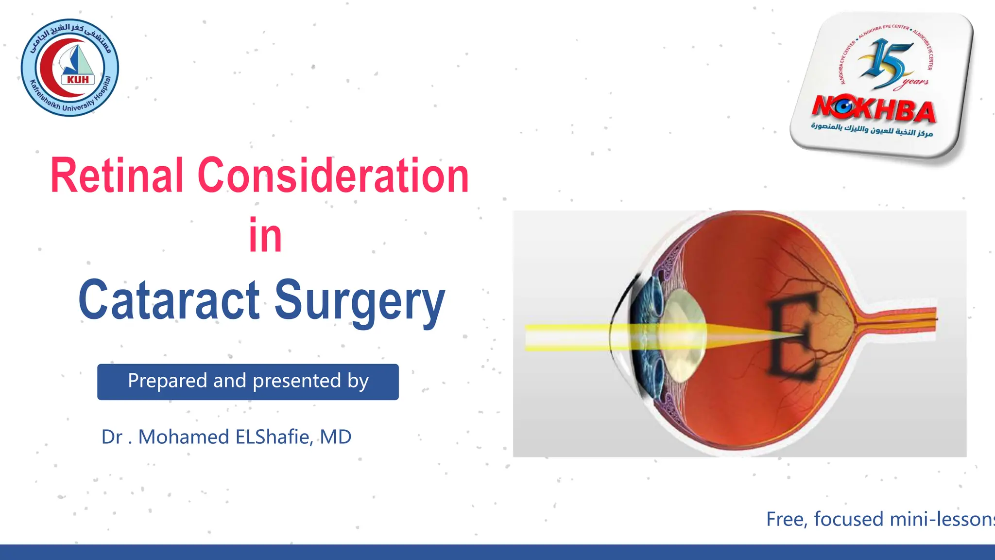
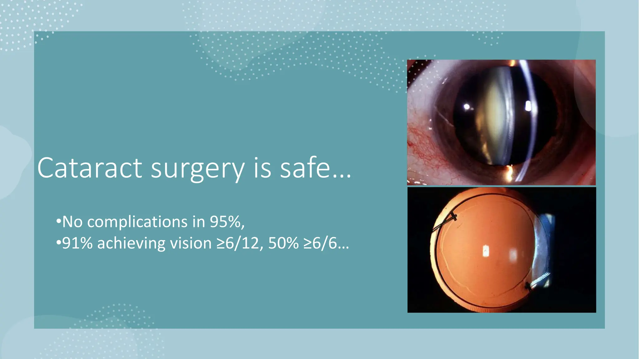
This document discusses considerations for cataract surgery in patients with various retinal conditions. It addresses diabetic macular edema, retinal vein occlusion, age-related macular degeneration, epiretinal membranes, peripheral retinal degenerations, and retinal detachment. For each condition, it outlines risks of cataract surgery exacerbating the underlying retinal disease and recommends treatments and monitoring to minimize risks and optimize visual outcomes following surgery. The document emphasizes the importance of carefully selecting patients and managing postoperative care based on their retinal status and pathology.
This document summarizes David R. Edmison's 25 years of experience at Focus Eye Centre from 1992 to 2016. It outlines the progression of refractive surgery technologies used at the practice over time, including starting with PRK in 1992, adopting LASIK and Intacs in 2000, and advancing to wavefront guided treatments with CustomVue in 2003 and iDesign in 2013. Patient expectations and outcomes have improved with these technological advances. The integrity and credibility of Focus Eye Centre is maintained through ISO certification, low employee turnover, and use of the most advanced equipment.
This study evaluated the outcomes of implanting intrastromal corneal ring segments (ICRS) in 25 eyes of 20 patients with corneal ectasia after refractive surgery. Post-operatively, uncorrected distance visual acuity significantly improved from 20/185 to 20/66 on average and corrected distance visual acuity significantly improved from 20/125 to 20/40 on average. Keratometry and corneal asphericity values also significantly improved. The study found that ICRS implantation can effectively treat corneal ectasia after refractive surgery by improving vision and corneal shape.
Premier IOL choices Technique & Decision Making do we really need femtosecond...presmedaustralia
This document discusses techniques for cataract surgery and considerations for improving patient outcomes and satisfaction. It recommends performing cataract surgery earlier based on symptoms to improve vision and quality of life. Achieving accurate refractive outcomes is emphasized through repeatable capsulorhexis, minimizing induced astigmatism, and using toric and multifocal IOLs. Sources of refractive surprise are examined. Markings and alignment of toric IOLs are covered. Stable lens position is important for predictable results. Multifocal IOLs are presented as an option for reducing glasses dependence.
- The document discusses complications that can occur after cataract surgery, specifically retinal detachment.
- It provides data from studies that show risk factors for retinal detachment include vitreous loss during surgery, posterior capsule tears, history of retinal detachment, high myopia, longer axial length, and Nd:YAG laser capsulotomy.
- The risk of retinal detachment is much higher if the eye is left aphakic after surgery rather than having an intraocular lens implanted, and increases further with additional anterior movement of the vitreous.
Principles and practice of corneal astigmatic surgeryLaurence Sullivan
This document contains a presentation by Dr. Laurie Sullivan on astigmatism correction techniques. It discusses various methods for measuring and quantifying astigmatism, including keratometry, topography, and wavefront analysis. Several options for correcting astigmatism are presented, such as spectacle lenses, contact lenses, refractive surgery techniques like excimer laser and astigmatic keratotomy, and toric intraocular lenses. Guidelines for performing astigmatic keratotomy and considerations for excimer laser treatment of astigmatism are provided.
ABSTRACT- The purpose of this study was to access the outcome of modified manual small incision cataract surgery (M-MSICS) in terms of postoperative visual recovery (Best Corrected Visual Acuity). In this prospective study, the patients having cataracts with nuclear sclerosis not more than early grade 3 were randomly assigned in 2-groups with 50- patients in each group [Group A (C-MSICS), Group B (M-MSICS)]. Both techniques were compared for each stage in terms of postoperative visual recovery (Best Corrected Visual Acuity). Follow ups in postoperative period were carried out on 1st and 3rd postoperative days, 2 weeks, 4 weeks and 6 weeks. Significant early postoperative visual recovery was observed in Modified manual small incision cataract surgery (M-MSICS) as compare to conventional technique. Postoperative surgical induced astigmatism at 6 weeks was significantly less in M-MSICS group (p<0.05%). So it can be concluded that M-MSICS is better technique than C-MSICS in terms of early postoperative visual recovery & less postoperative surgical induced astigmatism.
Key-words- Conventional manual small incision cataract surgery (C-MSICS), Modified manual small incision cataract surgery (M-MSICS), Postoperative visual outcome
Final clinical outcomes of laser refractive surgerySwetha Velpula
This document summarizes an optometrist conference that reviewed laser refractive surgery procedures. It discussed that LASIK can currently treat up to 11 diopters of myopic astigmatism and 4.5-5 diopters of hyperopic astigmatism. It is important to perform thorough preoperative evaluations including corneal topography and pachymetry. Results from the local eye center showed that 76% achieved 6/6 or better unaided vision after LASIK and PRK procedures. Complications were low, with only 2 eyes requiring enhancement procedures.
This document discusses cataracts and cataract surgery. It provides an overview of cataract anatomy, symptoms, and surgery. It describes the risks factors for developing cataracts and recommendations for cataract screening and treatment. The document then focuses on how using a femtosecond laser during cataract surgery can improve precision for steps like corneal incisions, anterior capsulotomy, and lens fragmentation compared to traditional manual techniques. This precision is said to allow for more consistent outcomes and reduced risks.
This document summarizes results from three studies comparing treatments for keratoconus: femtosecond laser-assisted circular keratotomy (FSCKT), corneal crosslinking (CXL), and iontophoresis-assisted corneal crosslinking (ICXL). FSCKT showed improvement in best-corrected visual acuity over 3 years with stable corneal thickness, keratometry, and ectasia values. CXL showed improvements in all outcomes over 5 years. ICXL results after 1 year were less clear, with no significant changes observed. The document concludes CXL may be most effective but longer-term ICXL data is needed, and direct comparative studies could improve comparisons between treatments.
An Optometrist S Personal Perspective Of Aspects Of Refractive SurgeryErin Taylor
- Refractive surgery has evolved significantly over the past century and become a viable alternative to contact lenses or glasses. Techniques like LASIK have improved outcomes and reduced complications compared to earlier methods.
- A prospective study of 104 eyes undergoing LASIK for high myopia found good outcomes with few complications. At 1 year post-op, most patients achieved 6/12 or better vision without aids. Residual error was greatest for those with extreme pre-op myopia.
- While results are encouraging, the author notes limitations remain for treating extreme myopia due to restrictions from minimum safe corneal thickness requirements. Subjective factors also impact patient satisfaction beyond objective visual outcomes.
This document discusses the various techniques used in refractive surgeries to correct vision, including steps for evaluating patients and determining suitability. It describes the history and evolution of refractive surgeries. The main techniques covered are corneal-based procedures like LASIK, PRK, and SMILE as well as lenticular procedures using phakic intraocular lenses. For LASIK specifically, it outlines the surgical technique and potential complications. Contraindications for refractive surgeries are also stated.
This document provides an overview of corneal transplantation techniques from their origins to recent advances. It discusses:
- The first corneal transplant in 1905 and subsequent developments like operating microscopes, finer sutures, and corticosteroids which improved outcomes.
- Why lamellar keratoplasty fell out of favor for decades due to interface irregularities causing poor vision, but has made a comeback as techniques improve vision.
- Recent advances like Descemet's Stripping Endothelial Keratoplasty (DSAEK) and Deep Anterior Lamellar Keratoplasty (DALK) which replace less of the cornea and have benefits like quicker recovery time and less rejection risk compared to
The document summarizes studies on the use of phakic intraocular lenses (ICL) to correct refractive errors in soldiers. A retrospective analysis of 206 ICL implantations at Fort Hood found high rates of visual acuity and few complications. A prospective study compared 95 eyes that received ICL implants to 95 that received LASIK, finding ICL provided better night vision and contrast sensitivity while maintaining high accuracy. Over 1,500 ICL procedures in the military have had no cases of serious complications like retinal detachment. The ICL appears to withstand trauma well in soldiers.
The document summarizes research on using phakic intraocular lenses (ICLs) to correct refractive errors in the military. A study at Fort Hood found the Visian ICL provided excellent visual outcomes for soldiers, with high predictability and safety. A prospective study found ICL patients had better low-light vision than LASIK patients, with greater improvement in low-light contrast sensitivity after ICL. The ICL has proven safe for soldiers over many cases and resistant to potential eye trauma, offering an option when LASIK is not suitable.
The document summarizes studies on the use of phakic intraocular lenses (ICL) to correct refractive errors in soldiers. A retrospective analysis of 206 ICL implantations at Fort Hood found high rates of visual acuity and few complications. A prospective study compared 95 eyes that received ICL implants to 48 eyes that received LASIK, finding ICL provided better night vision and contrast sensitivity while maintaining high accuracy. Over 1,500 ICL procedures at Army centers have shown the procedure withstands trauma well with no reported serious complications.
The Literature Review on Cataract Management in Children - PhdassistancePhD Assistance
PhD Assistance develops Medical coding systems using ICD-10-CM, CPT® framework and many more to support secure access control in Networking platforms. Hiring our experts, you are assured of quality and on-time delivery.The difference in frequency amongst populations is likely owing to higher detection rates in countries with screening programmes (for both cataracts and problems related to cataracts), lower rubella vaccination rates, and population genetic differences.
To Learn More: https://bit.ly/32u4mK2
For Enquiry:
India: +91 91769 66446
UK: +44 7537144372
Email: info@phdassistance.com
The document summarizes updates on corneal cross-linking (CXL) technology from Avedro, including:
1) In March 2015, Avedro received a letter from the FDA requesting further information about equivalency between study devices and commercial systems for CXL to treat keratoconus and ectasia.
2) Agreement was reached on additional measurements required to address the FDA's questions. The application was resubmitted in October 2015.
3) Preliminary data is presented on trials of a new CXL technique called PiXL for treating myopia, keratoconus, and post-cataract refractive error, showing safety and efficacy.
Comparison of the two treatments for Keratoconus shows: iCXL is only a treatment for grade I (and II)
Increase irradiation period: from 30 min to 40 min
- more time for oxygen diffusion
- compensates for epithelial UV-absorbance
The document discusses implantable contact lenses (ICL) for correcting high refractive errors. Key points include:
- ICL is preferred over LASIK for high myopia or thin corneas, as it has fewer risks of complications.
- ICL is made of a biocompatible collamer material and is implanted in the posterior chamber behind the iris.
- A clinical trial found nearly 60% of eyes achieved 20/20 vision and 95% achieved 20/40 vision or better after 3 years with ICL implantation.
- Complications are generally low but can include cataracts, increased eye pressure, damage to the natural lens, and other rare issues like infections. Most risks are
Resultados preliminares do implante de um novo anel associado ao PRK para pre...Ferrara Ophthalmics
Dr. Sandro Coscarelli, Dr. Pablo Rodrigues, Dr. Guilherme Rocha e Dr. Leonardo Torquetti compilaram e compartilham seus resultados com o uso de Segmentos de Anel de Ferrara HM associado ao PRK para a correção da miopia de pacientes com corneas finas e contra indicados para as técnicas de Excimer Laser apenas.
Indication of combined cataract & glaucoma surgery .pptxMdShahjahanSiraj2
Combined cataract and glaucoma surgery can provide benefits of reduced costs, stress, and risks compared to staged surgeries. However, it also carries risks like increased inflammation and complications affecting the success of both procedures. The choice depends on factors like glaucoma severity and damage, medications, prior surgeries, and surgeon expertise. Successful outcomes require minimizing these risks through techniques like two-site surgeries and addressing challenges like poor dilation.
This document provides an overview of a course on using femtosecond lasers for cataract and astigmatism surgery. It discusses the LenSx laser, which was the first femtosecond laser cleared by the FDA for use in cataract surgery. The LenSx laser allows for precise cuts to the anterior capsule, lens fragmentation, and corneal incisions through integrated OCT imaging and a curved patient interface. Studies show benefits like improved capsulotomy accuracy and consistency compared to manual techniques.
Similar to Retinal consideration in cataract surgery (20)
This document provides tips for performing and interpreting ultrasound B-scans of the posterior eye segment. It discusses the basic physics and principles of ultrasound imaging. It outlines indications for B-scans such as opaque ocular media, retinal conditions, and ocular trauma. Tips are provided on probe parts and orientation, different examination techniques, normal anatomical structures, and common pathologies. Examples of B-scan images from case studies are also presented to demonstrate clinical applications and findings. The goal is to make ultrasound imaging of the eye easier to understand.
This document provides information about the anatomy of the orbit and various orbital diseases. It describes the bones that make up each wall of the orbit, including the roof (frontal and sphenoid bones), lateral wall (zygomatic and sphenoid bones), medial wall (ethmoid, lacrimal, maxillary and sphenoid bones), and floor (maxillary, zygomatic and palatine bones). It then discusses dysthyroid orbitopathy as the most common cause of proptosis in adults and its main clinical manifestations. The document also covers orbital cellulitis, its causes including ethmoiditis, and appropriate treatment with antibiotics and drainage if needed. In summary, the document provides an anatomical overview of the
direct ophthalmoscope
fundoscopy course
fundus examination
medical students
ophthalmology
faculty of medicine
kafrelsheikh university
new mansoura university
delta university
Mohamed Ahmed ELShafie is a lecturer of ophthalmology at Harvard Medical School who is the director of training at his unit. The document provides information on viewing the optic nerve head, conditions like papilledema and optic disc cupping, and methods of examining the fundus including fundus biomicroscopy, indirect ophthalmoscopy, and direct ophthalmoscopy. It discusses the advantages and disadvantages of direct ophthalmoscopy, and provides instructions on preparing the ophthalmoscope and examining a patient's fundus directly through dilating the pupils, adjusting the focus and light, and systematically observing the optic disc, vessels, macula, and peripheral fundus.
The document discusses various conditions that affect the conjunctiva. It begins with the anatomy of the conjunctiva, identifying its main structures. It then discusses several infectious conditions that can affect the conjunctiva like acute bacterial conjunctivitis. It also discusses trachoma, an endemic chronic infectious disease of the conjunctiva and cornea in Egypt caused by Chlamydia trachomatis. Other conditions discussed include allergic conjunctivitis, vernal keratoconjunctivitis, phlyctenular keratoconjunctivitis, degenerative changes, subconjunctival hemorrhage, and xerosis.
Mohamed Ahmed ELShafie is a lecturer of ophthalmology at Kafrelsheikh University and alumni of Harvard Medical School. He directs the interns compulsory training program.
An 86-year-old widow, Mrs. Gladys, was referred to psychiatry by her GP for wandering her neighborhood at all hours asking neighbors to shop for her. She had issues with memory, orientation, and constructional praxis. She was found to have macular degeneration and was registered as blind.
A care plan meeting was held with Mrs. Gladys' GP, consultant, community psychiatric nurse, social worker, and day center manager to arrange attendance at a day center twice a week
Acute Limb Weakness
case presentation
PBL session
3rd year
neuro ophthalmology
new mansoura university
A 54-year-old man, Mr. Stephen Smith, was brought by ambulance to the Emergency Department. He had woken up from sleep with slurring of speech and weakness of his right arm and leg. His wife was extremely distressed as Mr Smith had been perfectly well the previous night when he went to sleep. Within 20 minutes after the initial call was made Mr. Smith was admitted to the Emergency Department and was reviewed by the SpR covering the Regional Specialist Stroke Unit. Mr. Smith had been on regular antihypertensive medication (lisinopril) for 8 years. He smoked 5-8 cigarettes a day and was a social drinker consuming about 6 units of alcohol a week. He was not diabetic.
His Serum lipids were checked and was advised to reduce weight and started on a Statin (Simvastatin). There was no family history of hyperlipidaemia but his grandfather died after a Stroke. Mr. Smith had an urgent appendectomy 1 week and made an uneventful recovery. He lives with his wife in a 4-bedroom detached house.
Neurological examination showed that Mr. Smith was fully conscious and alert. He had an upper motor neuron facial palsy on the right side. He had expressive dysphasia but appeared to comprehend speech. He was just able to lift his right arm off the bed for a short period but had no grip. His right leg was weak.
Reflexes on the right side were exaggerated and his right plantar was extensor. He responded to touch and pin prick equally on both sides. He either had visual inattention or a visual field defect on the right side. He had no papilloedema His blood pressure was 164/96, pulse 84 per min, regular. Other systems were entirely normal.
ECG and all routine blood tests were performed. An emergency CT scan was requested and even though the scan was normal the SpR ruled out emergency thrombolysis in this instance. Mr. Smith was admitted to the Acute Stroke Unit and was seen by the Stroke Consultant. Mr. Smith remained fully conscious and alert but had some difficulty in swallowing. Hence an intravenous infusion was commenced and an alternate strategy was adopted for providing his nutritional requirements. A carotid Doppler scan was requested.
After 3 weeks, Mr. Smith seemed to make good progress and the MDT meeting recorded a consistent improvement in his Barthel Index. The Stroke Team met him with his wife and discussed arrangements for discharge home. His wife was keen to know what support measures were available to them when Mr. Smith returned home. She also wanted to know about the risk of a future stroke and how this could be cut down.
ILOs:-
1- Consider the differential diagnosis of speech.
2- Discuss the risk factors for stroke and primary prevention of stroke.
3- Discuss the overall management of a patient with an acute stroke.
4- Complications of stroke
This document discusses the prevalence and grading of epiretinal membrane (ERM) as well as strategies for slowing the progression of diabetic retinopathy. It notes that stringent glycemic control can cause a rapid drop in HbA1c levels leading to deterioration of diabetic retinopathy. ERM is graded on a scale of 1 to 4. Maintaining stable blood sugar levels may help reduce the worsening of retinopathy in diabetes patients.
This document discusses various aspects of assessing and monitoring uveitis, an inflammatory eye condition, through different grading scales and biomarkers. It outlines several methods for clinically grading uveitis based on factors like anterior chamber cells, vitreous haze, and features seen on optical coherence tomography scans like epiretinal membrane thickness and vitreomacular interface changes. Integrity of the ellipsoid zone layer and measurement of choroidal thickness are mentioned as potential biomarkers for monitoring acute choroiditis. Maintaining control of inflammation and comorbidities can help prevent vision loss from conditions like uveitic macular edema.
This document contains contact information for Mohamed ELShafie, including that he is an MD and almuni of HMS, works at KSU Hospital, and encourages the reader to stay connected, updated, and never stop learning.
This document appears to be a presentation by Dr. Mohamed ELShafie, an MD and Harvard alumni, consisting of mini-lessons on various medical topics presented at KSU Hospital, as indicated by the repeated references to KSU Hospital on pages 2 through 18.
This document appears to be a presentation about COVID-19 in children. It includes slides with headings like "COVID19 IN CHILDREN Exposure (Observetional)" and "COVID19 IN CHILDREN Exposure (Experimental)". Other slides reference KSU Hospital and provide examples of how to write a research question in PICO (Population, Intervention, Comparison, Outcome) format. The final slide thanks the audience.
Travel Clinic Cardiff: Health Advice for International TravelersNX Healthcare
Travel Clinic Cardiff offers comprehensive travel health services, including vaccinations, travel advice, and preventive care for international travelers. Our expert team ensures you are well-prepared and protected for your journey, providing personalized consultations tailored to your destination. Conveniently located in Cardiff, we help you travel with confidence and peace of mind. Visit us: www.nxhealthcare.co.uk
5-hydroxytryptamine or 5-HT or Serotonin is a neurotransmitter that serves a range of roles in the human body. It is sometimes referred to as the happy chemical since it promotes overall well-being and happiness.
It is mostly found in the brain, intestines, and blood platelets.
5-HT is utilised to transport messages between nerve cells, is known to be involved in smooth muscle contraction, and adds to overall well-being and pleasure, among other benefits. 5-HT regulates the body's sleep-wake cycles and internal clock by acting as a precursor to melatonin.
It is hypothesised to regulate hunger, emotions, motor, cognitive, and autonomic processes.
Giloy in Ayurveda - Classical Categorization and SynonymsPlanet Ayurveda
Giloy, also known as Guduchi or Amrita in classical Ayurvedic texts, is a revered herb renowned for its myriad health benefits. It is categorized as a Rasayana, meaning it has rejuvenating properties that enhance vitality and longevity. Giloy is celebrated for its ability to boost the immune system, detoxify the body, and promote overall wellness. Its anti-inflammatory, antipyretic, and antioxidant properties make it a staple in managing conditions like fever, diabetes, and stress. The versatility and efficacy of Giloy in supporting health naturally highlight its importance in Ayurveda. At Planet Ayurveda, we provide a comprehensive range of health services and 100% herbal supplements that harness the power of natural ingredients like Giloy. Our products are globally available and affordable, ensuring that everyone can benefit from the ancient wisdom of Ayurveda. If you or your loved ones are dealing with health issues, contact Planet Ayurveda at 01725214040 to book an online video consultation with our professional doctors. Let us help you achieve optimal health and wellness naturally.
- Video recording of this lecture in English language: https://youtu.be/RvdYsTzgQq8
- Video recording of this lecture in Arabic language: https://youtu.be/ECILGWtgZko
- Link to download the book free: https://nephrotube.blogspot.com/p/nephrotube-nephrology-books.html
- Link to NephroTube website: www.NephroTube.com
- Link to NephroTube social media accounts: https://nephrotube.blogspot.com/p/join-nephrotube-on-social-media.html
Nano-gold for Cancer Therapy chemistry investigatory projectSIVAVINAYAKPK
chemistry investigatory project
The development of nanogold-based cancer therapy could revolutionize oncology by providing a more targeted, less invasive treatment option. This project contributes to the growing body of research aimed at harnessing nanotechnology for medical applications, paving the way for future clinical trials and potential commercial applications.
Cancer remains one of the leading causes of death worldwide, prompting the need for innovative treatment methods. Nanotechnology offers promising new approaches, including the use of gold nanoparticles (nanogold) for targeted cancer therapy. Nanogold particles possess unique physical and chemical properties that make them suitable for drug delivery, imaging, and photothermal therapy.
Gene therapy can be broadly defined as the transfer of genetic material to cure a disease or at least to improve the clinical status of a patient.
One of the basic concepts of gene therapy is to transform viruses into genetic shuttles, which will deliver the gene of interest into the target cells.
Safe methods have been devised to do this, using several viral and non-viral vectors.
In the future, this technique may allow doctors to treat a disorder by inserting a gene into a patient's cells instead of using drugs or surgery.
The biggest hurdle faced by medical research in gene therapy is the availability of effective gene-carrying vectors that meet all of the following criteria:
Protection of transgene or genetic cargo from degradative action of systemic and endonucleases,
Delivery of genetic material to the target site, i.e., either cell cytoplasm or nucleus,
Low potential of triggering unwanted immune responses or genotoxicity,
Economical and feasible availability for patients .
Viruses are naturally evolved vehicles that efficiently transfer their genes into host cells.
Choice of viral vector is dependent on gene transfer efficiency, capacity to carry foreign genes, toxicity, stability, immune responses towards viral antigens and potential viral recombination.
There are a wide variety of vectors used to deliver DNA or oligo nucleotides into mammalian cells, either in vitro or in vivo.
The most common vector system based on retroviruses, adenoviruses, herpes simplex viruses, adeno associated viruses.
The Children are very vulnerable to get affected with respiratory disease.
In our country, the respiratory Disease conditions are consider as major cause for mortality and Morbidity in Child.
8. 01
02
03
03
Reports on the effect of
cataract surgery on AMD
progression
but not proving causation
clearly states cataract surgery
did not increase risk of
developing advanced AMD up
to 10 years of follow-up
average gain in VA persisting
for at least 18 months
9. 9 COVID19 IN CHILDREN
Eyes with
mild AMD, significant
cataract
ARMD.
15. 1
4
5
3
2
compromise retinal
monitoring and treatment
Capsular opacification
and contraction
posterior subcapsular
cataracts
develop cataracts
earlier
need good glycaemic
and blood pressure
control before surgery
compromised blood-
aqueous barrier
increases
postoperative
inflammation
Diabetic
patients
16. 01
02
reports increased rates of new
onset retinopathy and worsening
of DME and existing retinopathy
after cataract surgery
double in the first postoperative year
diabetic patients who received
postoperative subconjunctival
injection with triamcinolone
acetonide had a lower macular
thickness and macular volume
at 6 and 12 weeks
postoperatively than patients
who did not.
20. 01
02
When considering cataract
surgery
completely free of retinal ischemia
macular oedema resolved and remains stable
for 2 to 3 months without intervention
03
04
unless the cataract precludes
visualization of the posterior pole
NSAID drops one week prior to surgery
and to continue
Anti- VEGF intraoperative minimise the risk.
05
25. COVID19 IN CHILDREN 25
KSU HOSPITAL
b C
c
proceed with cataract surgery alone.
increased risk of post-operative CMO
ERMs may progress more quickly
following cataract surgery
Treatment considerations
vision is affected primarily
by
Vitrectomy, ERM peeling cataract 3-12 m. later.
Cataract vitrectomy, ERM peeling 3-12 m. later.
phacovitrectomy, ERM peeling.
28. 01
02
When considering
cataract surgery major risk factors for RD
PCO after cataract surgery
03
IOL calculations are less reliable
esp AL > 30mm
04 no benefit in implanting zero power IOL !!!
cataracts and chronic symptomatic floaters.
05
33. 01
02
When considering
cataract surgery Long axial length affecting IOL power
Conjunctival scaring
03
Risk of scleral perforation during
anaesthesia injection
35. 01
02
When considering
cataract surgery Soft globe than normal
Unstable zonules
03 Deep anterior chamber
04 Intraoperative miosis, fluctuation of AC depth
Posterior capsule instability.
05
Cataract progression ?!
06
protects retina against blue light in visible spectrum. cataract protective against development of AMD, making removal of cataract a potential risk factor for AMD development
8.4% of patients who were diagnosed with non neovascular AMD prior to surgery were found to have neovascular changes that went undiagnosed until after cataract surgery due to lens opacity.
similar visual outcomes among advanced AMD sub-types (geographic atrophy/choroidal neovascularization,
recovery of vision when going from daylight to darkness,
defer cataract surgery until the patient has not required intervention for a 90-day period
IV injection 1-2 w before cataract to make sure work of anti vegf in postop period
They wont get back their central vision but appreciate brightness of peripheral vision after operation
dominant eye plano and the non-dominant eye myopic.
Decreased lens volume after cataract surgery causes a forward shift of vitreous and release of angiogenic factors and cytokines.
higher with extracapsular cataract surgery and in patients with combined diabetic retinopathy and hypertensive retinopathy
fluctuations in intraocular pressure (IOP) during the surgery contributed to RVO development
Blurred (cat) or distorted vision (erm)
Foveal contour persreved do cataract only
2 IOL does not stretch large capsular bag reduced posterior capsule adhesion to IOL optic, facilitating lens epithelial cell migration onto posterior capsule
4 physical barrier between anterior and posterior segments. prevent the vitreous from prolapsing into anterior chamber, generate peripheral vitreoretinal traction and cause RD, filter UV light prevent retinal phototoxicity
5 cataract surgery also promotes vitreous liquefaction leading to additional floaters, phacovit
5 Careful hydro dissection like post polar
6 vitreous absorbs oxygen released from retinal circulation and protects lens from oxidation. Following vitrectomy oxygen reaches lens more quickly, acceleration of nuclear cataract in 70% of patients within 3 years of surgery
5 Careful hydro dissection like post polar
6 vitreous absorbs oxygen released from retinal circulation and protects lens from oxidation. Following vitrectomy oxygen reaches lens more quickly, resulting in an acceleration of nuclear cataract in 70% of patients within three years of surgery