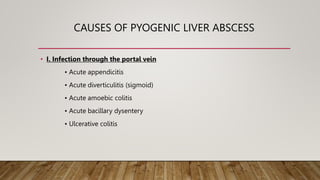
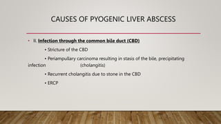
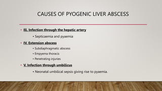
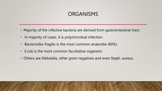
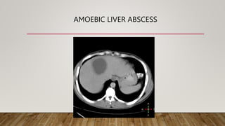
SlideShare a Scribd company logo
LIVER ABSCESS
BY :- DR. SIDDHI VINAYAK
PG RESIDENT
MODERATOR :- DR. SANJAY SHARMA
LIVER ABSCESS
• Collection of PURULENT MATERIAL in the liver parenchyma forming a
CAVITY.
• Most common type of visceral abscess.
• Can be:-
• Bacterial (Pyogenic)
• Parasitic (Amoebic)
PYOGENIC LIVER ABSCESS
• Liver abscess due to Bacteria such as –
• E Coli – Commonest.
• Klebsiella
• Proteus
• Pseudomonas
• Clostridia
• Enterococci, Streptococci
CAUSES OF PYOGENIC LIVER ABSCESS
• I. Infection through the portal vein
• Acute appendicitis
• Acute diverticulitis (sigmoid)
• Acute amoebic colitis
• Acute bacillary dysentery
• Ulcerative colitis
CAUSES OF PYOGENIC LIVER ABSCESS
• II. Infection through the common bile duct (CBD)
• Stricture of the CBD
• Periampullary carcinoma resulting in stasis of the bile, precipitating
infection (cholangitis)
• Recurrent cholangitis due to stone in the CBD
• ERCP
CAUSES OF PYOGENIC LIVER ABSCESS
• Ill. Infection through the hepatic artery
• Septicaemia and pyaemia
• IV. Extension abscess
• Subdiaphragmatic abscess
• Empyema thoracis
• Penetrating injuries
• V. Infection through umbilicus
• Neonatal umbilical sepsis giving rise to pyaemia.
ORGANISMS
• Majority of the infective bacteria are derived from gastrointestinal tract.
• In majority of cases, it is polymicrobial infection.
• Bacteroides fragilis is the most common anaerobe (60%).
• E.coli is the most common facultative organism.
• Others are Klebsiella, other gram negatives and even Staph. aureus.
SIGNS AND SYMPTOMS
• Fever, chills.
• Insidious onset of RUQ pain, epigastric pain
• Occasionally jaundice
• In Late stages, sepsis,
• Weight loss
• Hepatomegaly
• Amebic abscesses tend to have a more protracted course.
CLINICAL FEATURES
• Pain in the right hypochondrjum - 60%.
• High fever, with rigors - 90%.
• Weight loss.
• Jaundice occasionally - 20%.
• Intercostal tenderness.
• Tender, soft liver - 60%.
• Features of toxicity.
• Constitutional symptoms like malaise, lethargy, vomiting.
INVESTIGATIONS
• Total WBC count is raised.
• Stool routine examination: Amoebic cysts, culture and sensitivity for
typhoid bacilli.
• Abdominal ultrasound and ultrasound-guided aspiration establishes the
diagnosis.
• When in doubt, CT scan can be done, followed by FNAC which draws frank
pus. Pus is sent for Gram's stain, culture and sensitivity. CT also helps in
the diagnosis of associated conditions such as diverticulitis of the colon.
DIAGNOSIS
• Ultrasound abdomen, CT scan. Sensitivity is 90% for USG; 97% for contrast CT scan.
• Elevated LFT, low albumin, high PT, Total count.
• Ultrasound guided aspiration of pus after controlling PI.
• Chest X-ray shows elevated diaphragm often with right sided pleural effusion.
• Blood culture is very relevant.
DIFFERENTIAL DIAGNOSIS
TREATMENT
• Systemic antibiotics- combination of third generation cephalo-sporins and
metronidazole.
• Ultrasound guided aspiration/ Pigtail catheter - Percutaneous drainage is the
treatment of choice at present.
• Pus should be sent for culture and sensitivity.
• Follow-up USG,LFT assessment of quantity of daily drainage- should be done to
assess the response.
• 75% of pyogenic abscess is drained percutaneously.
• Occasionally, open drainage is required. Open drainage is indicated in recurrent
abscess, failure of percutaneous drainage, large abscess of size more than 5 cm. Open
drainage is becoming less common at present; but in selected patients it may be a
life-saving essential therapeutic modality.
• Treating the primary causes is very essential.
TREATMENT
• Open (surgical) method
• Laparotomy is required mainly to treat the primary causes, e.g. appendicectomy,
drainage of appendicular abscess.
• If liver shows a significant abscess, it is drained. Alternately, a pigtail catheter is
introduced into the abscess cavity and brought outside through a separate
opening. It helps to drain for a longer period of time.
• Laparoscopic drainage can also be done.
COMPLICATIONS
• Septicemia
• Liver failure
• Rupture (Rare) & Peritonitis can occur.
AMOEBIC LIVER ABSCESS
• Common in India and other tropical countries.
• Caused by parasite Entamoeba histolytica.
• More common in alcoholics and cirrhotic patients.
• Commonest extraintestinal presentation (8-10%) of amoebiasis.
• Infection commonly occurs from the caecum after an attack of amoebic typhlitis
through the superior mesenteric vein and portal vein. Infection from sigmoid
colon spreads through the inferior mesenteric vein and portal vein to liver.
AMOEBIC LIVER ABSCESS
• Right lobe is commonly involved over posterosuperior surface - because of streamline elect and larger size of the
right lobe.
• Trophozoites destroy the hepatocytes by releasing histolysin a cytolytic agent.
• It causes amoebic hepatitis with multiple micro abscesses formation.
• It leads into liquefaction necrosis, thrombosis of blood vessels. release and, breaking of red cells. It causes formation
of “Anchovy sauce” pus which is chocolate brown colored and odorless.
• Contains dead liver cells, RBCs, necrotic material (But no WBC)
• Secondary infection is common (30%).
• Amoebic abscess is usually sterile unless infected.
AMOEBIC LIVER ABSCESS
• In majority of the cases, pus is sterile.
• Secondary infection occurs in about 20 to 30% of the cases.
• Amoebae are rarely present in the pus but are present in the wall of the abscess
cavity. The wall contains monocytes, plasma cells, lymphocytes and fibroblasts.
• Abscesses are multiple which fuse to form a single large abscess cavity in about
70% of the cases. Due to perihepatitis, abscess gets fixed to the diaphragm
resulting in immobility of the diaphragm.
LIFE CYCLE OF ENTAMOEBA HISTOLYTICA
CLINICAL FEATURES
• It is common in males (10:1)
• H/o amoebic dysentery
• Fever
• Loss of weight
• Chills and rigors,
• Non- productive cough, shoulder pain.
• Pain in the right hypochondrium -30%. Soft, tender, smooth, liver with increased liver span--70%.
• Intercostal tenderness is elicited which is a useful clinical sign.
• Right sided pleural effusion may be evident. Mild jaundice
• In chronic amoebic liver abscess, smooth, firm/hard, nontender liver may be palpable.
INVESTIGATIONS
• Increased TLC.
• Low Hb% in 50% çases.
• Altered Liver function tests (altered bilirubin and albumin level).
• Prothrombin time may be widened and if it is so injection vitamin K 10 mg IM or IV for 5 days should be given.
• Even with this if P.T. remains widened then fresh frozen plasma (FFP) is needed to rectify the P.T. Serum alkaline
phosphatase, SGPT, SGOT levels are altered.
• US abdomen shows altered echogenicity (anechogenic, hypoechogenic), size, location, number of abscess, nature of
the liver- 90% sensitivity.
• Chest X-ray findings: Raised fixed diaphragm (tenting) ,Pleural effusion & Soft tissue shadow
INVESTIGATIONS
• CT scan shows raised diaphragm; abscess cavity (low density area) -its size, location, number;
presence of effusion; changes in the lung- -95% sensitivity.
• Sigmoidoscopy/colonoscopy are used to identify the active ulcers. Scrapings of the ulcer show
trophozoites.
TREATMENT
• I. Conservative line of management •
• It is indicated in amoebic hepatitis.
• Tab. Metronidazole 400-800 mg, 3 times a day is given for 14 days. The only
recognisable side-effect is metallic taste.
• If the condition does not improve, injection dryhydroemetine1.5 mg/kg body
weight to a total of 60 mg/day IM for 5 days is given.
• Choloroquine 250mg BD for 10-14 days.
TREATMENT
• II. USG-guided needle aspiration/pigtail catheter drainage
• It is indicated in cases of amoebic liver abscess (large abscess >10cm).
• Before it is aspirated, bleeding profile (BT, CT, PT) should be normal and injection
vitamin K 10 mg, IM should be given for at least 3 days.
• US-guided aspiration is also the treatment of choice where metronidazole is
contraindicated, e.g. 1st trimester of pregnancy.
• It can be easily done under local anaesthesia
• Can be repeated, if pus recollects.
• Typically it is anchovy sauce pus. Aspiration is followed by insertion of pig tail
catheter. Before removal of the catheter do a repeat ultrasound to check for residual
pus.
TREATMENT
• Ill. Surgery (open drainage) and laparoscopic:-
• Indications –
• 1. Failure of US-guided needle aspiration.
• 2. Ruptured amoebic liver abscess with amoebic peritonitis.
COMPLICATIONS
• Amoebic peritonitis, resulting in acute abdomen with shock. It has to be treated
like any peritonitis- laparotomy, drainage of pus and drain the abscess cavity to
outside (possibility of amebiasis cutis is still present but rare).
• Rupture into pleural space causing pleural effusion.
• Rupture into the bronchus resulting in coughing out anchovy sauce (may be a
natural cure) -bronchopleural fistula.
• Amoebic pericardial effusion occurs due to rupture of left liver lobe abscess into
pericardial space.
HYDATID CYST OF LIVER
HYDATID CYST OF THE LIVER
• The disease is caused by Echinococcus granulosus, transmitted by dogs which
are the chief mediators (host) and man is the intermediate host. After swallowing
the ova, they penetrate gastric mucosa , reach retroperitoneal structures,
penetrate portal vein directly and then enter into liver.
• Having reached liver, the organisms grow and develop their own protective layer
and form hydatid cyst.
LAYERS OF THE HYDATID CYST
• 1. The adventitia (pseudocyst) : This is the fibrous layer derived from the liver
tissue. It is the reaction of liver to the parasite. It is adherent to the liver and
cannot be separated, also known as pericyst.
• 2. The ectocyst (laminated membrane): It is white and elastic and is produced by
endocyst. It is this layer which gets peeled off at surgery.
• 3. The endocyst: This is called germinal epithelium and it is the innermost part of
hydatid cyst. It secretes hydatid fluid inside and ectocyst outside. Within the
hydatid fluid, scolices of Echinococcus granulosus develop.
CLINICAL FEATURES
• It can be silent-without any symptoms throughout life, accidentally discovered on
routine examination.
• Dragging pain in the upper abdomen due to hepatomegaly.
• Liver is enlarged, has a smooth surface, and is nontender.
• Typical hydatid thrill can be present on rare occasions. Hydatid thrill is demonstrated
by 3-finger method. Keep 3 fingers over the liver, percuss over the middle finger and
get the impulse by other 2 fingers (fluid thrill).
• Patient may present as an emergency with severe abdominal pain following minor
trauma.
• May present as an emergency with features of anaphylactic shock without any
obvious cause.
INVESTIGATIONS
• 1. USG can detect the cyst, localise the cyst and is used for aspiration purposes.
• 2. Plain X-ray abdomen may demonstrate speckled calcification.
• 3. CT scan may be necessary in selected cases. The cyst which is superficial and
has reached surface, should be operated upon.
• 4. ERCP if there is obstructive jaundice-in such cases, a wide sphincterotomy
should be given so as to allow free drainage of the hydatid contents into the
duodenum.
• 5. Cason's intradermal test: Sensitivity and specificity of this test is low and
hence, no longer used.
• 6. ELISA and immunoelectrophoresis may point towards the diagnosis.
HASSEN GHARBI'S ULTRASOUND BASED
CLASSIFICATION OF LIVER HYDATID CYSTS (1981)
• Type 1 - Pure fluid collection
• Type 2 - Fluid collection with split wall
• Type 3 - Fluid collection with septa
• Type 4 - Heterogeneous appearance
• Type 5- Reflecting thick walls
WHO CLASSIFICATION OF LIVER HYDATID
CYST
• Type - CL - Active; unilocular; no cyst wall; early stage; not fertile.
• Type - CE 1 - Active; cyst wall present; hydatid sand present; fertile.
• Type - CE 2 - Active; multivesicular rosette like cyst wall; fertile.
• Type-CE3 - Transitional; detaching laminated membrane; water-lily sign;
beginning of degeneration
• Type - CE 4 - Inactive; degenerative contents; no daughter cysts; not fertile
• Type - CE 5 - Inactive; thick calcified wall; not fertile
TREATMENT - CONSERVATIVE
• Calcified cysts are dead cysts. They are left alone.
• Symptomless, small hydatid cyst can be left alone. Once symptomatic, or if the
size is more than 5 cm, they may be treated.
• Medical treatment: • Albendazole 8 mg/kg or 400 mg BD is given for 21* days
followed by drug holiday for 2 weeks.
• If no improvement occurs maximum of 3 such cycles can be given.
TREATMENT - SURGERY
• 1. Laparotomy and isolation of the cyst from peritoneal cavity by packs dipped in
hypochlorite solution.
• 2. Aspirate the contents and inject scolicidal agents such as salon or hypertonic
saline.
• 3. Incise the cyst, peel off the laminated membrane by using sponge-holding forceps.
• 4. An attempt to remove adventitial layer may result in bleeding. It need not be
removed.
• 5. At surgery, all laminated membrane, endocyst has to be removed. Perfect
haemostasis must be achieved. Any bile leak to be sutured. Postoperative significant
bile leak is managed by ERCP stenting.
TREATMENT - PRECAUTIONS
• Albendazole should be given before surgery.
• Avoid spillage into the peritoneal cavity to avoid peritoneal hydatid.
• Injection hydrocortisone 100 mg IV before, during and after surgery to avoid
anaphylactic shock.
TREATMENT - PERCUTANEOUS DRAINAGE
• It is done after taking all necessary precautions/equipment with all emergency
drugs and with CT/US guidance.
• It is also called PAIR: Puncture, Aspiration, Injection and Reaspiration.

More Related Content

What's hot (20)

Choledochal cyst
Choledochal cystCholedochal cyst
Choledochal cyst
 
Volvulus in git
Volvulus in gitVolvulus in git
Volvulus in git
 
Bile duct injury
Bile duct injuryBile duct injury
Bile duct injury
 
Intestinal Fistula
Intestinal FistulaIntestinal Fistula
Intestinal Fistula
 
Intestinal obstruction by Dr.Usman Haqqani
Intestinal obstruction by Dr.Usman HaqqaniIntestinal obstruction by Dr.Usman Haqqani
Intestinal obstruction by Dr.Usman Haqqani
 
Liver trauma
Liver traumaLiver trauma
Liver trauma
 
Renal trauma
Renal traumaRenal trauma
Renal trauma
 
Lower GI bleed
Lower GI bleedLower GI bleed
Lower GI bleed
 
Shock
ShockShock
Shock
 
Acute peritonitis
Acute peritonitisAcute peritonitis
Acute peritonitis
 
Acute cholecystitis
Acute cholecystitisAcute cholecystitis
Acute cholecystitis
 
Enterocutaneous fistula
Enterocutaneous fistulaEnterocutaneous fistula
Enterocutaneous fistula
 
LOWER GI BLEEDING
LOWER GI BLEEDINGLOWER GI BLEEDING
LOWER GI BLEEDING
 
Bladder tumor
Bladder tumorBladder tumor
Bladder tumor
 
Biliary stricture ppt
Biliary stricture pptBiliary stricture ppt
Biliary stricture ppt
 
Bladder outlet obstruction
Bladder  outlet obstructionBladder  outlet obstruction
Bladder outlet obstruction
 
Acute abdomen
Acute abdomenAcute abdomen
Acute abdomen
 
Post gastrectomy syndrome
Post gastrectomy syndrome   Post gastrectomy syndrome
Post gastrectomy syndrome
 
Urinary bladder trauma.pptx
Urinary bladder trauma.pptxUrinary bladder trauma.pptx
Urinary bladder trauma.pptx
 
Choledocholithiasis
CholedocholithiasisCholedocholithiasis
Choledocholithiasis
 

Similar to LIVER ABSCESS.pptx

Similar to LIVER ABSCESS.pptx (20)

LIVER INFECTIONS.pptx
LIVER INFECTIONS.pptxLIVER INFECTIONS.pptx
LIVER INFECTIONS.pptx
 
AMOEBIC LIVER ABSCESS
AMOEBIC LIVER ABSCESSAMOEBIC LIVER ABSCESS
AMOEBIC LIVER ABSCESS
 
Liver abcess
Liver abcessLiver abcess
Liver abcess
 
Cystic diseases of liver
Cystic diseases of liverCystic diseases of liver
Cystic diseases of liver
 
Cystic diseases of liver
Cystic diseases of liverCystic diseases of liver
Cystic diseases of liver
 
Liver abscess
Liver abscessLiver abscess
Liver abscess
 
Short review of genitourinary tuberculosis
Short review of genitourinary tuberculosisShort review of genitourinary tuberculosis
Short review of genitourinary tuberculosis
 
LIVER & GALLBLADDER PATHOLOGY
LIVER & GALLBLADDER PATHOLOGYLIVER & GALLBLADDER PATHOLOGY
LIVER & GALLBLADDER PATHOLOGY
 
THE LIVER: LIVER ABSCESS
THE LIVER: LIVER ABSCESSTHE LIVER: LIVER ABSCESS
THE LIVER: LIVER ABSCESS
 
Tuberculosis in surgery
Tuberculosis in surgeryTuberculosis in surgery
Tuberculosis in surgery
 
Liver Cysts and Abscesses
Liver Cysts and AbscessesLiver Cysts and Abscesses
Liver Cysts and Abscesses
 
PYOGENIC LIVER ABSCESS.pptx
PYOGENIC LIVER ABSCESS.pptxPYOGENIC LIVER ABSCESS.pptx
PYOGENIC LIVER ABSCESS.pptx
 
Liver Abscess and Hydatid Cyst, Surgery
Liver Abscess and Hydatid Cyst, SurgeryLiver Abscess and Hydatid Cyst, Surgery
Liver Abscess and Hydatid Cyst, Surgery
 
Benign liver masses
Benign liver massesBenign liver masses
Benign liver masses
 
LIVER ABSCESS.pptx
LIVER ABSCESS.pptxLIVER ABSCESS.pptx
LIVER ABSCESS.pptx
 
Liver abscess .pptx
Liver abscess .pptxLiver abscess .pptx
Liver abscess .pptx
 
Diseases of the liver
Diseases of the liverDiseases of the liver
Diseases of the liver
 
lower git bleeding
lower git bleedinglower git bleeding
lower git bleeding
 
Liver abscess.pptx
Liver abscess.pptxLiver abscess.pptx
Liver abscess.pptx
 
AMOEBIASIS
AMOEBIASISAMOEBIASIS
AMOEBIASIS
 

More from SayanRDey

Anatomy of lower limb
Anatomy  of lower  limbAnatomy  of lower  limb
Anatomy of lower limbSayanRDey
 
abdomen_J.ppt
abdomen_J.pptabdomen_J.ppt
abdomen_J.pptSayanRDey
 
ulcer-190501174026.pdf
ulcer-190501174026.pdfulcer-190501174026.pdf
ulcer-190501174026.pdfSayanRDey
 
Final heart development.pptx
Final heart development.pptxFinal heart development.pptx
Final heart development.pptxSayanRDey
 
salivary glands-csu.ppt
salivary glands-csu.pptsalivary glands-csu.ppt
salivary glands-csu.pptSayanRDey
 

More from SayanRDey (7)

Anatomy of lower limb
Anatomy  of lower  limbAnatomy  of lower  limb
Anatomy of lower limb
 
abdomen_J.ppt
abdomen_J.pptabdomen_J.ppt
abdomen_J.ppt
 
Stomach
StomachStomach
Stomach
 
ulcer-190501174026.pdf
ulcer-190501174026.pdfulcer-190501174026.pdf
ulcer-190501174026.pdf
 
Final heart development.pptx
Final heart development.pptxFinal heart development.pptx
Final heart development.pptx
 
TPN.pptx
TPN.pptxTPN.pptx
TPN.pptx
 
salivary glands-csu.ppt
salivary glands-csu.pptsalivary glands-csu.ppt
salivary glands-csu.ppt
 

Recently uploaded

Presentació "Advancing Emergency Medicine Education through Virtual Reality"
Presentació "Advancing Emergency Medicine Education through Virtual Reality"Presentació "Advancing Emergency Medicine Education through Virtual Reality"
Presentació "Advancing Emergency Medicine Education through Virtual Reality"Badalona Serveis Assistencials
 
Book Trailer: PGMEE in a Nutshell (CEE MD/MS PG Entrance Examination)
Book Trailer: PGMEE in a Nutshell (CEE MD/MS PG Entrance Examination)Book Trailer: PGMEE in a Nutshell (CEE MD/MS PG Entrance Examination)
Book Trailer: PGMEE in a Nutshell (CEE MD/MS PG Entrance Examination)Dr. Aryan (Anish Dhakal)
 
180-hour Power Capsules For Men In Ghana
180-hour Power Capsules For Men In Ghana180-hour Power Capsules For Men In Ghana
180-hour Power Capsules For Men In Ghanahealthwatchghana
 
DECIPHERING COMMON ECG FINDINGS IN ED.pptx
DECIPHERING COMMON ECG FINDINGS IN ED.pptxDECIPHERING COMMON ECG FINDINGS IN ED.pptx
DECIPHERING COMMON ECG FINDINGS IN ED.pptxdrwaque
 
Final CAPNOCYTOPHAGA INFECTION by Gauri Gawande.pptx
Final CAPNOCYTOPHAGA INFECTION by Gauri Gawande.pptxFinal CAPNOCYTOPHAGA INFECTION by Gauri Gawande.pptx
Final CAPNOCYTOPHAGA INFECTION by Gauri Gawande.pptxgauripg8
 
Fundamental of Radiobiology -SABBU.pptx
Fundamental of Radiobiology  -SABBU.pptxFundamental of Radiobiology  -SABBU.pptx
Fundamental of Radiobiology -SABBU.pptxSabbu Khatoon
 
Arterial health throughout cancer treatment and exercise rehabilitation in wo...
Arterial health throughout cancer treatment and exercise rehabilitation in wo...Arterial health throughout cancer treatment and exercise rehabilitation in wo...
Arterial health throughout cancer treatment and exercise rehabilitation in wo...Catherine Liao
 
linearity concept of significance, standard deviation, chi square test, stude...
linearity concept of significance, standard deviation, chi square test, stude...linearity concept of significance, standard deviation, chi square test, stude...
linearity concept of significance, standard deviation, chi square test, stude...KavyasriPuttamreddy
 
TEST BANK For Wong’s Essentials of Pediatric Nursing, 11th Edition by Marilyn...
TEST BANK For Wong’s Essentials of Pediatric Nursing, 11th Edition by Marilyn...TEST BANK For Wong’s Essentials of Pediatric Nursing, 11th Edition by Marilyn...
TEST BANK For Wong’s Essentials of Pediatric Nursing, 11th Edition by Marilyn...kevinkariuki227
 
Alcohol_Dr. Jeenal Mistry MD Pharmacology.pdf
Alcohol_Dr. Jeenal Mistry MD Pharmacology.pdfAlcohol_Dr. Jeenal Mistry MD Pharmacology.pdf
Alcohol_Dr. Jeenal Mistry MD Pharmacology.pdfDr Jeenal Mistry
 
Effects of vaping e-cigarettes on arterial health
Effects of vaping e-cigarettes on arterial healthEffects of vaping e-cigarettes on arterial health
Effects of vaping e-cigarettes on arterial healthCatherine Liao
 
Lung Cancer: Artificial Intelligence, Synergetics, Complex System Analysis, S...
Lung Cancer: Artificial Intelligence, Synergetics, Complex System Analysis, S...Lung Cancer: Artificial Intelligence, Synergetics, Complex System Analysis, S...
Lung Cancer: Artificial Intelligence, Synergetics, Complex System Analysis, S...Oleg Kshivets
 
Blue Printing in medical education by Dr.Mumtaz Ali.pptx
Blue Printing in medical education by Dr.Mumtaz Ali.pptxBlue Printing in medical education by Dr.Mumtaz Ali.pptx
Blue Printing in medical education by Dr.Mumtaz Ali.pptxNeurosurgeon Mumtaz Ali Narejo
 
The hemodynamic and autonomic determinants of elevated blood pressure in obes...
The hemodynamic and autonomic determinants of elevated blood pressure in obes...The hemodynamic and autonomic determinants of elevated blood pressure in obes...
The hemodynamic and autonomic determinants of elevated blood pressure in obes...Catherine Liao
 
Anuman- An inference for helpful in diagnosis and treatment
Anuman- An inference for helpful in diagnosis and treatmentAnuman- An inference for helpful in diagnosis and treatment
Anuman- An inference for helpful in diagnosis and treatmentabdeli bhadarva
 
Antiplatelets in IHD, Dose Duration, DAPT vs SAPT
Antiplatelets in IHD, Dose Duration, DAPT vs SAPTAntiplatelets in IHD, Dose Duration, DAPT vs SAPT
Antiplatelets in IHD, Dose Duration, DAPT vs SAPTAkashGanganePatil1
 
Is preeclampsia and spontaneous preterm delivery associate with vascular and ...
Is preeclampsia and spontaneous preterm delivery associate with vascular and ...Is preeclampsia and spontaneous preterm delivery associate with vascular and ...
Is preeclampsia and spontaneous preterm delivery associate with vascular and ...Catherine Liao
 
Scientificity and feasibility study of non-invasive central arterial pressure...
Scientificity and feasibility study of non-invasive central arterial pressure...Scientificity and feasibility study of non-invasive central arterial pressure...
Scientificity and feasibility study of non-invasive central arterial pressure...Catherine Liao
 
Prix Galien International 2024 Forum Program
Prix Galien International 2024 Forum ProgramPrix Galien International 2024 Forum Program
Prix Galien International 2024 Forum ProgramLevi Shapiro
 

Recently uploaded (20)

Presentació "Advancing Emergency Medicine Education through Virtual Reality"
Presentació "Advancing Emergency Medicine Education through Virtual Reality"Presentació "Advancing Emergency Medicine Education through Virtual Reality"
Presentació "Advancing Emergency Medicine Education through Virtual Reality"
 
Book Trailer: PGMEE in a Nutshell (CEE MD/MS PG Entrance Examination)
Book Trailer: PGMEE in a Nutshell (CEE MD/MS PG Entrance Examination)Book Trailer: PGMEE in a Nutshell (CEE MD/MS PG Entrance Examination)
Book Trailer: PGMEE in a Nutshell (CEE MD/MS PG Entrance Examination)
 
180-hour Power Capsules For Men In Ghana
180-hour Power Capsules For Men In Ghana180-hour Power Capsules For Men In Ghana
180-hour Power Capsules For Men In Ghana
 
DECIPHERING COMMON ECG FINDINGS IN ED.pptx
DECIPHERING COMMON ECG FINDINGS IN ED.pptxDECIPHERING COMMON ECG FINDINGS IN ED.pptx
DECIPHERING COMMON ECG FINDINGS IN ED.pptx
 
Final CAPNOCYTOPHAGA INFECTION by Gauri Gawande.pptx
Final CAPNOCYTOPHAGA INFECTION by Gauri Gawande.pptxFinal CAPNOCYTOPHAGA INFECTION by Gauri Gawande.pptx
Final CAPNOCYTOPHAGA INFECTION by Gauri Gawande.pptx
 
Fundamental of Radiobiology -SABBU.pptx
Fundamental of Radiobiology  -SABBU.pptxFundamental of Radiobiology  -SABBU.pptx
Fundamental of Radiobiology -SABBU.pptx
 
Arterial health throughout cancer treatment and exercise rehabilitation in wo...
Arterial health throughout cancer treatment and exercise rehabilitation in wo...Arterial health throughout cancer treatment and exercise rehabilitation in wo...
Arterial health throughout cancer treatment and exercise rehabilitation in wo...
 
linearity concept of significance, standard deviation, chi square test, stude...
linearity concept of significance, standard deviation, chi square test, stude...linearity concept of significance, standard deviation, chi square test, stude...
linearity concept of significance, standard deviation, chi square test, stude...
 
TEST BANK For Wong’s Essentials of Pediatric Nursing, 11th Edition by Marilyn...
TEST BANK For Wong’s Essentials of Pediatric Nursing, 11th Edition by Marilyn...TEST BANK For Wong’s Essentials of Pediatric Nursing, 11th Edition by Marilyn...
TEST BANK For Wong’s Essentials of Pediatric Nursing, 11th Edition by Marilyn...
 
Alcohol_Dr. Jeenal Mistry MD Pharmacology.pdf
Alcohol_Dr. Jeenal Mistry MD Pharmacology.pdfAlcohol_Dr. Jeenal Mistry MD Pharmacology.pdf
Alcohol_Dr. Jeenal Mistry MD Pharmacology.pdf
 
Effects of vaping e-cigarettes on arterial health
Effects of vaping e-cigarettes on arterial healthEffects of vaping e-cigarettes on arterial health
Effects of vaping e-cigarettes on arterial health
 
Lung Cancer: Artificial Intelligence, Synergetics, Complex System Analysis, S...
Lung Cancer: Artificial Intelligence, Synergetics, Complex System Analysis, S...Lung Cancer: Artificial Intelligence, Synergetics, Complex System Analysis, S...
Lung Cancer: Artificial Intelligence, Synergetics, Complex System Analysis, S...
 
Blue Printing in medical education by Dr.Mumtaz Ali.pptx
Blue Printing in medical education by Dr.Mumtaz Ali.pptxBlue Printing in medical education by Dr.Mumtaz Ali.pptx
Blue Printing in medical education by Dr.Mumtaz Ali.pptx
 
The hemodynamic and autonomic determinants of elevated blood pressure in obes...
The hemodynamic and autonomic determinants of elevated blood pressure in obes...The hemodynamic and autonomic determinants of elevated blood pressure in obes...
The hemodynamic and autonomic determinants of elevated blood pressure in obes...
 
Anuman- An inference for helpful in diagnosis and treatment
Anuman- An inference for helpful in diagnosis and treatmentAnuman- An inference for helpful in diagnosis and treatment
Anuman- An inference for helpful in diagnosis and treatment
 
Antiplatelets in IHD, Dose Duration, DAPT vs SAPT
Antiplatelets in IHD, Dose Duration, DAPT vs SAPTAntiplatelets in IHD, Dose Duration, DAPT vs SAPT
Antiplatelets in IHD, Dose Duration, DAPT vs SAPT
 
Is preeclampsia and spontaneous preterm delivery associate with vascular and ...
Is preeclampsia and spontaneous preterm delivery associate with vascular and ...Is preeclampsia and spontaneous preterm delivery associate with vascular and ...
Is preeclampsia and spontaneous preterm delivery associate with vascular and ...
 
Contact dermaititis (irritant and allergic).pdf
Contact dermaititis (irritant and allergic).pdfContact dermaititis (irritant and allergic).pdf
Contact dermaititis (irritant and allergic).pdf
 
Scientificity and feasibility study of non-invasive central arterial pressure...
Scientificity and feasibility study of non-invasive central arterial pressure...Scientificity and feasibility study of non-invasive central arterial pressure...
Scientificity and feasibility study of non-invasive central arterial pressure...
 
Prix Galien International 2024 Forum Program
Prix Galien International 2024 Forum ProgramPrix Galien International 2024 Forum Program
Prix Galien International 2024 Forum Program
 

LIVER ABSCESS.pptx

  • 1. LIVER ABSCESS BY :- DR. SIDDHI VINAYAK PG RESIDENT MODERATOR :- DR. SANJAY SHARMA
  • 2. LIVER ABSCESS • Collection of PURULENT MATERIAL in the liver parenchyma forming a CAVITY. • Most common type of visceral abscess. • Can be:- • Bacterial (Pyogenic) • Parasitic (Amoebic)
  • 4. PYOGENIC LIVER ABSCESS • Liver abscess due to Bacteria such as – • E Coli – Commonest. • Klebsiella • Proteus • Pseudomonas • Clostridia • Enterococci, Streptococci
  • 5. CAUSES OF PYOGENIC LIVER ABSCESS • I. Infection through the portal vein • Acute appendicitis • Acute diverticulitis (sigmoid) • Acute amoebic colitis • Acute bacillary dysentery • Ulcerative colitis
  • 6. CAUSES OF PYOGENIC LIVER ABSCESS • II. Infection through the common bile duct (CBD) • Stricture of the CBD • Periampullary carcinoma resulting in stasis of the bile, precipitating infection (cholangitis) • Recurrent cholangitis due to stone in the CBD • ERCP
  • 7. CAUSES OF PYOGENIC LIVER ABSCESS • Ill. Infection through the hepatic artery • Septicaemia and pyaemia • IV. Extension abscess • Subdiaphragmatic abscess • Empyema thoracis • Penetrating injuries • V. Infection through umbilicus • Neonatal umbilical sepsis giving rise to pyaemia.
  • 8. ORGANISMS • Majority of the infective bacteria are derived from gastrointestinal tract. • In majority of cases, it is polymicrobial infection. • Bacteroides fragilis is the most common anaerobe (60%). • E.coli is the most common facultative organism. • Others are Klebsiella, other gram negatives and even Staph. aureus.
  • 9. SIGNS AND SYMPTOMS • Fever, chills. • Insidious onset of RUQ pain, epigastric pain • Occasionally jaundice • In Late stages, sepsis, • Weight loss • Hepatomegaly • Amebic abscesses tend to have a more protracted course.
  • 10. CLINICAL FEATURES • Pain in the right hypochondrjum - 60%. • High fever, with rigors - 90%. • Weight loss. • Jaundice occasionally - 20%. • Intercostal tenderness. • Tender, soft liver - 60%. • Features of toxicity. • Constitutional symptoms like malaise, lethargy, vomiting.
  • 11. INVESTIGATIONS • Total WBC count is raised. • Stool routine examination: Amoebic cysts, culture and sensitivity for typhoid bacilli. • Abdominal ultrasound and ultrasound-guided aspiration establishes the diagnosis. • When in doubt, CT scan can be done, followed by FNAC which draws frank pus. Pus is sent for Gram's stain, culture and sensitivity. CT also helps in the diagnosis of associated conditions such as diverticulitis of the colon.
  • 12. DIAGNOSIS • Ultrasound abdomen, CT scan. Sensitivity is 90% for USG; 97% for contrast CT scan. • Elevated LFT, low albumin, high PT, Total count. • Ultrasound guided aspiration of pus after controlling PI. • Chest X-ray shows elevated diaphragm often with right sided pleural effusion. • Blood culture is very relevant.
  • 14. TREATMENT • Systemic antibiotics- combination of third generation cephalo-sporins and metronidazole. • Ultrasound guided aspiration/ Pigtail catheter - Percutaneous drainage is the treatment of choice at present. • Pus should be sent for culture and sensitivity. • Follow-up USG,LFT assessment of quantity of daily drainage- should be done to assess the response. • 75% of pyogenic abscess is drained percutaneously. • Occasionally, open drainage is required. Open drainage is indicated in recurrent abscess, failure of percutaneous drainage, large abscess of size more than 5 cm. Open drainage is becoming less common at present; but in selected patients it may be a life-saving essential therapeutic modality. • Treating the primary causes is very essential.
  • 15. TREATMENT • Open (surgical) method • Laparotomy is required mainly to treat the primary causes, e.g. appendicectomy, drainage of appendicular abscess. • If liver shows a significant abscess, it is drained. Alternately, a pigtail catheter is introduced into the abscess cavity and brought outside through a separate opening. It helps to drain for a longer period of time. • Laparoscopic drainage can also be done.
  • 16. COMPLICATIONS • Septicemia • Liver failure • Rupture (Rare) & Peritonitis can occur.
  • 18. AMOEBIC LIVER ABSCESS • Common in India and other tropical countries. • Caused by parasite Entamoeba histolytica. • More common in alcoholics and cirrhotic patients. • Commonest extraintestinal presentation (8-10%) of amoebiasis. • Infection commonly occurs from the caecum after an attack of amoebic typhlitis through the superior mesenteric vein and portal vein. Infection from sigmoid colon spreads through the inferior mesenteric vein and portal vein to liver.
  • 19. AMOEBIC LIVER ABSCESS • Right lobe is commonly involved over posterosuperior surface - because of streamline elect and larger size of the right lobe. • Trophozoites destroy the hepatocytes by releasing histolysin a cytolytic agent. • It causes amoebic hepatitis with multiple micro abscesses formation. • It leads into liquefaction necrosis, thrombosis of blood vessels. release and, breaking of red cells. It causes formation of “Anchovy sauce” pus which is chocolate brown colored and odorless. • Contains dead liver cells, RBCs, necrotic material (But no WBC) • Secondary infection is common (30%). • Amoebic abscess is usually sterile unless infected.
  • 20. AMOEBIC LIVER ABSCESS • In majority of the cases, pus is sterile. • Secondary infection occurs in about 20 to 30% of the cases. • Amoebae are rarely present in the pus but are present in the wall of the abscess cavity. The wall contains monocytes, plasma cells, lymphocytes and fibroblasts. • Abscesses are multiple which fuse to form a single large abscess cavity in about 70% of the cases. Due to perihepatitis, abscess gets fixed to the diaphragm resulting in immobility of the diaphragm.
  • 21. LIFE CYCLE OF ENTAMOEBA HISTOLYTICA
  • 22. CLINICAL FEATURES • It is common in males (10:1) • H/o amoebic dysentery • Fever • Loss of weight • Chills and rigors, • Non- productive cough, shoulder pain. • Pain in the right hypochondrium -30%. Soft, tender, smooth, liver with increased liver span--70%. • Intercostal tenderness is elicited which is a useful clinical sign. • Right sided pleural effusion may be evident. Mild jaundice • In chronic amoebic liver abscess, smooth, firm/hard, nontender liver may be palpable.
  • 23. INVESTIGATIONS • Increased TLC. • Low Hb% in 50% çases. • Altered Liver function tests (altered bilirubin and albumin level). • Prothrombin time may be widened and if it is so injection vitamin K 10 mg IM or IV for 5 days should be given. • Even with this if P.T. remains widened then fresh frozen plasma (FFP) is needed to rectify the P.T. Serum alkaline phosphatase, SGPT, SGOT levels are altered. • US abdomen shows altered echogenicity (anechogenic, hypoechogenic), size, location, number of abscess, nature of the liver- 90% sensitivity. • Chest X-ray findings: Raised fixed diaphragm (tenting) ,Pleural effusion & Soft tissue shadow
  • 24. INVESTIGATIONS • CT scan shows raised diaphragm; abscess cavity (low density area) -its size, location, number; presence of effusion; changes in the lung- -95% sensitivity. • Sigmoidoscopy/colonoscopy are used to identify the active ulcers. Scrapings of the ulcer show trophozoites.
  • 25. TREATMENT • I. Conservative line of management • • It is indicated in amoebic hepatitis. • Tab. Metronidazole 400-800 mg, 3 times a day is given for 14 days. The only recognisable side-effect is metallic taste. • If the condition does not improve, injection dryhydroemetine1.5 mg/kg body weight to a total of 60 mg/day IM for 5 days is given. • Choloroquine 250mg BD for 10-14 days.
  • 26. TREATMENT • II. USG-guided needle aspiration/pigtail catheter drainage • It is indicated in cases of amoebic liver abscess (large abscess >10cm). • Before it is aspirated, bleeding profile (BT, CT, PT) should be normal and injection vitamin K 10 mg, IM should be given for at least 3 days. • US-guided aspiration is also the treatment of choice where metronidazole is contraindicated, e.g. 1st trimester of pregnancy. • It can be easily done under local anaesthesia • Can be repeated, if pus recollects. • Typically it is anchovy sauce pus. Aspiration is followed by insertion of pig tail catheter. Before removal of the catheter do a repeat ultrasound to check for residual pus.
  • 27. TREATMENT • Ill. Surgery (open drainage) and laparoscopic:- • Indications – • 1. Failure of US-guided needle aspiration. • 2. Ruptured amoebic liver abscess with amoebic peritonitis.
  • 28. COMPLICATIONS • Amoebic peritonitis, resulting in acute abdomen with shock. It has to be treated like any peritonitis- laparotomy, drainage of pus and drain the abscess cavity to outside (possibility of amebiasis cutis is still present but rare). • Rupture into pleural space causing pleural effusion. • Rupture into the bronchus resulting in coughing out anchovy sauce (may be a natural cure) -bronchopleural fistula. • Amoebic pericardial effusion occurs due to rupture of left liver lobe abscess into pericardial space.
  • 30. HYDATID CYST OF THE LIVER • The disease is caused by Echinococcus granulosus, transmitted by dogs which are the chief mediators (host) and man is the intermediate host. After swallowing the ova, they penetrate gastric mucosa , reach retroperitoneal structures, penetrate portal vein directly and then enter into liver. • Having reached liver, the organisms grow and develop their own protective layer and form hydatid cyst.
  • 31. LAYERS OF THE HYDATID CYST • 1. The adventitia (pseudocyst) : This is the fibrous layer derived from the liver tissue. It is the reaction of liver to the parasite. It is adherent to the liver and cannot be separated, also known as pericyst. • 2. The ectocyst (laminated membrane): It is white and elastic and is produced by endocyst. It is this layer which gets peeled off at surgery. • 3. The endocyst: This is called germinal epithelium and it is the innermost part of hydatid cyst. It secretes hydatid fluid inside and ectocyst outside. Within the hydatid fluid, scolices of Echinococcus granulosus develop.
  • 32. CLINICAL FEATURES • It can be silent-without any symptoms throughout life, accidentally discovered on routine examination. • Dragging pain in the upper abdomen due to hepatomegaly. • Liver is enlarged, has a smooth surface, and is nontender. • Typical hydatid thrill can be present on rare occasions. Hydatid thrill is demonstrated by 3-finger method. Keep 3 fingers over the liver, percuss over the middle finger and get the impulse by other 2 fingers (fluid thrill). • Patient may present as an emergency with severe abdominal pain following minor trauma. • May present as an emergency with features of anaphylactic shock without any obvious cause.
  • 33. INVESTIGATIONS • 1. USG can detect the cyst, localise the cyst and is used for aspiration purposes. • 2. Plain X-ray abdomen may demonstrate speckled calcification. • 3. CT scan may be necessary in selected cases. The cyst which is superficial and has reached surface, should be operated upon. • 4. ERCP if there is obstructive jaundice-in such cases, a wide sphincterotomy should be given so as to allow free drainage of the hydatid contents into the duodenum. • 5. Cason's intradermal test: Sensitivity and specificity of this test is low and hence, no longer used. • 6. ELISA and immunoelectrophoresis may point towards the diagnosis.
  • 34. HASSEN GHARBI'S ULTRASOUND BASED CLASSIFICATION OF LIVER HYDATID CYSTS (1981) • Type 1 - Pure fluid collection • Type 2 - Fluid collection with split wall • Type 3 - Fluid collection with septa • Type 4 - Heterogeneous appearance • Type 5- Reflecting thick walls
  • 35. WHO CLASSIFICATION OF LIVER HYDATID CYST • Type - CL - Active; unilocular; no cyst wall; early stage; not fertile. • Type - CE 1 - Active; cyst wall present; hydatid sand present; fertile. • Type - CE 2 - Active; multivesicular rosette like cyst wall; fertile. • Type-CE3 - Transitional; detaching laminated membrane; water-lily sign; beginning of degeneration • Type - CE 4 - Inactive; degenerative contents; no daughter cysts; not fertile • Type - CE 5 - Inactive; thick calcified wall; not fertile
  • 36. TREATMENT - CONSERVATIVE • Calcified cysts are dead cysts. They are left alone. • Symptomless, small hydatid cyst can be left alone. Once symptomatic, or if the size is more than 5 cm, they may be treated. • Medical treatment: • Albendazole 8 mg/kg or 400 mg BD is given for 21* days followed by drug holiday for 2 weeks. • If no improvement occurs maximum of 3 such cycles can be given.
  • 37. TREATMENT - SURGERY • 1. Laparotomy and isolation of the cyst from peritoneal cavity by packs dipped in hypochlorite solution. • 2. Aspirate the contents and inject scolicidal agents such as salon or hypertonic saline. • 3. Incise the cyst, peel off the laminated membrane by using sponge-holding forceps. • 4. An attempt to remove adventitial layer may result in bleeding. It need not be removed. • 5. At surgery, all laminated membrane, endocyst has to be removed. Perfect haemostasis must be achieved. Any bile leak to be sutured. Postoperative significant bile leak is managed by ERCP stenting.
  • 38. TREATMENT - PRECAUTIONS • Albendazole should be given before surgery. • Avoid spillage into the peritoneal cavity to avoid peritoneal hydatid. • Injection hydrocortisone 100 mg IV before, during and after surgery to avoid anaphylactic shock.
  • 39. TREATMENT - PERCUTANEOUS DRAINAGE • It is done after taking all necessary precautions/equipment with all emergency drugs and with CT/US guidance. • It is also called PAIR: Puncture, Aspiration, Injection and Reaspiration.