Downloaded 147 times


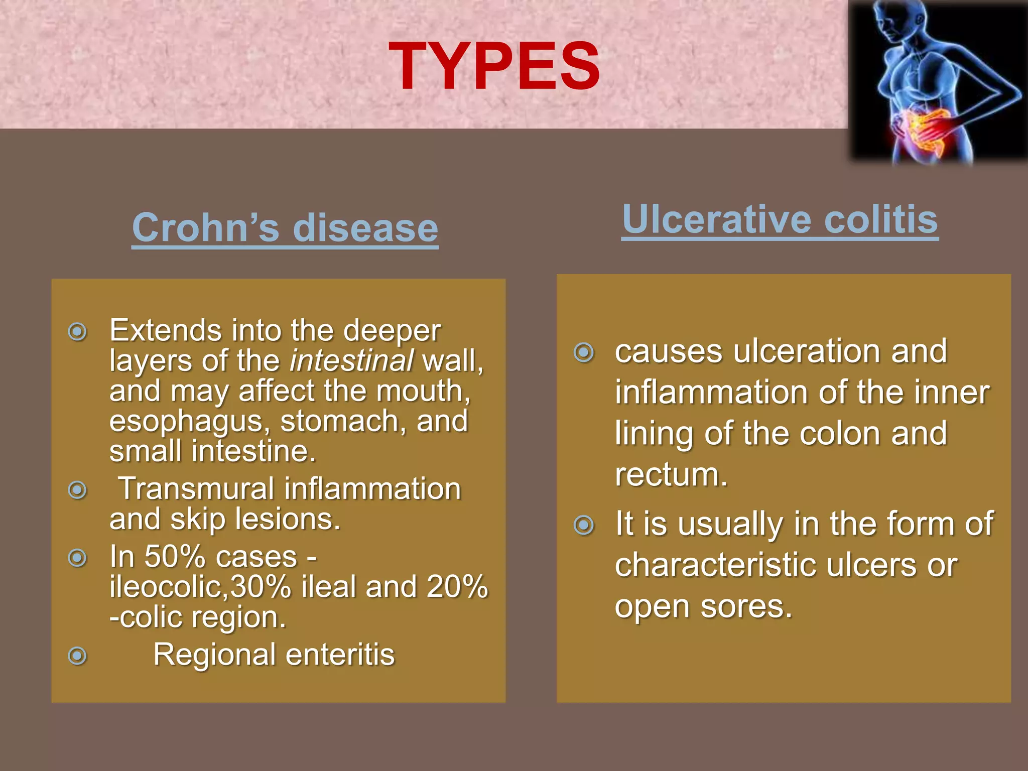


This document provides information on Inflammatory Bowel Disease (IBD), which includes Crohn's Disease and Ulcerative Colitis. It discusses the types of IBD, symptoms, investigations, complications, and treatments. The two main types are Crohn's Disease, which causes transmural inflammation of the GI tract, and Ulcerative Colitis, which causes mucosal inflammation confined to the colon and rectum. Symptoms and complications are described for each condition. Investigations include blood tests, endoscopy, and imaging. Treatments aim to induce and maintain remission, and include medications like aminosalicylates, corticosteroids, immunosuppressants, biologics, and antibiotics


