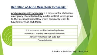
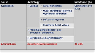
Acute mesenteric ischemia is a life-threatening condition caused by interrupted blood flow to the intestines. It has a high mortality rate of 60-80% if not treated promptly. The document discusses the definition, causes, clinical presentation, diagnostic tests and management of acute mesenteric ischemia. Key diagnostic tests include bloodwork, abdominal imaging like CT angiography and angiography. Treatment involves fluid resuscitation, antibiotics, stopping vasoconstrictors, and often emergency surgery to revascularize the intestines or resect non-viable bowel segments. Prompt diagnosis and treatment are critical given the rapid progression of intestinal tissue damage from ischemia.
in this presentation me & my colleagues discuss briefly the types of mesenteric ischemia ( acute , chronic , venous ) and its related syndromes (superior mesenteric artery syndrome , celiac trunk syndrome and supply it by good radiologic images ..
Approach to Management of Upper Gastrointestinal (GI) BleedingArun Vasireddy
Upper gastrointestinal bleeding is gastrointestinal bleeding in the upper gastrointestinal tract, commonly defined as bleeding arising from the esophagus, stomach, or duodenum. Blood may be observed in vomit (hematemesis) or in altered form in the stool (melena). Depending on the severity of the blood loss, there may be symptoms of insufficient circulating blood volume and shock. As a result, upper gastrointestinal bleeding is considered a medical emergency and typically requires hospital care for urgent diagnosis and treatment. Upper gastrointestinal bleeding can be caused by peptic ulcers, gastric erosions, esophageal varices, and some rarer causes such as gastric cancer.
The initial assessment includes measurement of the blood pressure and heart rate, as well as blood tests to determine hemoglobin concentration. In significant bleeding, fluid replacement is often required, as well as blood transfusion, before the source of bleeding can be determined by endoscopy of the upper digestive tract with an esophagogastroduodenoscopy. Depending on the source, endoscopic therapy can be applied to reduce rebleeding risk. Specific medical treatments (such as proton pump inhibitors for peptic ulcer disease) or procedures (such as TIPS for variceal hemorrhage) may be used. Recurrent or refractory bleeding may lead to need for surgery, although this has become uncommon as a result of improved endoscopic and medical treatment.
Sites of the highest risk are the duodenum, for adenocarcinomas, and the ileum, for carcinoids and lymphomas.
In industrialized countries, small bowel cancers are predominantly adenocarcinomas;
In developing countries, lymphomas are much more common.
The incidence of small bowel cancer rises with age and has generally been higher among males than among females.
The risk factors for small bowel cancer include
Dietary factor
Cigarette smoking,
Alcohol intake,
Medical conditions -Crohn's disease, familial adenomatous polyposis, cholecystectomy, peptic ulcer disease, and cystic fibrosis.
The protective factors may include rapid cell turnover, a general absence of bacteria, an alkaline environment, and low levels of activating enzymes of precarcinogens.
MBBS STUDENTS UNDER GRADUATES ..WITH USES PROPERTIE IMAGES......FOLEYS CATHETER MALECOTS CATHETER ARTERY FORCEPS SMALL MEDIUM LARGE RETRACTOR KELLYS SURGERY SURGICAL INSTRUMENTS MBBS STUDENTS UNDERGRADUATES SUTURE MATERIALS MAYOS SCISSORS MBBS STUDENTS UNDER GRADUATES .......LISTERS SINUS FORCEPS....RIGHT ANGLED FORCEPS....KOCHERS CLAMP...suture material suture removal Jolls thyroid retractor .....WITH USES PROPERTIE IMAGES......FOLEYS CATHETER MALECOTS CATHETER ARTERY FORCEPS SMALL MEDIUM LARGE RETRACTOR KELLYS SURGERY SURGICAL INSTRUMENTS MBBS STUDENTS UNDERGRADUATES SUTURE MATERIALS MAYOS SCISSORS ..FOLEYS CATHETER ....3 WAY 2WAY RYLES TUBE.........HILTONS METHOD ..ALLIS .... MOSQUITO . LANES FORCEPS ....Lanes twin anastomosis clamp......MALECOTS CATHETER.. ARTERY FORCEPS..... SMALL MEDIUM LARGE ......RETRACTOR ....KELLYS ....SURGERY SURGICAL INSTRUMENTS MBBS STUDENTS UNDERGRADUATES..... SUTURE MATERIALS.... MAYOS SCISSORS
...........................MBBS STUDENTS UNDER GRADUATES ..COMPARISON WITH IMAGES NOTES FROM LECTURE CLASSES.............................................................................................................................................................................................................................................................................................................................................................................................................................................................................................................................................................................................................................................................................................................................................................................................
Vascular disease is a condition that develops when the arteries that supply the intestines with blood become narrowed due to the build-up of plaque. This results in a lack of blood supply to the intestines.
Ischemic colitis is the most common form of intestinal ischemia. It manifests as a spectrum of injury from transient self-limited ischemia involving the mucosa and submucosa to acute fulminant ischemia with transmural infarction that may progress to necrosis and death. Although there are a variety of causes, the most common mechanism is an acute, self-limited compromise in intestinal blood flow.
MESENTERIC ISCHEMIA- GENERALISED ABDOMINAL PAIN
#surgicaleducator #epigastricabdominalpain #pepticulcerdisease #usmle #babysurgeon #surgicaltutor
• Dear Viewers,
• Greetings from “Surgical Educator”
• Today I have uploaded a video on Mesenteric Ischemia- a didactic lecture.
• It is one of the uncommon but life-threatening surgical problems you see in surgical wards.
• I have discussed the various causes for Generalised Abdominal Pain, epidemiology, etiology, pathology, clinical features, investigations, and treatment of Mesenteric Ischemia.
• I have also included a mind map and a treatment algorithm for Mesenteric Ischemia.
• I hope the video will be very useful and you will enjoy it.
• You can watch all my surgical teaching videos in the following link:
• youtube.com/c/surgicaleducator
• Thank you for watching the video.
G I bleeding with radiological interventions(ACR Appropriateness Criteria).Tc-99m RBC scintigraphy,Catheter-directed Angiography,Pharmacological control,Embolization,Arterial interventions,Endoscopy,CT Angiography
2 cases of colorectal trauma - one due to blunt trauma abdomen and one due to penetrating trauma to rectum are discussed in the light of colorectal trauma
These lecture slides, by Dr Sidra Arshad, offer a quick overview of physiological basis of a normal electrocardiogram.
Learning objectives:
1. Define an electrocardiogram (ECG) and electrocardiography
2. Describe how dipoles generated by the heart produce the waveforms of the ECG
3. Describe the components of a normal electrocardiogram of a typical bipolar leads (limb II)
4. Differentiate between intervals and segments
5. Enlist some common indications for obtaining an ECG
Study Resources:
1. Chapter 11, Guyton and Hall Textbook of Medical Physiology, 14th edition
2. Chapter 9, Human Physiology - From Cells to Systems, Lauralee Sherwood, 9th edition
3. Chapter 29, Ganong’s Review of Medical Physiology, 26th edition
4. Electrocardiogram, StatPearls - https://www.ncbi.nlm.nih.gov/books/NBK549803/
5. ECG in Medical Practice by ABM Abdullah, 4th edition
6. ECG Basics, http://www.nataliescasebook.com/tag/e-c-g-basics
Title: Sense of Taste
Presenter: Dr. Faiza, Assistant Professor of Physiology
Qualifications:
MBBS (Best Graduate, AIMC Lahore)
FCPS Physiology
ICMT, CHPE, DHPE (STMU)
MPH (GC University, Faisalabad)
MBA (Virtual University of Pakistan)
Learning Objectives:
Describe the structure and function of taste buds.
Describe the relationship between the taste threshold and taste index of common substances.
Explain the chemical basis and signal transduction of taste perception for each type of primary taste sensation.
Recognize different abnormalities of taste perception and their causes.
Key Topics:
Significance of Taste Sensation:
Differentiation between pleasant and harmful food
Influence on behavior
Selection of food based on metabolic needs
Receptors of Taste:
Taste buds on the tongue
Influence of sense of smell, texture of food, and pain stimulation (e.g., by pepper)
Primary and Secondary Taste Sensations:
Primary taste sensations: Sweet, Sour, Salty, Bitter, Umami
Chemical basis and signal transduction mechanisms for each taste
Taste Threshold and Index:
Taste threshold values for Sweet (sucrose), Salty (NaCl), Sour (HCl), and Bitter (Quinine)
Taste index relationship: Inversely proportional to taste threshold
Taste Blindness:
Inability to taste certain substances, particularly thiourea compounds
Example: Phenylthiocarbamide
Structure and Function of Taste Buds:
Composition: Epithelial cells, Sustentacular/Supporting cells, Taste cells, Basal cells
Features: Taste pores, Taste hairs/microvilli, and Taste nerve fibers
Location of Taste Buds:
Found in papillae of the tongue (Fungiform, Circumvallate, Foliate)
Also present on the palate, tonsillar pillars, epiglottis, and proximal esophagus
Mechanism of Taste Stimulation:
Interaction of taste substances with receptors on microvilli
Signal transduction pathways for Umami, Sweet, Bitter, Sour, and Salty tastes
Taste Sensitivity and Adaptation:
Decrease in sensitivity with age
Rapid adaptation of taste sensation
Role of Saliva in Taste:
Dissolution of tastants to reach receptors
Washing away the stimulus
Taste Preferences and Aversions:
Mechanisms behind taste preference and aversion
Influence of receptors and neural pathways
Impact of Sensory Nerve Damage:
Degeneration of taste buds if the sensory nerve fiber is cut
Abnormalities of Taste Detection:
Conditions: Ageusia, Hypogeusia, Dysgeusia (parageusia)
Causes: Nerve damage, neurological disorders, infections, poor oral hygiene, adverse drug effects, deficiencies, aging, tobacco use, altered neurotransmitter levels
Neurotransmitters and Taste Threshold:
Effects of serotonin (5-HT) and norepinephrine (NE) on taste sensitivity
Supertasters:
25% of the population with heightened sensitivity to taste, especially bitterness
Increased number of fungiform papillae
New Directions in Targeted Therapeutic Approaches for Older Adults With Mantl...i3 Health
i3 Health is pleased to make the speaker slides from this activity available for use as a non-accredited self-study or teaching resource.
This slide deck presented by Dr. Kami Maddocks, Professor-Clinical in the Division of Hematology and
Associate Division Director for Ambulatory Operations
The Ohio State University Comprehensive Cancer Center, will provide insight into new directions in targeted therapeutic approaches for older adults with mantle cell lymphoma.
STATEMENT OF NEED
Mantle cell lymphoma (MCL) is a rare, aggressive B-cell non-Hodgkin lymphoma (NHL) accounting for 5% to 7% of all lymphomas. Its prognosis ranges from indolent disease that does not require treatment for years to very aggressive disease, which is associated with poor survival (Silkenstedt et al, 2021). Typically, MCL is diagnosed at advanced stage and in older patients who cannot tolerate intensive therapy (NCCN, 2022). Although recent advances have slightly increased remission rates, recurrence and relapse remain very common, leading to a median overall survival between 3 and 6 years (LLS, 2021). Though there are several effective options, progress is still needed towards establishing an accepted frontline approach for MCL (Castellino et al, 2022). Treatment selection and management of MCL are complicated by the heterogeneity of prognosis, advanced age and comorbidities of patients, and lack of an established standard approach for treatment, making it vital that clinicians be familiar with the latest research and advances in this area. In this activity chaired by Michael Wang, MD, Professor in the Department of Lymphoma & Myeloma at MD Anderson Cancer Center, expert faculty will discuss prognostic factors informing treatment, the promising results of recent trials in new therapeutic approaches, and the implications of treatment resistance in therapeutic selection for MCL.
Target Audience
Hematology/oncology fellows, attending faculty, and other health care professionals involved in the treatment of patients with mantle cell lymphoma (MCL).
Learning Objectives
1.) Identify clinical and biological prognostic factors that can guide treatment decision making for older adults with MCL
2.) Evaluate emerging data on targeted therapeutic approaches for treatment-naive and relapsed/refractory MCL and their applicability to older adults
3.) Assess mechanisms of resistance to targeted therapies for MCL and their implications for treatment selection
Pulmonary Thromboembolism - etilogy, types, medical- Surgical and nursing man...VarunMahajani
Disruption of blood supply to lung alveoli due to blockage of one or more pulmonary blood vessels is called as Pulmonary thromboembolism. In this presentation we will discuss its causes, types and its management in depth.
micro teaching on communication m.sc nursing.pdfAnurag Sharma
Microteaching is a unique model of practice teaching. It is a viable instrument for the. desired change in the teaching behavior or the behavior potential which, in specified types of real. classroom situations, tends to facilitate the achievement of specified types of objectives.
The prostate is an exocrine gland of the male mammalian reproductive system
It is a walnut-sized gland that forms part of the male reproductive system and is located in front of the rectum and just below the urinary bladder
Function is to store and secrete a clear, slightly alkaline fluid that constitutes 10-30% of the volume of the seminal fluid that along with the spermatozoa, constitutes semen
A healthy human prostate measures (4cm-vertical, by 3cm-horizontal, 2cm ant-post ).
It surrounds the urethra just below the urinary bladder. It has anterior, median, posterior and two lateral lobes
It’s work is regulated by androgens which are responsible for male sex characteristics
Generalised disease of the prostate due to hormonal derangement which leads to non malignant enlargement of the gland (increase in the number of epithelial cells and stromal tissue)to cause compression of the urethra leading to symptoms (LUTS
Lung Cancer: Artificial Intelligence, Synergetics, Complex System Analysis, S...Oleg Kshivets
RESULTS: Overall life span (LS) was 2252.1±1742.5 days and cumulative 5-year survival (5YS) reached 73.2%, 10 years – 64.8%, 20 years – 42.5%. 513 LCP lived more than 5 years (LS=3124.6±1525.6 days), 148 LCP – more than 10 years (LS=5054.4±1504.1 days).199 LCP died because of LC (LS=562.7±374.5 days). 5YS of LCP after bi/lobectomies was significantly superior in comparison with LCP after pneumonectomies (78.1% vs.63.7%, P=0.00001 by log-rank test). AT significantly improved 5YS (66.3% vs. 34.8%) (P=0.00000 by log-rank test) only for LCP with N1-2. Cox modeling displayed that 5YS of LCP significantly depended on: phase transition (PT) early-invasive LC in terms of synergetics, PT N0—N12, cell ratio factors (ratio between cancer cells- CC and blood cells subpopulations), G1-3, histology, glucose, AT, blood cell circuit, prothrombin index, heparin tolerance, recalcification time (P=0.000-0.038). Neural networks, genetic algorithm selection and bootstrap simulation revealed relationships between 5YS and PT early-invasive LC (rank=1), PT N0—N12 (rank=2), thrombocytes/CC (3), erythrocytes/CC (4), eosinophils/CC (5), healthy cells/CC (6), lymphocytes/CC (7), segmented neutrophils/CC (8), stick neutrophils/CC (9), monocytes/CC (10); leucocytes/CC (11). Correct prediction of 5YS was 100% by neural networks computing (area under ROC curve=1.0; error=0.0).
CONCLUSIONS: 5YS of LCP after radical procedures significantly depended on: 1) PT early-invasive cancer; 2) PT N0--N12; 3) cell ratio factors; 4) blood cell circuit; 5) biochemical factors; 6) hemostasis system; 7) AT; 8) LC characteristics; 9) LC cell dynamics; 10) surgery type: lobectomy/pneumonectomy; 11) anthropometric data. Optimal diagnosis and treatment strategies for LC are: 1) screening and early detection of LC; 2) availability of experienced thoracic surgeons because of complexity of radical procedures; 3) aggressive en block surgery and adequate lymph node dissection for completeness; 4) precise prediction; 5) adjuvant chemoimmunoradiotherapy for LCP with unfavorable prognosis.
Factory Supply Best Quality Pmk Oil CAS 28578–16–7 PMK Powder in Stockrebeccabio
Factory Supply Best Quality Pmk Oil CAS 28578–16–7 PMK Powder in Stock
Telegram: bmksupplier
signal: +85264872720
threema: TUD4A6YC
You can contact me on Telegram or Threema
Communicate promptly and reply
Free of customs clearance, Double Clearance 100% pass delivery to USA, Canada, Spain, Germany, Netherland, Poland, Italy, Sweden, UK, Czech Republic, Australia, Mexico, Russia, Ukraine, Kazakhstan.Door to door service
Hot Selling Organic intermediates
Title: Sense of Smell
Presenter: Dr. Faiza, Assistant Professor of Physiology
Qualifications:
MBBS (Best Graduate, AIMC Lahore)
FCPS Physiology
ICMT, CHPE, DHPE (STMU)
MPH (GC University, Faisalabad)
MBA (Virtual University of Pakistan)
Learning Objectives:
Describe the primary categories of smells and the concept of odor blindness.
Explain the structure and location of the olfactory membrane and mucosa, including the types and roles of cells involved in olfaction.
Describe the pathway and mechanisms of olfactory signal transmission from the olfactory receptors to the brain.
Illustrate the biochemical cascade triggered by odorant binding to olfactory receptors, including the role of G-proteins and second messengers in generating an action potential.
Identify different types of olfactory disorders such as anosmia, hyposmia, hyperosmia, and dysosmia, including their potential causes.
Key Topics:
Olfactory Genes:
3% of the human genome accounts for olfactory genes.
400 genes for odorant receptors.
Olfactory Membrane:
Located in the superior part of the nasal cavity.
Medially: Folds downward along the superior septum.
Laterally: Folds over the superior turbinate and upper surface of the middle turbinate.
Total surface area: 5-10 square centimeters.
Olfactory Mucosa:
Olfactory Cells: Bipolar nerve cells derived from the CNS (100 million), with 4-25 olfactory cilia per cell.
Sustentacular Cells: Produce mucus and maintain ionic and molecular environment.
Basal Cells: Replace worn-out olfactory cells with an average lifespan of 1-2 months.
Bowman’s Gland: Secretes mucus.
Stimulation of Olfactory Cells:
Odorant dissolves in mucus and attaches to receptors on olfactory cilia.
Involves a cascade effect through G-proteins and second messengers, leading to depolarization and action potential generation in the olfactory nerve.
Quality of a Good Odorant:
Small (3-20 Carbon atoms), volatile, water-soluble, and lipid-soluble.
Facilitated by odorant-binding proteins in mucus.
Membrane Potential and Action Potential:
Resting membrane potential: -55mV.
Action potential frequency in the olfactory nerve increases with odorant strength.
Adaptation Towards the Sense of Smell:
Rapid adaptation within the first second, with further slow adaptation.
Psychological adaptation greater than receptor adaptation, involving feedback inhibition from the central nervous system.
Primary Sensations of Smell:
Camphoraceous, Musky, Floral, Pepperminty, Ethereal, Pungent, Putrid.
Odor Detection Threshold:
Examples: Hydrogen sulfide (0.0005 ppm), Methyl-mercaptan (0.002 ppm).
Some toxic substances are odorless at lethal concentrations.
Characteristics of Smell:
Odor blindness for single substances due to lack of appropriate receptor protein.
Behavioral and emotional influences of smell.
Transmission of Olfactory Signals:
From olfactory cells to glomeruli in the olfactory bulb, involving lateral inhibition.
Primitive, less old, and new olfactory systems with different path
1. Acute Mesenteric Ischemia
Dr. Debayan Chowdhury
22.02.2017
Malda Medical Collegewww.surgical-tutor.org.uk
An Account of:
2. Interesting Fact
On 3rd January 2017, The Mesentery has been declared as
a New Organ and has been published in The Lancet Medical
Journal(The Lancet Gastroenterology & Hepatology) by J
Calvin Coffey, a researcher at the University Hospital
Limerick, Ireland.
Gray’s Anatomy has already been
updated with the definition.
3. Definition of Acute Mesenteric Ischaemia:
Acute Mesenteric Ischaemia is a catastrophic abdominal
emergency characterized by sudden critical interruption
to the intestinal blood flow which commonly leads to
bowel infarction and death.
It is uncommon but life-threatening disease
Incidence ~1 in every 1000 hospital admissions1
Mortality remains as high as 60-80%
Prognosis is poor
1. Mark et al Semin Vasc Surg 23:9-20 ,2010
5. Acute SMA Occlusion
SMA Embolism
Aortic ostium
~15%
Around
Middle colic artery
~40%
Distal branches
~45%
SMA Thrombosis
Aortic ostium
~60-80%
Distal branches
~5%
Around
Middle colic artery
~15%
6. Acute Mesenteric Ischemia due to Embolism
Embolism - commonest cause of acute mesenteric ischaemia.
Majority of emboli arise from the heart, most commonly the left atrium
in patients of atrial fibrillation.
SMA is most commonly affected – acute angle of origin from abdominal
aorta.
7. Acute Mesenteric Ischemia due to thrombosis
Commonly involves the Aortic ostium.
Thrombosis occurs on top of atherosclerosis.
Prognosis - worse than embolic ischaemia
Often previous history of
• intestinal angina
• Sitophobia – fear of eating
• significant wt loss
8. Acute Mesenteric Ischemia due to nonocclusive disease
Results from systemic hypoperfusion, or low flow states – CCF, Shock,
critically ill patients following surgery
Cause - Intense vasospasm and Sympathetic-induced vasoconstriction.
Most Lethal - Because once arterial vasospasm is initiated, it may persist
even after correction of the initiating event.
Prognosis is very poor
9. Acute Mesenteric Ischemia due to venous thrombosis
Least common
Typically affects superior mesenteric vein and rarely
inferior mesenteric vein
10. Cause Aetiology Incidence (%)
1.Embolism Cardiac • Atrial fibrillation Commonest (40-
50%)
• Mural Thrombus following
Myocardial Infarction
• Left atrial myxoma
• Prosthetic heart valves
Proximal aortic disease, e.g.
aneurysm, atheromas
Iatrogenic, e.g. arteriography
2.Thrombosis Mesenteric Atherosclerosis 25-30%
11. Cause Aetiology Incidence (%)
3.Non-occlusive
mesenteric
ischaemia
• Low-flow states, e.g. shock 15-20%
• Drugs, e.g. digitalis, vasopressors
4.Mesenteric vein
thrombosis
Inherited
hypercoagulable
states
• Factor V Leiden mutation Least
common (5-
10%)
• Protein C,S, antithrombin
III deficiency
Acquired
hypercoagulable
states
• Malignancy
• Oral contraceptives
• Portal Hypertension
• Intra-abdominal sepsis,
e.g. acute pancreatitis
• Postoperative states, e.g.
abdominal surgery
13. Presentation
Early
Prominent symptoms
of GI emptying
( nausea, vomiting ,
diarrhea )
Late
Bloody diarrhea
Abdominal distension
Features of Peritonitis-
Fever
Shock
TachycardiaEarly diagnosis
requires high index
of suspicion
15. 15 mins
- Structural changes to intestinal villi
3 hours
- Mucosal sloughing
- Still reversible
6 hours
- Transmural necrosis
- Gangrene
- Perforation
15 mins 3 hours 6 hours
Udassin R, et al. J Surg Res 1994;56:221-5
Absolute ischaemia
What happens to bowel during absolute ischaemia?
Time is crucial !
Signs of Peritonitis
appear
16. Investigation (Preliminary)
Blood test:
Most common laboratory abnormalities are:
Haemoconcentration
Leukocytosis (Neutrophilic)
Metabolic acidosis
Lactic acidosis (in more advanced case)
Other serum markers
Raised
amylase
ALP
Neither sensitive nor specific.
But Ix help exclude other DDx
21. Doppler USG
Able to identify severe stenosis or total or partial
occlusion and velocity of blood flowing through the
vessels
Unable to detect
emboli beyond the proximal
main vessel
Non-obstructive mesenteric
ischaemia
Colour Doppler USG showing partially occluded Artery
37. Patient presents with severe abdominal pain consistent with ischemic bowel
Obtain history and perform physical examination.
Pain is out of proportion to physical findings is a significant clue.
Look for risk factors for acute mesenteric ischemia.
Order investigative studies:
Laboratory tests: WBC count, lactate, AST
Imaging: abdominal X-ray, Doppler USG, CT-Angiography, MRA
Peritoneal sign is present
Peritoneal sign is absent
Management
Acute mesenteric ischemia established
Treat with: Moist O2 , Fluid Resuscitation, Naso-
Gastric decompression, Broad Spectrum Antibiotics,
Bowel rest,Stop Vasopressor drugs/Digitalis, Invasive
haemodynamic monitoring, Treat Arrhythmia or Heart
failure, IV HEPARIN 5000IU
Laparotomy
+/- Revascularisation
+/- Bowel Resection
38. Definitive surgical exploration
1. Assessment of bowel viability
2. Determination of underlying cause
3. Mesenteric revascularization
4. Resection of necrotic bowel
5. Second look laparotomy
Midline laparotomy
39. Assessment of bowel viability
1. Clinical Judgment
- pink serosa
- visible peristalsis
- positive pulsations
- bleeding from cut edges
2. Doppler USG
- hand-held Doppler(Detects anti-mesenteric
blood flow)
3. Fluorescein
-Injection of IV Sodium fluorescein(1gm) and
inspection under Wood’s lamp
(Viable bowel has smooth, uniform fluorescence)
40. Assessment of bowel viability
Necrotic bowel
(Gangrenous)
Extensive
Infarction
Or
Frankly Necrotic
Limited
infarction
Equivocal viability
Or
Marginally-viable bowel
Revascularization
procedures
Bowel Resection
Allow 30 mins
intraoperatively to
assess bowel
viability
41. Determination of underlying Pathology:
Thrombosis or embolism?
Palpate the main trunk of SMA
(at the base of small bowel mesentery)
Normal pulse
Proximal jejunum and
transverse colon
are spared from ischemia
Diffuse midgut bowel
ischemia is noted
SMA Embolism
SMA thrombosis
Non-occlusive
mesenteric ischemia
Mesenteric
Venous thrombosis
Weak pulseNo pulsePulse present proximally
but not distally
45. Who should have second look
laparotomy?
Some surgeons advocate routine second-look
laparotomy at 24-48hr
Claimed reduced mortality rate
Other adopt a selective approach and perform a
second laparotomy when patient deterioates
clinically.
Can avoid unnecessary second operation if patient remains
well
46. Alternative to surgery…
Endovascular therapy
Acute SMA thrombosis NOMI
Percutaneous transluminal
Balloon angioplasty ± stenting
Transarterial
Thrombolysis
Transarterial infusion
of vasodilator
Limited use in acute situations
Cannot assess bowel viability
Only indicated in early cases without
bowel infarction
47.
48. Management of non-occlusive
mesenteric ischemia
Correct underlying condition.
Optimize fluid status, improve cardiac output,
and eliminate vasopressors (alpha-blocker)
Consider catheter-directed intra-arterial
infusion of vasodilator (papaverine 30-60mg/hr)
Laparotomy if peritoneal signs develop
Bradbury et al The British Journal of Surgery Vol 82(11), November 1995
ACS surgery : principles and practice
49. Management of Mesenteric venous
thrombosis
Anticoagulation with Heparin is mainstay of treatment
Workup for hypercoagulability .
Laparotomy if peritoneal signs develop.
50. Summary
Acute Mesenteric Ischaemia is an abdominal emergency both if physical
signs are present or absent.
We have very less time for investigation, so assessing clinically is
important.
Every minute we waste is every centimeter of small bowel we loose.
Angiography is diagnostic as well as therapeutic.
Preoperative heparin infusion and postoperative papaverine infusion is
must.
Still Prognosis is Poor & Mortality is High as 80%