NUCLEIC ACID
HYBERDIZATION:
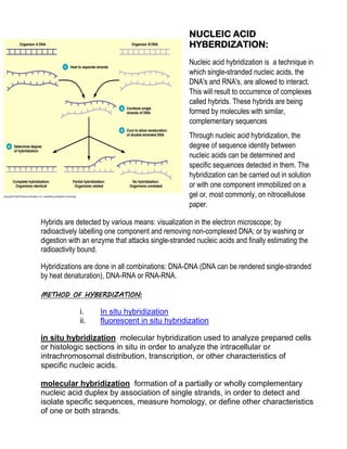
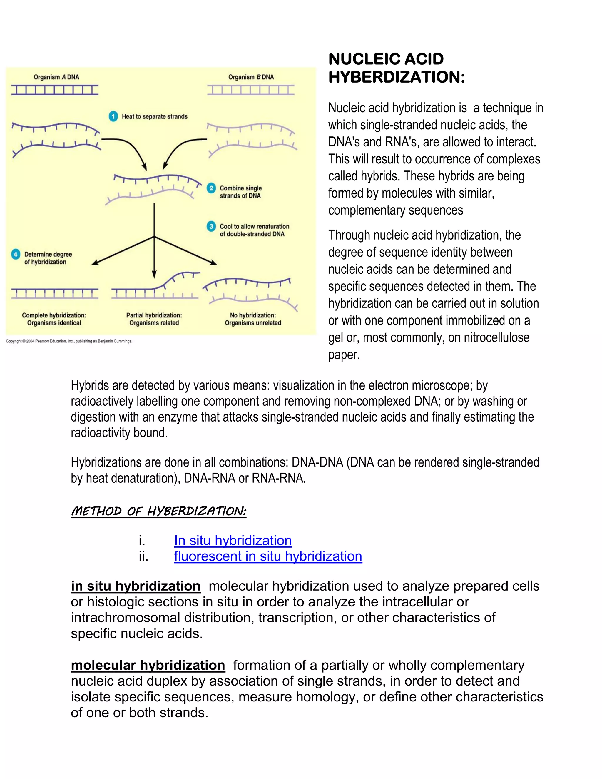
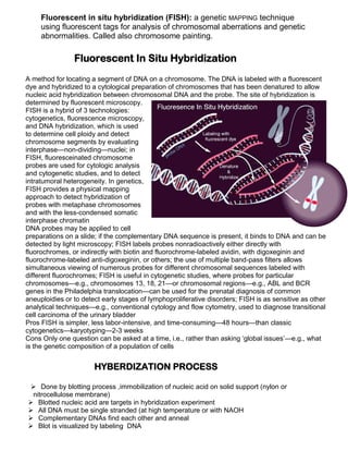
Nucleic acid hybridization is a technique in
which single-stranded nucleic acids, the
DNA's and RNA's, are allowed to interact.
This will result to occurrence of complexes
called hybrids. These hybrids are being
formed by molecules with similar,
complementary sequences
Through nucleic acid hybridization, the
degree of sequence identity between
nucleic acids can be determined and
specific sequences detected in them. The
hybridization can be carried out in solution
or with one component immobilized on a
gel or, most commonly, on nitrocellulose
paper.
Hybrids are detected by various means: visualization in the electron microscope; by
radioactively labelling one component and removing non-complexed DNA; or by washing or
digestion with an enzyme that attacks single-stranded nucleic acids and finally estimating the
radioactivity bound.
Hybridizations are done in all combinations: DNA-DNA (DNA can be rendered single-stranded
by heat denaturation), DNA-RNA or RNA-RNA.
METHOD OF HYBERDIZATION:

i.
ii.

In situ hybridization
fluorescent in situ hybridization

in situ hybridization molecular hybridization used to analyze prepared cells
or histologic sections in situ in order to analyze the intracellular or
intrachromosomal distribution, transcription, or other characteristics of
specific nucleic acids.
molecular hybridization formation of a partially or wholly complementary
nucleic acid duplex by association of single strands, in order to detect and
isolate specific sequences, measure homology, or define other characteristics
of one or both strands.
Fluorescent in situ hybridization (FISH): a genetic MAPPING technique
using fluorescent tags for analysis of chromosomal aberrations and genetic
abnormalities. Called also chromosome painting.

Fluorescent In Situ Hybridization
A method for locating a segment of DNA on a chromosome. The DNA is labeled with a fluorescent
dye and hybridized to a cytological preparation of chromosomes that has been denatured to allow
nucleic acid hybridization between chromosomal DNA and the probe. The site of hybridization is
determined by fluorescent microscopy.
FISH is a hybrid of 3 technologies:
cytogenetics, fluorescence microscopy,
and DNA hybridization, which is used
to determine cell ploidy and detect
chromosome segments by evaluating
interphase—non-dividing—nuclei; in
FISH, fluoresceinated chromosome
probes are used for cytologic analysis
and cytogenetic studies, and to detect
intratumoral heterogeneity. In genetics,
FISH provides a physical mapping
approach to detect hybridization of
probes with metaphase chromosomes
and with the less-condensed somatic
interphase chromatin
DNA probes may be applied to cell
preparations on a slide; if the complementary DNA sequence is present, it binds to DNA and can be
detected by light microscopy; FISH labels probes nonradioactively either directly with
fluorochromes, or indirectly with biotin and fluorochrome-labeled avidin, with digoxeginin and
fluorochrome-labeled anti-digoxeginin, or others; the use of multiple band-pass filters allows
simultaneous viewing of numerous probes for different chromosomal sequences labeled with
different fluorochromes; FISH is useful in cytogenetic studies, where probes for particular
chromosomes—e.g., chromosomes 13, 18, 21—or chromosomal regions—e.g., ABL and BCR
genes in the Philadelphia translocation—can be used for the prenatal diagnosis of common
aneuploidies or to detect early stages of lymphoproliferative disorders; FISH is as sensitive as other
analytical techniques—e.g., conventional cytology and flow cytometry, used to diagnose transitional
cell carcinoma of the urinary bladder
Pros FISH is simpler, less labor-intensive, and time-consuming—48 hours—than classic
cytogenetics—karyotyping—2-3 weeks
Cons Only one question can be asked at a time, i.e., rather than asking ‘global issues’—e.g., what
is the genetic composition of a population of cells

HYBERDIZATION PROCESS
 Done by blotting process ,immobilization of nucleic acid on solid support (nylon or
nitrocellulose membrane)
 Blotted nucleic acid are targets in hybridization experiment
 All DNA must be single stranded (at high temperature or with NAOH
 Complementary DNAs find each other and anneal
 Blot is visualized by labeling DNA
Main blotting procedures are:

–
–
–
–
–

Southern blot: DNA digested by a restriction enzyme then separated on an
electrophoresis gel
Northern blot: use RNA on the gel instead
In situ hybridization: probing a chromosomes or tissue
Colony hybridization: detection of clones
Microarrays

Applications
a) Isolation and quantification of specific nucleic acid sequences
b) Intracellular localization: presence and absence of a particular gene and its
copy number in the genome of an organism
c) Degree of similarity between chromosomal gene and the probe sequence.
d) Presence and absence of recognition sites for particular restriction
endonucleases in the gene.
e) Expression and regulation of a particular gene.
f) Diagnosis of infectious and inherited diseases.

Mutation
Mutation is a change in the genetic material. This means changes to the DNA or to the
chromosomes which carry the DNA. All such changes are heritable (can be passed on to
the next generation) unless they have lethal effects.

DNA mutations
When DNA is copied mistakes are
sometimes made – these are called
mutations. There are four main types of
mutations:






Deletion, where one or more bases
are left out.
Insertion, where one or more extra
base is put in.
Substitution, where one or more
bases are substituted for another
base in the sequence.
Duplication, where whole genes are
duplicated.

IRTRODUCING OF
MUTATION IN DNA
is a process by which the genetic information of an
organism is changed in a stable manner, resulting in a mutation., or as a result of
exposure to mutagens.this process is also called mutagenesis .

Mutagenesis laboratory technique
Mutagenesis in the laboratory is an important technique whereby DNA mutations are
deliberately engineered to produce
mutant genes, proteins, or strains
of organism. Various constituents
of a gene, such as its control
elements and its gene product,
may be mutated so that the
functioning of a gene or protein
can be examined in detail. The
mutation may also produce mutant
proteins with interesting properties,
or enhanced or novel functions
that may be of commercial use.
Mutants strains may also be
produced that have practical
application or allow the molecular
basis of particular cell function to
be investigated.
Early methods of mutagenesis
produces entirely random
mutations, later methods of
mutagenesis however may
produce site-specific mutation.

Types of mutagenesis







Directed mutagenesis
Site-directed mutagenesis
PCR mutagenesis
Insertional mutagenesis
Signature tagged mutagenesis
Transposon mutagenesis

Site-directed mutagenesis
Site-directed mutagenesis is a molecular biology method that is used to make specific and
intentional changes to the DNA sequence of a gene and any gene products. Also called
site-specific mutagenesis or oligonucleotide-directed mutagenesis, it is used for
investigating the structure and biological activity of DNA, RNA, and protein molecules, and
for protein engineering. With decreasing costs of oligonucleotide synthesis, artificial gene
synthesis is now occasionally used as an alternative to site-directed mutagenesis.
Basic mechanism of Mutagenesis
The basic procedure requires the synthesis of a short DNA primer. This synthetic primer
contains the desired mutation and is complementary to the template DNA around the
mutation site so it can hybridize with the DNA in the gene of interest. The mutation may be
a single base change (a point mutation), multiple base changes, deletion, or insertion. The
single-strand primer is then extended using a DNA polymerase, which copies the rest of
the gene. The gene thus copied contains the mutated site, and is then introduced into a
host cell as a vector and cloned. Finally, mutants are selected
The original method using single-primer extension was inefficient due to a low yield of
mutants. The resulting mixture contains both the original un-mutated template as well as
the mutant strand, producing a mixed population of mutant and non-mutant progenies. The
mutants may also be counter-selected due to presence of mismatch repair system that
favors the methylated template DNA, resulting in fewer mutants. Many approaches have
since been developed to improve the efficiency of mutagenesis.

Advantages of Mutagenesis
By this process we can get benefits in plants and animal by following
way
•

Plants:





•

More disease-resistant
Larger yields
More transportable
More nutritious

Animals:
 Make proteins for medicinal purposes
 Make organs for transplant to humans

Hybrdization

  • 1.
    NUCLEIC ACID HYBERDIZATION: Nucleic acidhybridization is a technique in which single-stranded nucleic acids, the DNA's and RNA's, are allowed to interact. This will result to occurrence of complexes called hybrids. These hybrids are being formed by molecules with similar, complementary sequences Through nucleic acid hybridization, the degree of sequence identity between nucleic acids can be determined and specific sequences detected in them. The hybridization can be carried out in solution or with one component immobilized on a gel or, most commonly, on nitrocellulose paper. Hybrids are detected by various means: visualization in the electron microscope; by radioactively labelling one component and removing non-complexed DNA; or by washing or digestion with an enzyme that attacks single-stranded nucleic acids and finally estimating the radioactivity bound. Hybridizations are done in all combinations: DNA-DNA (DNA can be rendered single-stranded by heat denaturation), DNA-RNA or RNA-RNA. METHOD OF HYBERDIZATION: i. ii. In situ hybridization fluorescent in situ hybridization in situ hybridization molecular hybridization used to analyze prepared cells or histologic sections in situ in order to analyze the intracellular or intrachromosomal distribution, transcription, or other characteristics of specific nucleic acids. molecular hybridization formation of a partially or wholly complementary nucleic acid duplex by association of single strands, in order to detect and isolate specific sequences, measure homology, or define other characteristics of one or both strands.
  • 2.
    Fluorescent in situhybridization (FISH): a genetic MAPPING technique using fluorescent tags for analysis of chromosomal aberrations and genetic abnormalities. Called also chromosome painting. Fluorescent In Situ Hybridization A method for locating a segment of DNA on a chromosome. The DNA is labeled with a fluorescent dye and hybridized to a cytological preparation of chromosomes that has been denatured to allow nucleic acid hybridization between chromosomal DNA and the probe. The site of hybridization is determined by fluorescent microscopy. FISH is a hybrid of 3 technologies: cytogenetics, fluorescence microscopy, and DNA hybridization, which is used to determine cell ploidy and detect chromosome segments by evaluating interphase—non-dividing—nuclei; in FISH, fluoresceinated chromosome probes are used for cytologic analysis and cytogenetic studies, and to detect intratumoral heterogeneity. In genetics, FISH provides a physical mapping approach to detect hybridization of probes with metaphase chromosomes and with the less-condensed somatic interphase chromatin DNA probes may be applied to cell preparations on a slide; if the complementary DNA sequence is present, it binds to DNA and can be detected by light microscopy; FISH labels probes nonradioactively either directly with fluorochromes, or indirectly with biotin and fluorochrome-labeled avidin, with digoxeginin and fluorochrome-labeled anti-digoxeginin, or others; the use of multiple band-pass filters allows simultaneous viewing of numerous probes for different chromosomal sequences labeled with different fluorochromes; FISH is useful in cytogenetic studies, where probes for particular chromosomes—e.g., chromosomes 13, 18, 21—or chromosomal regions—e.g., ABL and BCR genes in the Philadelphia translocation—can be used for the prenatal diagnosis of common aneuploidies or to detect early stages of lymphoproliferative disorders; FISH is as sensitive as other analytical techniques—e.g., conventional cytology and flow cytometry, used to diagnose transitional cell carcinoma of the urinary bladder Pros FISH is simpler, less labor-intensive, and time-consuming—48 hours—than classic cytogenetics—karyotyping—2-3 weeks Cons Only one question can be asked at a time, i.e., rather than asking ‘global issues’—e.g., what is the genetic composition of a population of cells HYBERDIZATION PROCESS  Done by blotting process ,immobilization of nucleic acid on solid support (nylon or nitrocellulose membrane)  Blotted nucleic acid are targets in hybridization experiment  All DNA must be single stranded (at high temperature or with NAOH  Complementary DNAs find each other and anneal  Blot is visualized by labeling DNA
  • 3.
    Main blotting proceduresare: – – – – – Southern blot: DNA digested by a restriction enzyme then separated on an electrophoresis gel Northern blot: use RNA on the gel instead In situ hybridization: probing a chromosomes or tissue Colony hybridization: detection of clones Microarrays Applications a) Isolation and quantification of specific nucleic acid sequences b) Intracellular localization: presence and absence of a particular gene and its copy number in the genome of an organism c) Degree of similarity between chromosomal gene and the probe sequence. d) Presence and absence of recognition sites for particular restriction endonucleases in the gene. e) Expression and regulation of a particular gene. f) Diagnosis of infectious and inherited diseases. Mutation Mutation is a change in the genetic material. This means changes to the DNA or to the chromosomes which carry the DNA. All such changes are heritable (can be passed on to the next generation) unless they have lethal effects. DNA mutations When DNA is copied mistakes are sometimes made – these are called mutations. There are four main types of mutations:     Deletion, where one or more bases are left out. Insertion, where one or more extra base is put in. Substitution, where one or more bases are substituted for another base in the sequence. Duplication, where whole genes are duplicated. IRTRODUCING OF MUTATION IN DNA
  • 4.
    is a processby which the genetic information of an organism is changed in a stable manner, resulting in a mutation., or as a result of exposure to mutagens.this process is also called mutagenesis . Mutagenesis laboratory technique Mutagenesis in the laboratory is an important technique whereby DNA mutations are deliberately engineered to produce mutant genes, proteins, or strains of organism. Various constituents of a gene, such as its control elements and its gene product, may be mutated so that the functioning of a gene or protein can be examined in detail. The mutation may also produce mutant proteins with interesting properties, or enhanced or novel functions that may be of commercial use. Mutants strains may also be produced that have practical application or allow the molecular basis of particular cell function to be investigated. Early methods of mutagenesis produces entirely random mutations, later methods of mutagenesis however may produce site-specific mutation. Types of mutagenesis       Directed mutagenesis Site-directed mutagenesis PCR mutagenesis Insertional mutagenesis Signature tagged mutagenesis Transposon mutagenesis Site-directed mutagenesis Site-directed mutagenesis is a molecular biology method that is used to make specific and intentional changes to the DNA sequence of a gene and any gene products. Also called site-specific mutagenesis or oligonucleotide-directed mutagenesis, it is used for investigating the structure and biological activity of DNA, RNA, and protein molecules, and for protein engineering. With decreasing costs of oligonucleotide synthesis, artificial gene synthesis is now occasionally used as an alternative to site-directed mutagenesis.
  • 5.
    Basic mechanism ofMutagenesis The basic procedure requires the synthesis of a short DNA primer. This synthetic primer contains the desired mutation and is complementary to the template DNA around the mutation site so it can hybridize with the DNA in the gene of interest. The mutation may be a single base change (a point mutation), multiple base changes, deletion, or insertion. The single-strand primer is then extended using a DNA polymerase, which copies the rest of the gene. The gene thus copied contains the mutated site, and is then introduced into a host cell as a vector and cloned. Finally, mutants are selected The original method using single-primer extension was inefficient due to a low yield of mutants. The resulting mixture contains both the original un-mutated template as well as the mutant strand, producing a mixed population of mutant and non-mutant progenies. The mutants may also be counter-selected due to presence of mismatch repair system that favors the methylated template DNA, resulting in fewer mutants. Many approaches have since been developed to improve the efficiency of mutagenesis. Advantages of Mutagenesis By this process we can get benefits in plants and animal by following way • Plants:     • More disease-resistant Larger yields More transportable More nutritious Animals:  Make proteins for medicinal purposes  Make organs for transplant to humans