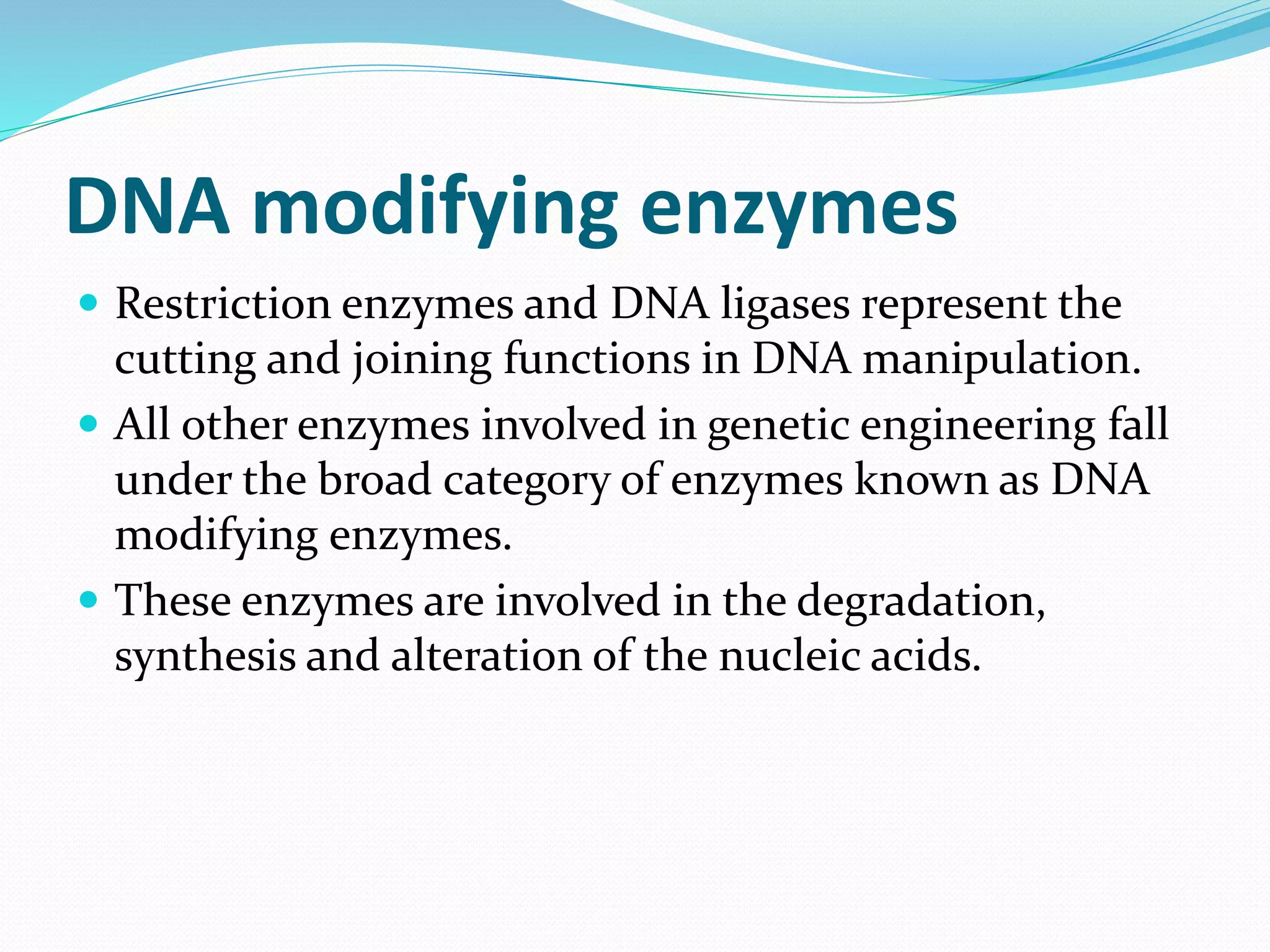
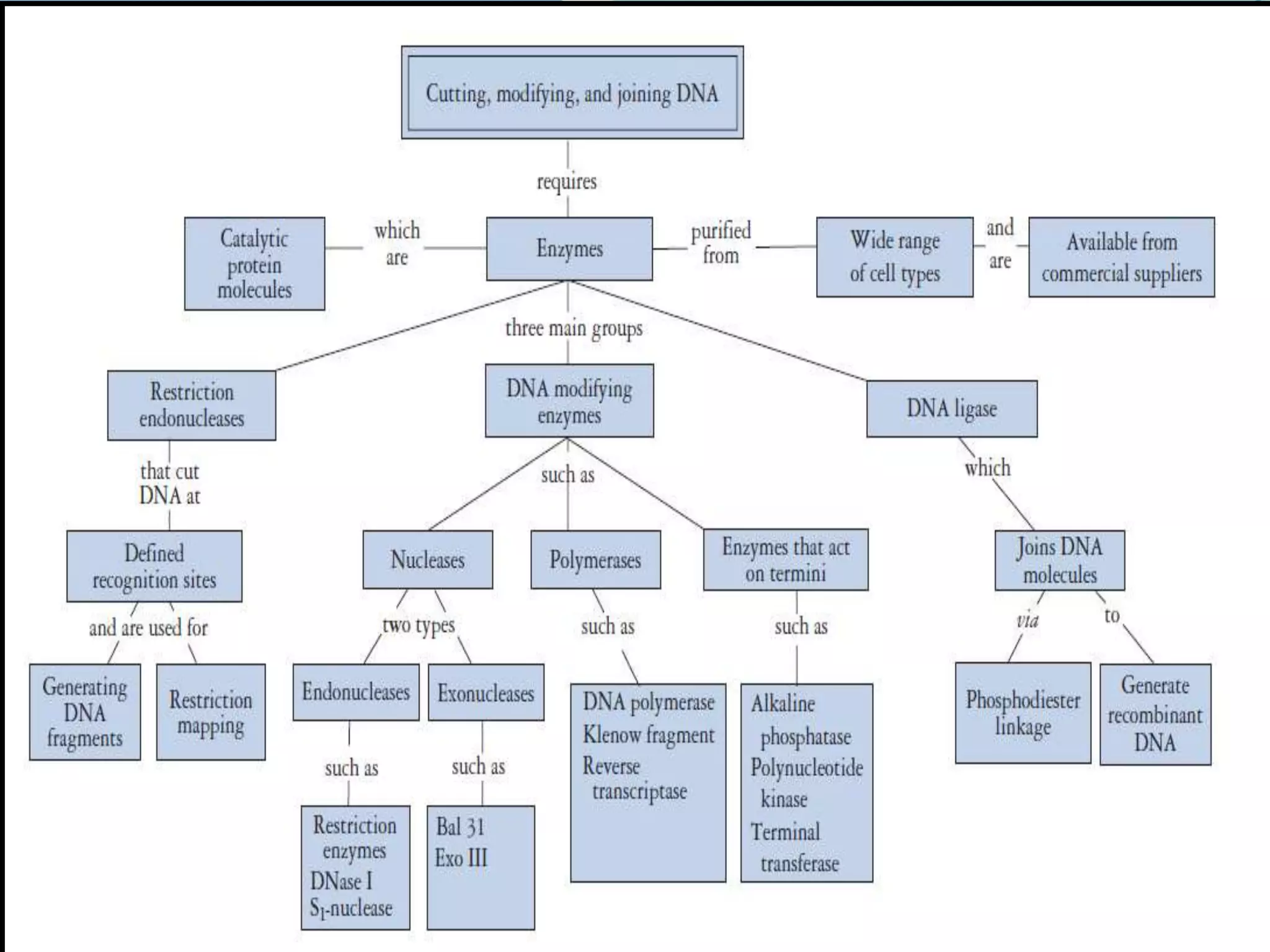
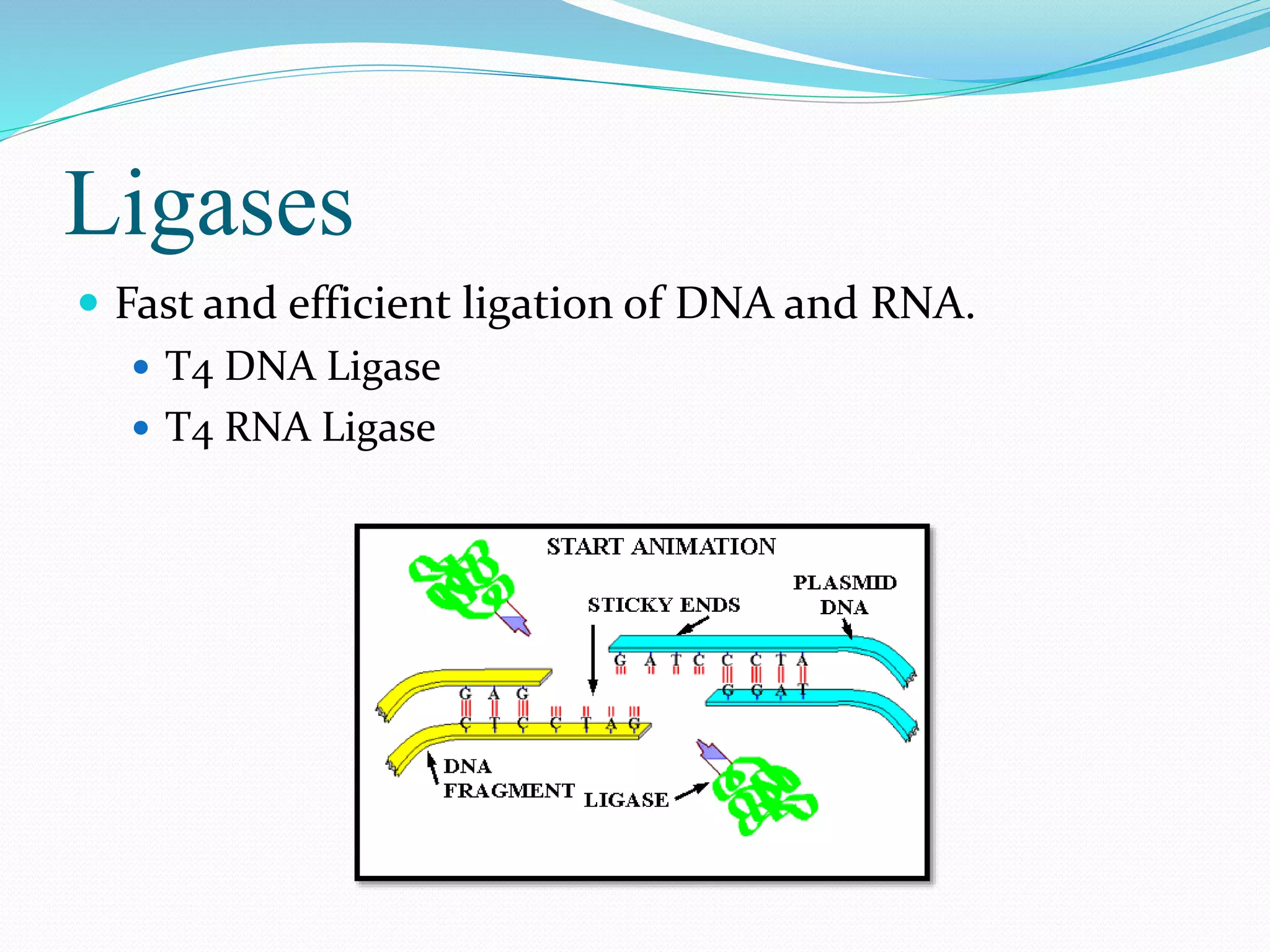
This document discusses various enzymes used for genetic engineering and DNA manipulation. It describes restriction endonucleases and DNA ligase which cut and join DNA fragments. It also discusses other DNA modifying enzymes like nucleases which degrade DNA, and polymerases which synthesize DNA copies. Specific enzymes covered in detail include DNA polymerase I, T4 DNA polymerase, T7 DNA polymerase, terminal transferase, T4 DNA ligase, and T4 RNA ligase.
MBB 501 PLANT BIOTECHNOLOGY
INFORMATION ABOUT DIFFERENT DNA MODIFYING ENZYMES
WHAT IS AN ENZYME?
Alkaline Phosphatase
Polynucleotide kinase
Terminal deoxyneucleotidyl transferase
Nucleases
Exonuclease
Bal31 Exonuclease III
Endonuclease
S1 endonulease
Deoxyribonuclease 1 (Dnase 1)
RNase A
RNase H
Restriction Endonuclease
PvuI
PvuII
Different types of endonuclease enzymes
The recognition sequences for some of the most frequently used restriction endonucleases.
Categorization of enzymes
Isoschizomers
Neoschizomers
Isocaudomers
BAC & YAC are artificially prepared chromosomes to clone DNA sequences.yeast artificial chromosome is capable of carrying upto 1000 kbp of inserted DNA sequence
Creation of a cDNA library starts with mRNA instead of DNA. Messenger RNA carries encoded information from DNA to ribosomes for translation into protein. To create a cDNA library, these mRNA molecules are treated with the enzyme reverse transcriptase, which is used to make a DNA copy of an mRNA (i.e., cDNA). A cDNA library represents a sampling of the transcribed genes, but a genomic library includes untranscribed regions.
Genomic library and shotgun sequencing. It includes the topics about genomic library,construction method, its uses and applications, shotgun sequencing, difference between random and whole genome sequencing, its advantages and disadvantages etc.
Techniques based on the principle of selectively amplifying a subset of restriction fragments from a complex mixture of DNA fragments obtained after digestion of genomic DNA with restriction endonucleases.
The enzymes that effect change in the DNA chemical constitution or topology are generally referred to as DNA modifying enzymes.
All enzymes involved in genetic engineering (the degradation, synthesis and alteration of the nucleic acids) fall under the broad category of enzymes known as DNA modifying enzymes.
List and define the significance of the following proteinsstructures.pdfsivakumar19831
List and define the significance of the following proteins/structures involved in DNA
replication: DNA polymerase III, DNA polymerase I, Primase, Single stranded binding proteins
(SSBPs), Helicase, Ligase, Primers, Topoisomerase, Anti-parallel ds DNA, Leading strand of
DNA, Lagging Strand of DNA, replication bubble, and deoxyribonucleotide tri-phosphates
(dNTPs). Describe the process of DNA replication using all of the terms that were listed (and
defined) above. Make sure you include the difference in how the leading strand and lagging
strand are synthesized. Also, include any differences (covered in class) between bacterial DNA
replication and eukaryotic DNA replication. Lastly, include a hand drawn diagram with each of
these structures and enzymes in different colors to illustrate how this process occurs.
Solution
DNA Replication: it is the biological process in which two identical replicas of DNA are
produced. It involves many steps and components.
Protein/ structures involved in DNA replication are as follows:
DNA polymerase III: it was discovered by Thomas Kornberg and Malcolm Gefter. It is an
enzyme which has its role to play in prokaryotic DNA replication. It has high processivity which
is the number of nucleotides added per binding event. It can also perform proof reading which is
the correction of wrongly added nucleotides is
DNA polymerase I: it was first discovered polymerase enzyme and Arthur Korenberg discovered
it. It does not have a role in replication but poses exonuclease activity that is removal of
nucleotides during DNA repair.
Primase: it is a type of RNA polymerase. This is very important in initiation of DNA replication
because no polymerase can synthesise a DNA without small single stranded RNA. Thus a
primase help in synthesising such small single stranded RNA complementary to the template
DNA. After initiation, this RNA piece is removed.
Single-stranded binding proteins (SSBPs): these are the class of proteins that prevent premature
binding of nucleotides to template DNA. They protect single stranded DNA from being digested
by nucleases and also prevents looping of single stranded DNA, so that other enzymes can
perform their function.
Helicases: these are the group of enzymes which help in separating two strands of DNA for the
process of replication.
Ligases: another class of enzymes which help in binding of two strands of DNA by catalysing
formation of phosphodiester bond between the two strands.
Primer: a short single stranded DNA/RNA that serves as a starting point for the DNA synthesis.
Primase helps in the formation of primer.
Topoisomerases: during replication DNA over wounds ahead the replication point making
further replication difficult, in such instance topoisomerase help in unwinding supercoiled DNA.
Anti-parallel double strand: DNA is made of two strands running in opposite directions and
hence anti parallel one runs from 5’ to 3’ and the other from 3’ to 5’.
Leading strand: DNA replication on leading stra.
Observation of Io’s Resurfacing via Plume Deposition Using Ground-based Adapt...Sérgio Sacani
Since volcanic activity was first discovered on Io from Voyager images in 1979, changes
on Io’s surface have been monitored from both spacecraft and ground-based telescopes.
Here, we present the highest spatial resolution images of Io ever obtained from a groundbased telescope. These images, acquired by the SHARK-VIS instrument on the Large
Binocular Telescope, show evidence of a major resurfacing event on Io’s trailing hemisphere. When compared to the most recent spacecraft images, the SHARK-VIS images
show that a plume deposit from a powerful eruption at Pillan Patera has covered part
of the long-lived Pele plume deposit. Although this type of resurfacing event may be common on Io, few have been detected due to the rarity of spacecraft visits and the previously low spatial resolution available from Earth-based telescopes. The SHARK-VIS instrument ushers in a new era of high resolution imaging of Io’s surface using adaptive
optics at visible wavelengths.
Richard's entangled aventures in wonderlandRichard Gill
Since the loophole-free Bell experiments of 2020 and the Nobel prizes in physics of 2022, critics of Bell's work have retreated to the fortress of super-determinism. Now, super-determinism is a derogatory word - it just means "determinism". Palmer, Hance and Hossenfelder argue that quantum mechanics and determinism are not incompatible, using a sophisticated mathematical construction based on a subtle thinning of allowed states and measurements in quantum mechanics, such that what is left appears to make Bell's argument fail, without altering the empirical predictions of quantum mechanics. I think however that it is a smoke screen, and the slogan "lost in math" comes to my mind. I will discuss some other recent disproofs of Bell's theorem using the language of causality based on causal graphs. Causal thinking is also central to law and justice. I will mention surprising connections to my work on serial killer nurse cases, in particular the Dutch case of Lucia de Berk and the current UK case of Lucy Letby.
Cancer cell metabolism: special Reference to Lactate PathwayAADYARAJPANDEY1
Normal Cell Metabolism:
Cellular respiration describes the series of steps that cells use to break down sugar and other chemicals to get the energy we need to function.
Energy is stored in the bonds of glucose and when glucose is broken down, much of that energy is released.
Cell utilize energy in the form of ATP.
The first step of respiration is called glycolysis. In a series of steps, glycolysis breaks glucose into two smaller molecules - a chemical called pyruvate. A small amount of ATP is formed during this process.
Most healthy cells continue the breakdown in a second process, called the Kreb's cycle. The Kreb's cycle allows cells to “burn” the pyruvates made in glycolysis to get more ATP.
The last step in the breakdown of glucose is called oxidative phosphorylation (Ox-Phos).
It takes place in specialized cell structures called mitochondria. This process produces a large amount of ATP. Importantly, cells need oxygen to complete oxidative phosphorylation.
If a cell completes only glycolysis, only 2 molecules of ATP are made per glucose. However, if the cell completes the entire respiration process (glycolysis - Kreb's - oxidative phosphorylation), about 36 molecules of ATP are created, giving it much more energy to use.
IN CANCER CELL:
Unlike healthy cells that "burn" the entire molecule of sugar to capture a large amount of energy as ATP, cancer cells are wasteful.
Cancer cells only partially break down sugar molecules. They overuse the first step of respiration, glycolysis. They frequently do not complete the second step, oxidative phosphorylation.
This results in only 2 molecules of ATP per each glucose molecule instead of the 36 or so ATPs healthy cells gain. As a result, cancer cells need to use a lot more sugar molecules to get enough energy to survive.
introduction to WARBERG PHENOMENA:
WARBURG EFFECT Usually, cancer cells are highly glycolytic (glucose addiction) and take up more glucose than do normal cells from outside.
Otto Heinrich Warburg (; 8 October 1883 – 1 August 1970) In 1931 was awarded the Nobel Prize in Physiology for his "discovery of the nature and mode of action of the respiratory enzyme.
WARNBURG EFFECT : cancer cells under aerobic (well-oxygenated) conditions to metabolize glucose to lactate (aerobic glycolysis) is known as the Warburg effect. Warburg made the observation that tumor slices consume glucose and secrete lactate at a higher rate than normal tissues.
Multi-source connectivity as the driver of solar wind variability in the heli...Sérgio Sacani
The ambient solar wind that flls the heliosphere originates from multiple
sources in the solar corona and is highly structured. It is often described
as high-speed, relatively homogeneous, plasma streams from coronal
holes and slow-speed, highly variable, streams whose source regions are
under debate. A key goal of ESA/NASA’s Solar Orbiter mission is to identify
solar wind sources and understand what drives the complexity seen in the
heliosphere. By combining magnetic feld modelling and spectroscopic
techniques with high-resolution observations and measurements, we show
that the solar wind variability detected in situ by Solar Orbiter in March
2022 is driven by spatio-temporal changes in the magnetic connectivity to
multiple sources in the solar atmosphere. The magnetic feld footpoints
connected to the spacecraft moved from the boundaries of a coronal hole
to one active region (12961) and then across to another region (12957). This
is refected in the in situ measurements, which show the transition from fast
to highly Alfvénic then to slow solar wind that is disrupted by the arrival of
a coronal mass ejection. Our results describe solar wind variability at 0.5 au
but are applicable to near-Earth observatories.
This presentation explores a brief idea about the structural and functional attributes of nucleotides, the structure and function of genetic materials along with the impact of UV rays and pH upon them.
Deep Behavioral Phenotyping in Systems Neuroscience for Functional Atlasing a...Ana Luísa Pinho
Functional Magnetic Resonance Imaging (fMRI) provides means to characterize brain activations in response to behavior. However, cognitive neuroscience has been limited to group-level effects referring to the performance of specific tasks. To obtain the functional profile of elementary cognitive mechanisms, the combination of brain responses to many tasks is required. Yet, to date, both structural atlases and parcellation-based activations do not fully account for cognitive function and still present several limitations. Further, they do not adapt overall to individual characteristics. In this talk, I will give an account of deep-behavioral phenotyping strategies, namely data-driven methods in large task-fMRI datasets, to optimize functional brain-data collection and improve inference of effects-of-interest related to mental processes. Key to this approach is the employment of fast multi-functional paradigms rich on features that can be well parametrized and, consequently, facilitate the creation of psycho-physiological constructs to be modelled with imaging data. Particular emphasis will be given to music stimuli when studying high-order cognitive mechanisms, due to their ecological nature and quality to enable complex behavior compounded by discrete entities. I will also discuss how deep-behavioral phenotyping and individualized models applied to neuroimaging data can better account for the subject-specific organization of domain-general cognitive systems in the human brain. Finally, the accumulation of functional brain signatures brings the possibility to clarify relationships among tasks and create a univocal link between brain systems and mental functions through: (1) the development of ontologies proposing an organization of cognitive processes; and (2) brain-network taxonomies describing functional specialization. To this end, tools to improve commensurability in cognitive science are necessary, such as public repositories, ontology-based platforms and automated meta-analysis tools. I will thus discuss some brain-atlasing resources currently under development, and their applicability in cognitive as well as clinical neuroscience.
Seminar of U.V. Spectroscopy by SAMIR PANDASAMIR PANDA
Spectroscopy is a branch of science dealing the study of interaction of electromagnetic radiation with matter.
Ultraviolet-visible spectroscopy refers to absorption spectroscopy or reflect spectroscopy in the UV-VIS spectral region.
Ultraviolet-visible spectroscopy is an analytical method that can measure the amount of light received by the analyte.
Introduction:
RNA interference (RNAi) or Post-Transcriptional Gene Silencing (PTGS) is an important biological process for modulating eukaryotic gene expression.
It is highly conserved process of posttranscriptional gene silencing by which double stranded RNA (dsRNA) causes sequence-specific degradation of mRNA sequences.
dsRNA-induced gene silencing (RNAi) is reported in a wide range of eukaryotes ranging from worms, insects, mammals and plants.
This process mediates resistance to both endogenous parasitic and exogenous pathogenic nucleic acids, and regulates the expression of protein-coding genes.
What are small ncRNAs?
micro RNA (miRNA)
short interfering RNA (siRNA)
Properties of small non-coding RNA:
Involved in silencing mRNA transcripts.
Called “small” because they are usually only about 21-24 nucleotides long.
Synthesized by first cutting up longer precursor sequences (like the 61nt one that Lee discovered).
Silence an mRNA by base pairing with some sequence on the mRNA.
Discovery of siRNA?
The first small RNA:
In 1993 Rosalind Lee (Victor Ambros lab) was studying a non- coding gene in C. elegans, lin-4, that was involved in silencing of another gene, lin-14, at the appropriate time in the
development of the worm C. elegans.
Two small transcripts of lin-4 (22nt and 61nt) were found to be complementary to a sequence in the 3' UTR of lin-14.
Because lin-4 encoded no protein, she deduced that it must be these transcripts that are causing the silencing by RNA-RNA interactions.
Types of RNAi ( non coding RNA)
MiRNA
Length (23-25 nt)
Trans acting
Binds with target MRNA in mismatch
Translation inhibition
Si RNA
Length 21 nt.
Cis acting
Bind with target Mrna in perfect complementary sequence
Piwi-RNA
Length ; 25 to 36 nt.
Expressed in Germ Cells
Regulates trnasposomes activity
MECHANISM OF RNAI:
First the double-stranded RNA teams up with a protein complex named Dicer, which cuts the long RNA into short pieces.
Then another protein complex called RISC (RNA-induced silencing complex) discards one of the two RNA strands.
The RISC-docked, single-stranded RNA then pairs with the homologous mRNA and destroys it.
THE RISC COMPLEX:
RISC is large(>500kD) RNA multi- protein Binding complex which triggers MRNA degradation in response to MRNA
Unwinding of double stranded Si RNA by ATP independent Helicase
Active component of RISC is Ago proteins( ENDONUCLEASE) which cleave target MRNA.
DICER: endonuclease (RNase Family III)
Argonaute: Central Component of the RNA-Induced Silencing Complex (RISC)
One strand of the dsRNA produced by Dicer is retained in the RISC complex in association with Argonaute
ARGONAUTE PROTEIN :
1.PAZ(PIWI/Argonaute/ Zwille)- Recognition of target MRNA
2.PIWI (p-element induced wimpy Testis)- breaks Phosphodiester bond of mRNA.)RNAse H activity.
MiRNA:
The Double-stranded RNAs are naturally produced in eukaryotic cells during development, and they have a key role in regulating gene expression .
This pdf is about the Schizophrenia.
For more details visit on YouTube; @SELF-EXPLANATORY;
https://www.youtube.com/channel/UCAiarMZDNhe1A3Rnpr_WkzA/videos
Thanks...!
2. Such manipulations of DNA are
conducted by a toolkit of enzymes:
restriction endonucleases are used as molecular scissors,
DNA ligase functions to bond pieces of DNA together, and
a variety of additional enzymes that modify DNA are used to
facilitate the process.
3. DNA modifying enzymes
Restriction enzymes and DNA ligases represent the
cutting and joining functions in DNA manipulation.
All other enzymes involved in genetic engineering fall
under the broad category of enzymes known as DNA
modifying enzymes.
These enzymes are involved in the degradation,
synthesis and alteration of the nucleic acids.
4.
5.
6. Nucleases
Nuclease enzymes degrade nucleic acids by breaking
the phosphodiester bond that holds the nucleotides
together.
Restriction enzymes are good examples of
endonucleases, which cut within a DNA strand.
A second group of nucleases, which degrade DNA
from the termini of the molecule, are known as
exonucleases.
8. Polymerases
Polymerase enzymes synthesise copies of nucleic acid
molecules and are used in many genetic engineering
procedures.
When describing a polymerase enzyme, the terms ‘DNA-
dependent’ or ‘RNA-dependent’ may be used to indicate
the type of nucleic acid template that the enzyme uses.
Thus, a
DNA-dependent DNA polymerase copies DNA into DNA,
an RNA-dependent DNA polymerase copies RNA into DNA,
and
a DNA-dependent RNA polymerase transcribes DNA into
RNA.
9. DNA Polymerases
Mesophilic and thermophilic DNA
polymerases for different polymerization
reactions, DNA end blunting and
amplification, labeling and others.
DNA Polymerase, Large Fragment
DNA Polymerase I
T4 DNA Polymerase
T7 DNA Polymerase
Terminal Transferase (TdT)
10. DNA Polymerase, Large Fragment
•DNA Polymerase, Large Fragment, is a portion of
DNA polymerase of Bacillus smithii, which catalyzes
5'=>3' synthesis of DNA and lacks 5'→3' and 3'→5'
exonuclease activities.
Highlights
Thermophilic DNA polymerase with strong strand
displacement activity
11. DNA Polymerase I
DNA Polymerase I, a template-dependent DNA
polymerase, catalyzes 5'→3' synthesis of DNA.
The enzyme also exhibits 3'→5' exonuclease
(proofreading) activity, 5'→3' exonuclease activity, and
ribonuclease H activity.
12. Highlights
Incorporates modified nucleotides
Active in multiple buffers, including restriction
enzyme, PCR, and RT buffers
Applications
DNA labeling
Second-strand synthesis of cDNA in
conjunction with RNaseH
13. T4 DNA Polymerase
T4 DNA Polymerase, a template-dependent DNA
polymerase, catalyzes 5'-3' synthesis from primed
single-stranded DNA.
The enzyme has a 3'-5' exonuclease activity, but lacks
5'-3' exonuclease activity.
14. T7 DNA Polymerase
T7 DNA Polymerase, a template dependent DNA
polymerase.
It catalyzes DNA synthesis in the 5'=>3' direction.
It is a highly processive DNA polymerase allowing
continuous synthesis of long stretches of DNA.
15. Applications
Purification of covalently closed circular DNA by
removal of residual genomic DNA
Primer extension reactions on long templates
DNA 3'-end labeling
Strand extensions in site-directed mutagenesis
16. Terminal Transferase (TdT)
Protruding, recessed or blunt ended double or single
stranded DNA molecules serve as a substrate for TdT.
TdT is isolated and purified from an E. coli strain
carrying the cloned terminal transferase gene from calf
thymus.
17. DNA ligase
DNA ligase is an important cellular enzyme, as its
function is to repair broken phosphodiester bonds that
may occur at random or as a consequence of DNA
replication or recombination.
It can therefore be thought of as molecular glue, which
is used to stick pieces of DNA together.
18. Ligases
Fast and efficient ligation of DNA and RNA.
T4 DNA Ligase
T4 RNA Ligase
19. T4 DNA Ligase
The enzyme repairs single-strand nicks in duplex
DNA, RNA, or DNA/RNA hybrids.
It also joins DNA fragments with either cohesive or
blunt termini, but has no activity on single-stranded
nucleic acids.
The T4 DNA Ligase requires ATP as a cofactor.
20. Applications
Cloning of restriction enzyme generated DNA
fragments
Cloning of PCR products
Joining of double-stranded oligonucleotide
linkers or adaptors to DNA
Site-directed mutagenesis
21. T4 RNA Ligase
T4 RNA Ligase catalyzes the ATP-dependent intra-
and intermolecular formation of phosphodiester
bonds between 5'-phosphate and 3'-hydroxyl termini
of oligonucleotides, single-stranded RNA and DNA.
Applications
Joining RNA to RNA
Specific modifications of tRNAs
Site-specific generation of composite primers for PCR
22. CONCLUSION:
These are the modifying enzymes represent the
cutting and joining functions in DNA manipulation
and genetic engineering.
A mesophile is an organism that grows best in moderate temperature, neither too hot nor too cold, typically between 20 and 45 °C (68 and 113 °F).[1] The term is mainly applied tomicroorganisms.
A thermophile is an organism — a type of extremophile — that thrives at relatively high temperatures, between 45 and 122 °C (113 and 252 °F).