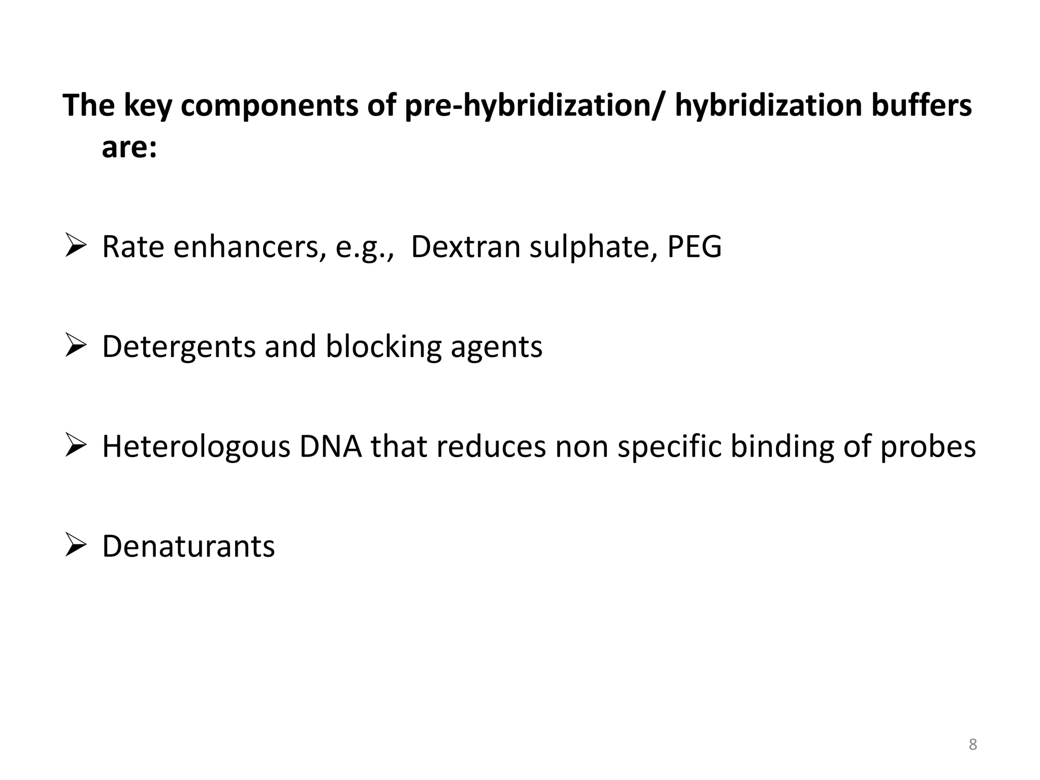
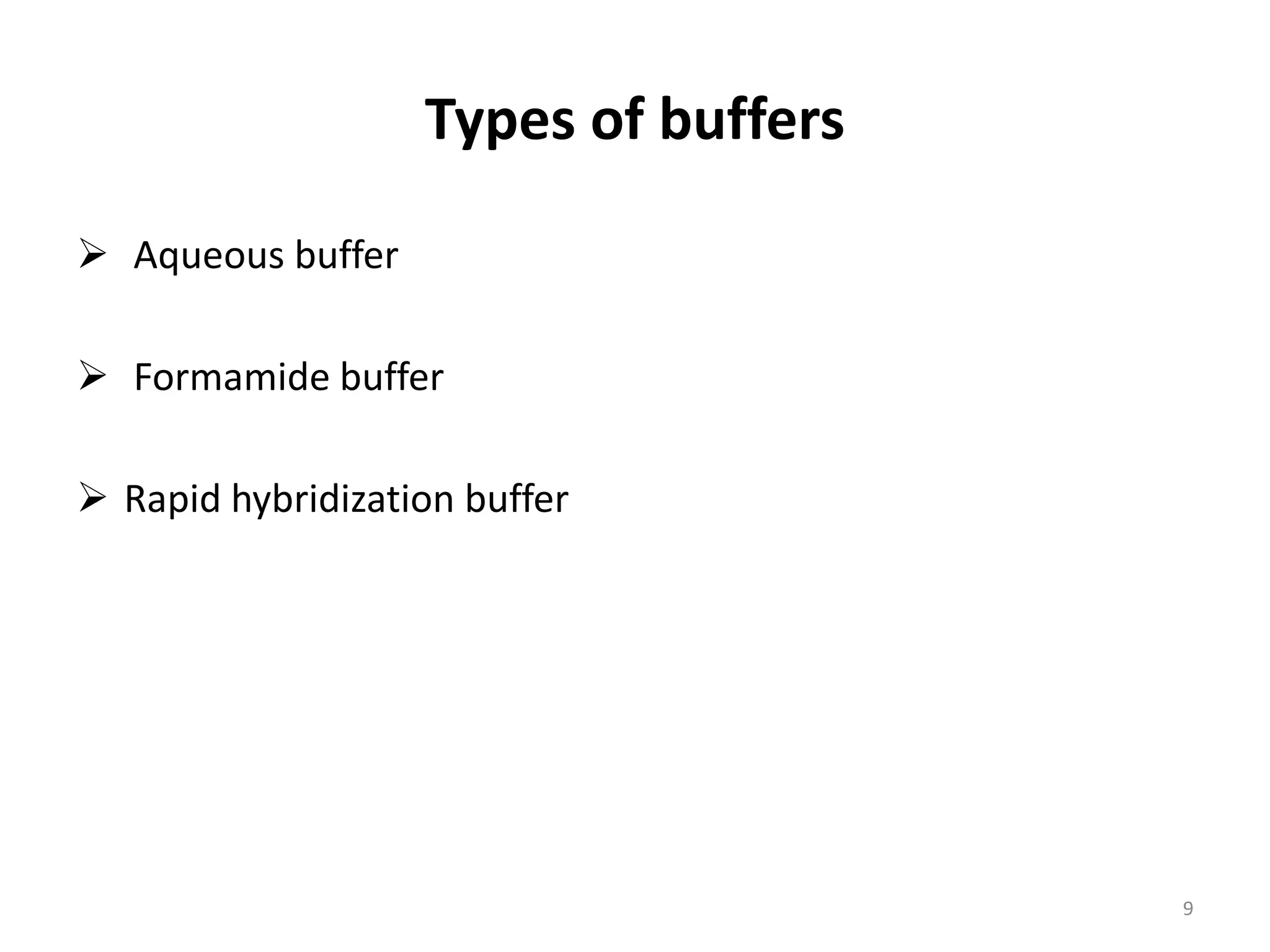
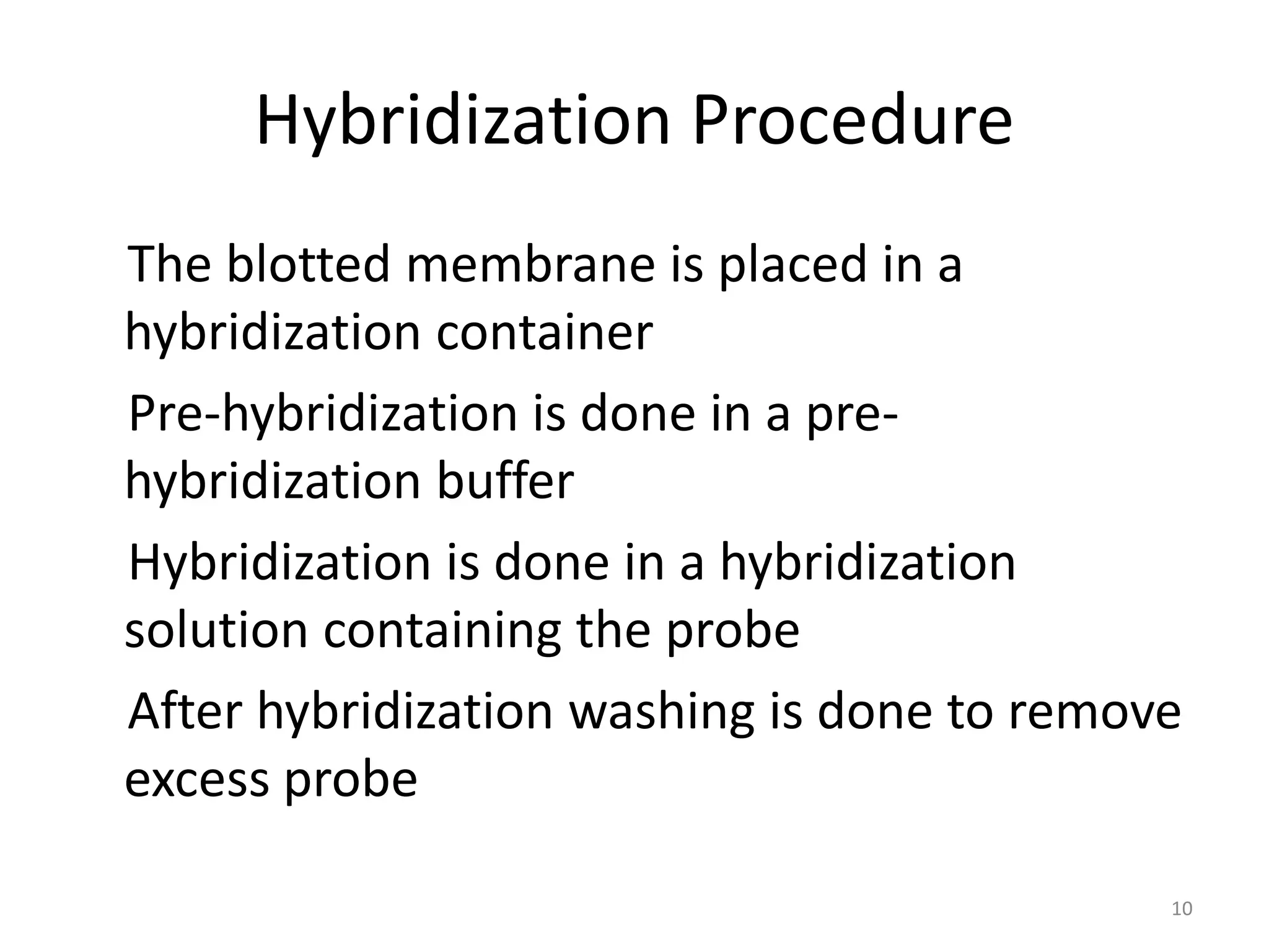
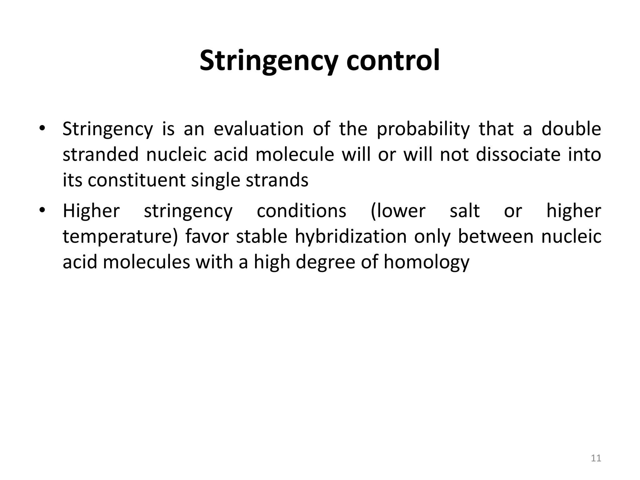
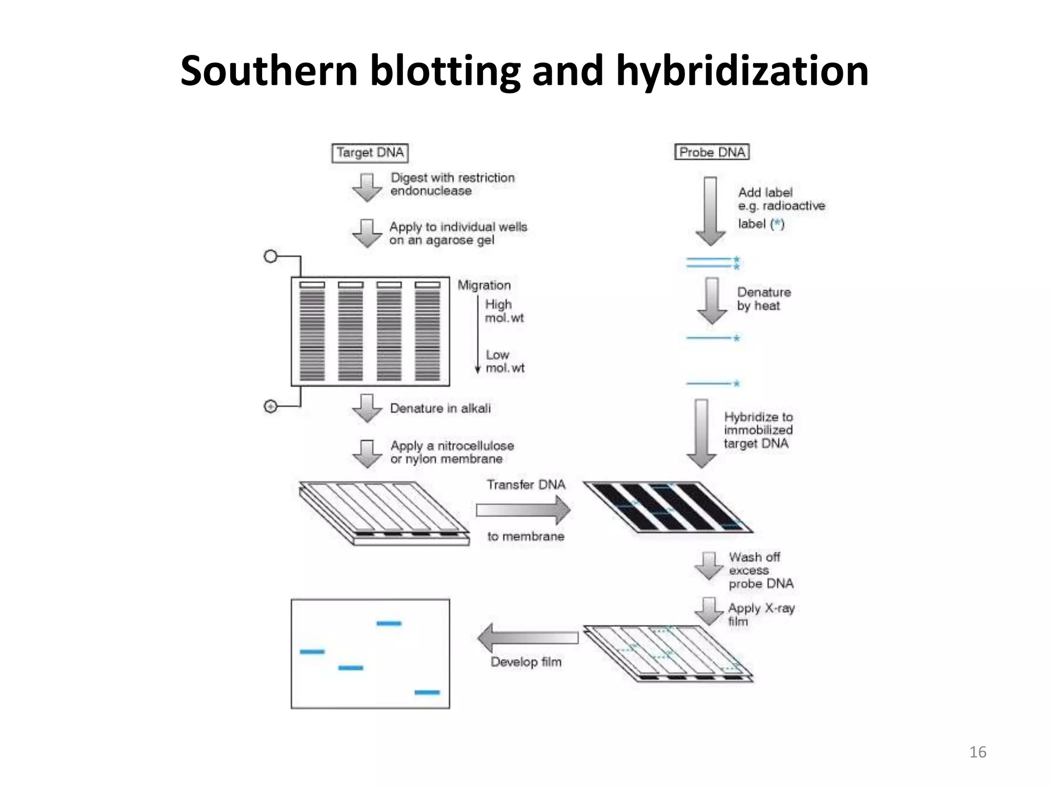
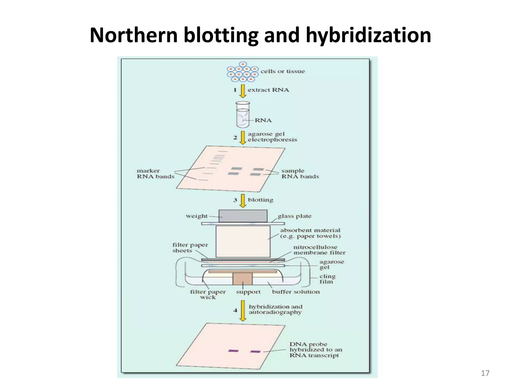
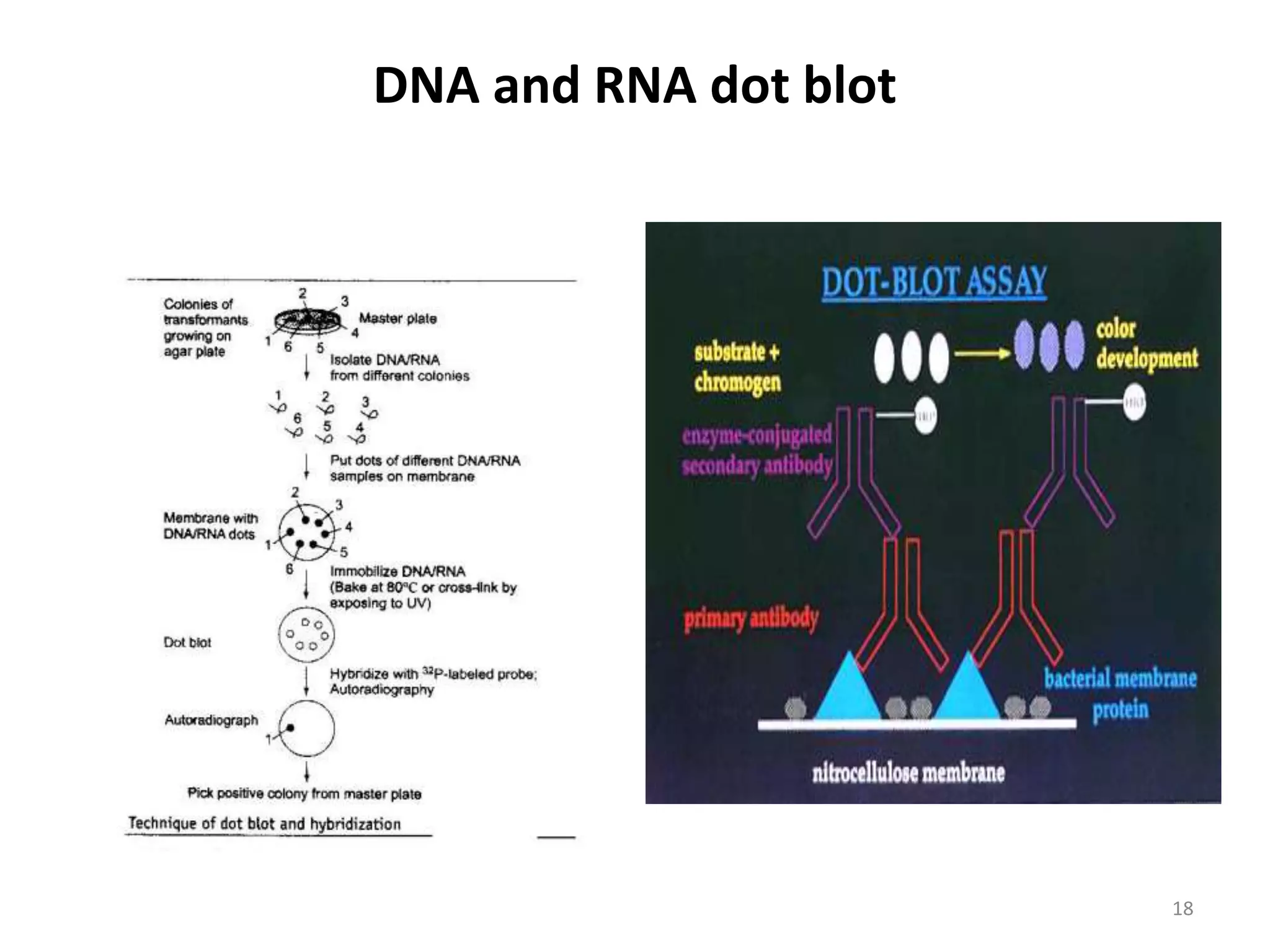
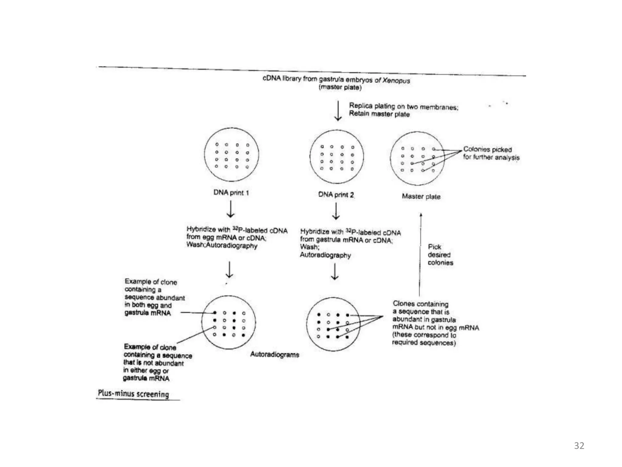
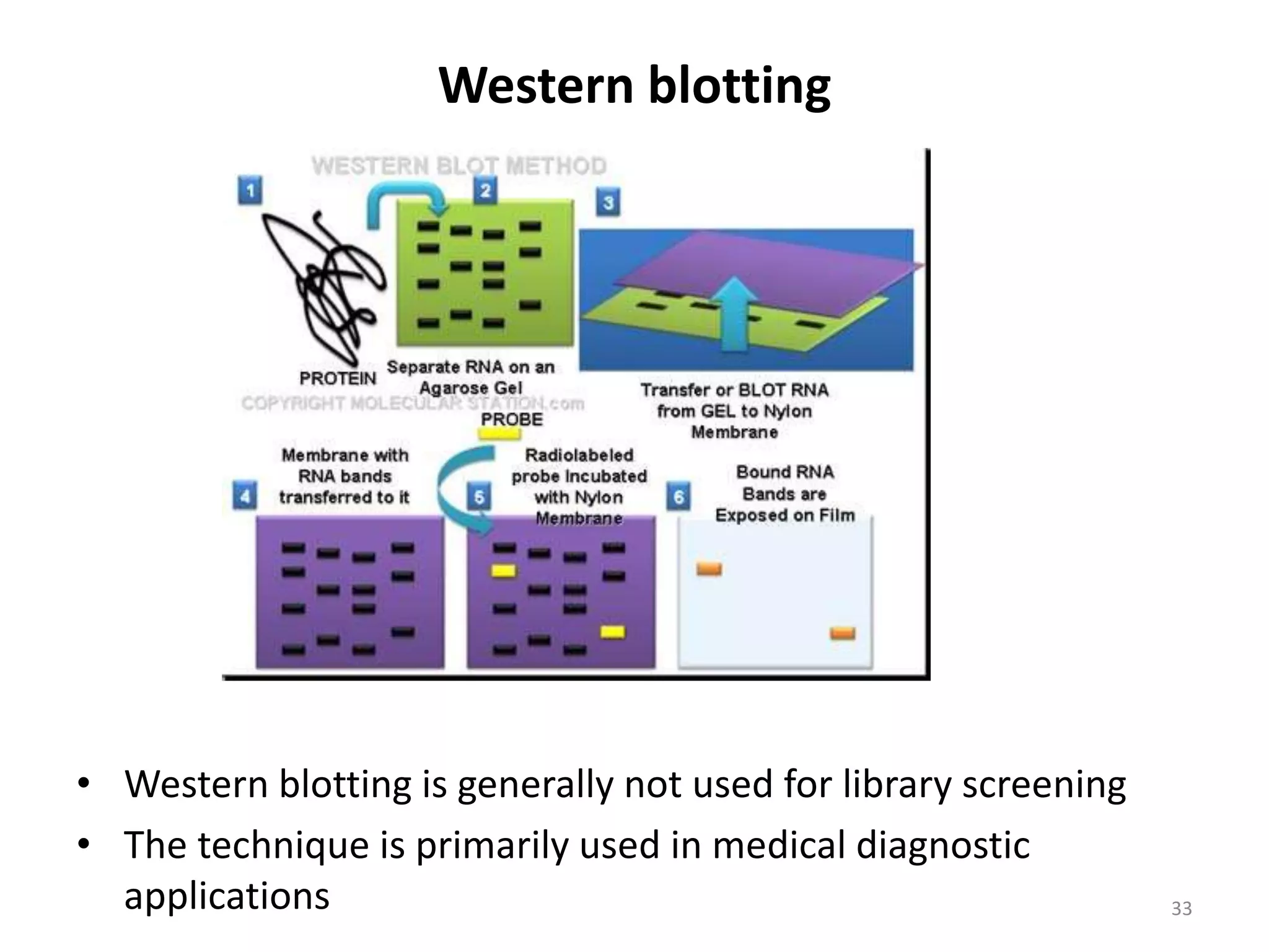
Hybridization Procedures
1
Lecture- 22
2
In these techniques nucleic acid hybridization is used to detect and quantify specific
DNAs/RNAs. The DNAs/RNAs to be probed are immobilized on a membrane and
hybridized with a labeled DNA/RNA probe. The probe hybridizes to its
complementary sequence and the hybridization product is detected with a detection
method. A nucleic acid hybridization technique has the following steps:
• Immobilization of the target DNA/RNA
The target nucleic acid to be probed is isolated and immobilized on a
nitrocellulose or nylon membrane. Nitrocellulose membranes have low
capacity to bind nucleic acids and cannot be used for repeated hybridizations
since these become brittle during the baking step of immobilization.
These limitations of the nitrocellulose membranes have been overcome
with the introduction of various types of nylon membranes. The nucleic acids
are either blotted to the membrane from a gel or spotted directly on the
membrane.
3
If the nucleic acid is double stranded DNA, it is denatured just before
blotting/spotting. The bacterial colonies can be transferred to the membranes and lysed
to liberate DNA which is immobilized subsequently to the membrane. The nucleic acid
blotted to the membrane is immobilized by baking in a vacuum/microwave oven or
cross-linking by UV irradiation, depending upon the membrane used.
The transfer of DNA from an agarose gel to a membrane is called as Southern
blotting after the name of the scientist Edwin M. Southern who introduced this
technique in 1975. The RNA transfer from the gel to the membrane has been given the
name northern blotting.
•Preparation of the labeled probes
A labeled DNA or RNA molecule having sequence complementary to a target nucleic
acid molecule is used as a probe. A probe may have complete or partial homology to
the nucleic acid molecule to be probed and it could be end-labeled or continuously
labeled.
An end-labelled probe is made by adding a label at the 5′ or 3′-end. A continuously
labeled probe is produced by nick translation, random priming, PCR, in vitro
transcription or cross-linking with a label. A label on DNA or RNA could be
radioisotopic or non-isotopic. The radioisotopic labels used are 32P, 3H and 35S. Among
these 32P is the most commonly used radioisotopic label.
4
A nucleoside triphosphate precursor, labeled at the α or γ position, is used to
label nucleic acids. A commonly used technique for radiolabelling of DNA is the
nick translation technique which was discovered by Paul Berg and his coworkers in
1977.
In this technique random nicks are introduced in a double stranded DNA
molecule by DNase I enzyme and the normal nucleotides near the nick are
replaced by radio-labeled nucleotides by the joint action of 5′- 3′ exonuclease and
5′- 3′ polymerase activities of E. coli DNA polymerase I enzyme in the presence of
one radiolabeled dNTP and four normal dNTPs.
The commonly used non-isotopic labels for making nucleic acid probes are
biotin, digoxigenin and fluorescein. The radioisotopic probes are highly sensitive
and compatible with several labeling techniques, however, these probes suffer from
the disadvantages of being potential health hazards and having short half life of the
isotope.
Moreover these are not suitable for repeated probing of the target nucleic acid
molecules. Non-isotopic labels are quite safe to use and can be stored for long
periods.
5
Hybridization of the probe with the target nucleic acid
The membrane on which target nucleic acid is immobilized is placed in a
hybridization container. A pre-hybridization buffer is added to the hybridization
container to prevent non-specific binding of the probe to the membrane.
After treatment for 1.5 hrs to 12 hrs, the pre-hybridization buffer is removed and
the hybridization solution (pre-hybridization buffer containing the denatured
probe) is added to the hybridization container.
The main components of the pre-hybridization/hybridization buffer are: rate
enhancer, detergent, blocking agent, heterologous DNA and denaturant. The
hybridization container is placed on a roller. After overnight incubation, the
hybridization solution is discarded into a proper liquid waste container and the
membrane is given a series of washes with a suitable washing solution.
6
• Detection of the probe
The probe on the washed membrane is detected by an appropriate method.
The selection of the method is made based on the type of probe used. The
radioisotopic probes are detected by autoradiography.
Chemiluminescence, chemifluorescence or chromogenic techniques are
used for the detection of non-isotopic probes. Biotinylated probes are detected
with streptavidin conjugated to a reporter enzyme.
Enzyme-linked immunoassay using an antibody conjugated alkaline
phosphatase is used to detect digoxigenin labeled probes.
The detection of fluorescein labeled probes is carried out using
antifluorescein antibodies conjugated to alkaline phosphatase or horseradish
peroxidase.
Membrane Hybridization
• Unlike solution hybridization, membrane
hybridization does not proceed to completion as
some of the bound nucleic acids are embedded in
the membrane and are inaccessible to the probe
• The composition of pre-hybridization/hybridization
buffers can greatly affect the speed of reaction and
sensitivity of detection
7
The key components of pre-hybridization/ hybridization buffers
are:
 Rate enhancers, e.g., Dextran sulphate, PEG
 Detergents and blocking agents
 Heterologous DNA that reduces non specific binding of probes
 Denaturants
8
Types of buffers
 Aqueous buffer
 Formamide buffer
 Rapid hybridization buffer
9
Hybridization Procedure
The blotted membrane is placed in a
hybridization container
Pre-hybridization is done in a pre-
hybridization buffer
Hybridization is done in a hybridization
solution containing the probe
After hybridization washing is done to remove
excess probe
10
Stringency control
• Stringency is an evaluation of the probability that a double
stranded nucleic acid molecule will or will not dissociate into
its constituent single strands
• Higher stringency conditions (lower salt or higher
temperature) favor stable hybridization only between nucleic
acid molecules with a high degree of homology
11
Probe Stripping
• After detection, a probe is removed from a nylon blot
to allow subsequent hybridization with a different
probe by heating at 65 o C in presence of 0.1 % SDS
12
Techniques based on nucleic acid blotting
and hybridization
• Colony blotting and hybridization
• Plaque lift and hybridization
• Southern blotting and hybridization
• Northern blotting and hybridization
• DNA and RNA dot blot
• Zoo blot hybridization
13
Colony blotting and hybridization
•Applied to nucleic acid released from blotted microbial colonies
•The most commonly used and rapid method of library screening as it can be
applied to very large number of clones
14
Plaque lift and hybridization
15
Southern blotting and hybridization
16
Northern blotting and hybridization
17
DNA and RNA dot blot
18
Zoo blot hybridization
• Zoo blot hybridization is a type of an assay for DNA
sequences that are highly conserved between two species
• It is not a distinct method but variant of Southern blot that
can be used for analysis of DNA of different species
19
Protein Structure/Function Based
Techniques
• When a DNA library is constructed using expression vectors, it
permits a range of alternative techniques to be employed,
each of which exploits some structural or functional property
of the gene product as each individual clone can be expressed
to yield a polypeptide
• These approaches are very important in cases where the DNA
sequences of target clone is completely unknown and enough
information is not available to design a suitable probe or set
of primers
20
Screening by determination of protein activity
• If the target gene produces an enzyme that is not normally
made by the host cell, an in situ (direct plate) assay can be
developed to identify members of a library that carry the
particular gene encoding that enzyme
• For example, the gene for α-amylase, β-glucosidase and many
other enzymes from various organisms have been isolated in
this way
21
Screening by functional complementation
• The process by which a particular DNA sequence
compensates for a missing function in a mutant cell and thus
restores the wild type phenotype
• The functional complementation can be homologous or
heterologous, which means that the cloned gene may come
from either the same or a different species, respectively
22
Functional complementation
• In case the cloning is done within
the antibiotic resistance gene, the
recombinants become antibiotic
sensitive, while nonrecombinants
are antibiotic resistant
• Examples are:
 Blue white screening (α-
complementation of β–galactosidase)
 Blue white screening
Blue white screening
23
Functional complementation
• Most commonly, a pool system is employed where cells are
transfected with a complex mixture of up to 1,00,000 clones
• Thereafter, the pools that successfully complement the
mutant phenotype are then subdivided for a further round of
transfection and the procedure is repeated until the individual
cDNA is isolated
• The advantage of using this screening procedure is that no
sequence information is required; without the functional
assay, a mouse or a human gene is only identified through a
laborious chromosome walk from a linked marker
24
Screening by gain of function
• In many cases, the function of the target gene is too specific,
which does not work in bacterial or yeast expression host and
even in a higher eukaryotic system
• Therefore an alternative method has been devised in which
positive clones can be identified on the basis of gain of
function conferred on the host cell
• This gain of function may be a selectable phenotype that
permits cells having the corresponding clone to be positively
selected
• Gain of function strategy of screening can also be employed
for analysis of transgenes
25
Phage display
• In phage display system, a library of proteins or peptides is
expressed as fusion proteins with a surface-displayed coat
protein of a filamentous phage such as M13
• This technique is called phage display because it involves the
display of proteins on the surface of M13 bacteriophage
• Proteins expressed in this manner can be probed using
strategies similar to those utilized for screening conventional
bacterial expression libraries
26
Phage display
Phage display is mainly used:
• To identify and analyze features on the surface of proteins
• To isolate new ligands that bind to particular amino
sequences
• To improve the affinity and specificity of the interaction
between ligands and their target structures
27
Protein blotting techniques
• Similar to nucleic acid blotting, protein blotting techniques are
also used for detection of proteins
• These include :-
 Immunological screening
 Plus-minus screening
 Western blotting
 Southwestern blotting
 Northwestern blotting
28
Immunological screening
• It involves the use of antibodies that specifically recognize
antigenic determinants on the polypeptide synthesized by a
target clone
• This is one of the most versatile expression cloning strategies
because it can be used for any protein provided an antibody is
available
29
30
Plus-minus screening
• Also known as differential screening, involves construction of
cDNA library from cells expressing the gene of interest
• Duplicate copies of the library are then screened separately
with labeled cDNA probes synthesized from two preparations
of mRNA
• First, mRNAs are isolated from a cell type or tissue that
expresses the gene(s) of interest in high abundance and the
second from a cell type or tissue with low abundance of the
target gene
31
32
Western blotting
• Western blotting is generally not used for library screening
• The technique is primarily used in medical diagnostic
applications 33
Southwestern and Northwestern blotting
• South western blotting : This technique combine the principles
of southern and western blots and has been efficiently used for
screening and isolation of clones expressing sequence specific
DNA-binding proteins by using ss DNA probe
• North western blotting : This technique is devised to isolate
sequence specific RNA-binding proteins by using ss RNA probe
• Both of these techniques are most efficient when the
oligonucleotides contain the binding sequence in multimeric
form
34

Lectut btn-202-ppt-l22. hybridization procedures

  • 1.
  • 2.
    2 In these techniquesnucleic acid hybridization is used to detect and quantify specific DNAs/RNAs. The DNAs/RNAs to be probed are immobilized on a membrane and hybridized with a labeled DNA/RNA probe. The probe hybridizes to its complementary sequence and the hybridization product is detected with a detection method. A nucleic acid hybridization technique has the following steps: • Immobilization of the target DNA/RNA The target nucleic acid to be probed is isolated and immobilized on a nitrocellulose or nylon membrane. Nitrocellulose membranes have low capacity to bind nucleic acids and cannot be used for repeated hybridizations since these become brittle during the baking step of immobilization. These limitations of the nitrocellulose membranes have been overcome with the introduction of various types of nylon membranes. The nucleic acids are either blotted to the membrane from a gel or spotted directly on the membrane.
  • 3.
    3 If the nucleicacid is double stranded DNA, it is denatured just before blotting/spotting. The bacterial colonies can be transferred to the membranes and lysed to liberate DNA which is immobilized subsequently to the membrane. The nucleic acid blotted to the membrane is immobilized by baking in a vacuum/microwave oven or cross-linking by UV irradiation, depending upon the membrane used. The transfer of DNA from an agarose gel to a membrane is called as Southern blotting after the name of the scientist Edwin M. Southern who introduced this technique in 1975. The RNA transfer from the gel to the membrane has been given the name northern blotting. •Preparation of the labeled probes A labeled DNA or RNA molecule having sequence complementary to a target nucleic acid molecule is used as a probe. A probe may have complete or partial homology to the nucleic acid molecule to be probed and it could be end-labeled or continuously labeled. An end-labelled probe is made by adding a label at the 5′ or 3′-end. A continuously labeled probe is produced by nick translation, random priming, PCR, in vitro transcription or cross-linking with a label. A label on DNA or RNA could be radioisotopic or non-isotopic. The radioisotopic labels used are 32P, 3H and 35S. Among these 32P is the most commonly used radioisotopic label.
  • 4.
    4 A nucleoside triphosphateprecursor, labeled at the α or γ position, is used to label nucleic acids. A commonly used technique for radiolabelling of DNA is the nick translation technique which was discovered by Paul Berg and his coworkers in 1977. In this technique random nicks are introduced in a double stranded DNA molecule by DNase I enzyme and the normal nucleotides near the nick are replaced by radio-labeled nucleotides by the joint action of 5′- 3′ exonuclease and 5′- 3′ polymerase activities of E. coli DNA polymerase I enzyme in the presence of one radiolabeled dNTP and four normal dNTPs. The commonly used non-isotopic labels for making nucleic acid probes are biotin, digoxigenin and fluorescein. The radioisotopic probes are highly sensitive and compatible with several labeling techniques, however, these probes suffer from the disadvantages of being potential health hazards and having short half life of the isotope. Moreover these are not suitable for repeated probing of the target nucleic acid molecules. Non-isotopic labels are quite safe to use and can be stored for long periods.
  • 5.
    5 Hybridization of theprobe with the target nucleic acid The membrane on which target nucleic acid is immobilized is placed in a hybridization container. A pre-hybridization buffer is added to the hybridization container to prevent non-specific binding of the probe to the membrane. After treatment for 1.5 hrs to 12 hrs, the pre-hybridization buffer is removed and the hybridization solution (pre-hybridization buffer containing the denatured probe) is added to the hybridization container. The main components of the pre-hybridization/hybridization buffer are: rate enhancer, detergent, blocking agent, heterologous DNA and denaturant. The hybridization container is placed on a roller. After overnight incubation, the hybridization solution is discarded into a proper liquid waste container and the membrane is given a series of washes with a suitable washing solution.
  • 6.
    6 • Detection ofthe probe The probe on the washed membrane is detected by an appropriate method. The selection of the method is made based on the type of probe used. The radioisotopic probes are detected by autoradiography. Chemiluminescence, chemifluorescence or chromogenic techniques are used for the detection of non-isotopic probes. Biotinylated probes are detected with streptavidin conjugated to a reporter enzyme. Enzyme-linked immunoassay using an antibody conjugated alkaline phosphatase is used to detect digoxigenin labeled probes. The detection of fluorescein labeled probes is carried out using antifluorescein antibodies conjugated to alkaline phosphatase or horseradish peroxidase.
  • 7.
    Membrane Hybridization • Unlikesolution hybridization, membrane hybridization does not proceed to completion as some of the bound nucleic acids are embedded in the membrane and are inaccessible to the probe • The composition of pre-hybridization/hybridization buffers can greatly affect the speed of reaction and sensitivity of detection 7
  • 8.
    The key componentsof pre-hybridization/ hybridization buffers are:  Rate enhancers, e.g., Dextran sulphate, PEG  Detergents and blocking agents  Heterologous DNA that reduces non specific binding of probes  Denaturants 8
  • 9.
    Types of buffers Aqueous buffer  Formamide buffer  Rapid hybridization buffer 9
  • 10.
    Hybridization Procedure The blottedmembrane is placed in a hybridization container Pre-hybridization is done in a pre- hybridization buffer Hybridization is done in a hybridization solution containing the probe After hybridization washing is done to remove excess probe 10
  • 11.
    Stringency control • Stringencyis an evaluation of the probability that a double stranded nucleic acid molecule will or will not dissociate into its constituent single strands • Higher stringency conditions (lower salt or higher temperature) favor stable hybridization only between nucleic acid molecules with a high degree of homology 11
  • 12.
    Probe Stripping • Afterdetection, a probe is removed from a nylon blot to allow subsequent hybridization with a different probe by heating at 65 o C in presence of 0.1 % SDS 12
  • 13.
    Techniques based onnucleic acid blotting and hybridization • Colony blotting and hybridization • Plaque lift and hybridization • Southern blotting and hybridization • Northern blotting and hybridization • DNA and RNA dot blot • Zoo blot hybridization 13
  • 14.
    Colony blotting andhybridization •Applied to nucleic acid released from blotted microbial colonies •The most commonly used and rapid method of library screening as it can be applied to very large number of clones 14
  • 15.
    Plaque lift andhybridization 15
  • 16.
    Southern blotting andhybridization 16
  • 17.
    Northern blotting andhybridization 17
  • 18.
    DNA and RNAdot blot 18
  • 19.
    Zoo blot hybridization •Zoo blot hybridization is a type of an assay for DNA sequences that are highly conserved between two species • It is not a distinct method but variant of Southern blot that can be used for analysis of DNA of different species 19
  • 20.
    Protein Structure/Function Based Techniques •When a DNA library is constructed using expression vectors, it permits a range of alternative techniques to be employed, each of which exploits some structural or functional property of the gene product as each individual clone can be expressed to yield a polypeptide • These approaches are very important in cases where the DNA sequences of target clone is completely unknown and enough information is not available to design a suitable probe or set of primers 20
  • 21.
    Screening by determinationof protein activity • If the target gene produces an enzyme that is not normally made by the host cell, an in situ (direct plate) assay can be developed to identify members of a library that carry the particular gene encoding that enzyme • For example, the gene for α-amylase, β-glucosidase and many other enzymes from various organisms have been isolated in this way 21
  • 22.
    Screening by functionalcomplementation • The process by which a particular DNA sequence compensates for a missing function in a mutant cell and thus restores the wild type phenotype • The functional complementation can be homologous or heterologous, which means that the cloned gene may come from either the same or a different species, respectively 22
  • 23.
    Functional complementation • Incase the cloning is done within the antibiotic resistance gene, the recombinants become antibiotic sensitive, while nonrecombinants are antibiotic resistant • Examples are:  Blue white screening (α- complementation of β–galactosidase)  Blue white screening Blue white screening 23
  • 24.
    Functional complementation • Mostcommonly, a pool system is employed where cells are transfected with a complex mixture of up to 1,00,000 clones • Thereafter, the pools that successfully complement the mutant phenotype are then subdivided for a further round of transfection and the procedure is repeated until the individual cDNA is isolated • The advantage of using this screening procedure is that no sequence information is required; without the functional assay, a mouse or a human gene is only identified through a laborious chromosome walk from a linked marker 24
  • 25.
    Screening by gainof function • In many cases, the function of the target gene is too specific, which does not work in bacterial or yeast expression host and even in a higher eukaryotic system • Therefore an alternative method has been devised in which positive clones can be identified on the basis of gain of function conferred on the host cell • This gain of function may be a selectable phenotype that permits cells having the corresponding clone to be positively selected • Gain of function strategy of screening can also be employed for analysis of transgenes 25
  • 26.
    Phage display • Inphage display system, a library of proteins or peptides is expressed as fusion proteins with a surface-displayed coat protein of a filamentous phage such as M13 • This technique is called phage display because it involves the display of proteins on the surface of M13 bacteriophage • Proteins expressed in this manner can be probed using strategies similar to those utilized for screening conventional bacterial expression libraries 26
  • 27.
    Phage display Phage displayis mainly used: • To identify and analyze features on the surface of proteins • To isolate new ligands that bind to particular amino sequences • To improve the affinity and specificity of the interaction between ligands and their target structures 27
  • 28.
    Protein blotting techniques •Similar to nucleic acid blotting, protein blotting techniques are also used for detection of proteins • These include :-  Immunological screening  Plus-minus screening  Western blotting  Southwestern blotting  Northwestern blotting 28
  • 29.
    Immunological screening • Itinvolves the use of antibodies that specifically recognize antigenic determinants on the polypeptide synthesized by a target clone • This is one of the most versatile expression cloning strategies because it can be used for any protein provided an antibody is available 29
  • 30.
  • 31.
    Plus-minus screening • Alsoknown as differential screening, involves construction of cDNA library from cells expressing the gene of interest • Duplicate copies of the library are then screened separately with labeled cDNA probes synthesized from two preparations of mRNA • First, mRNAs are isolated from a cell type or tissue that expresses the gene(s) of interest in high abundance and the second from a cell type or tissue with low abundance of the target gene 31
  • 32.
  • 33.
    Western blotting • Westernblotting is generally not used for library screening • The technique is primarily used in medical diagnostic applications 33
  • 34.
    Southwestern and Northwesternblotting • South western blotting : This technique combine the principles of southern and western blots and has been efficiently used for screening and isolation of clones expressing sequence specific DNA-binding proteins by using ss DNA probe • North western blotting : This technique is devised to isolate sequence specific RNA-binding proteins by using ss RNA probe • Both of these techniques are most efficient when the oligonucleotides contain the binding sequence in multimeric form 34