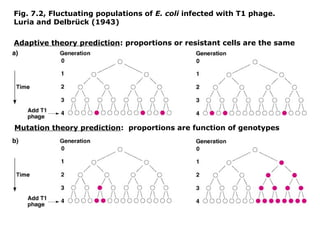
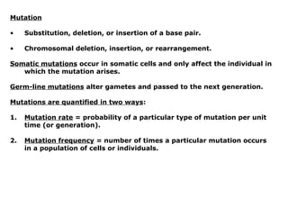
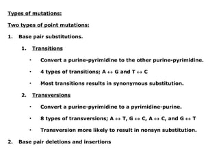
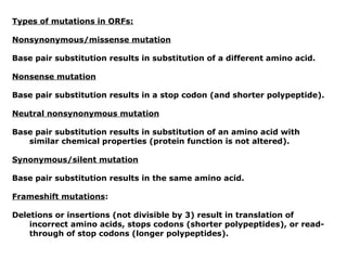
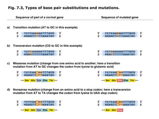
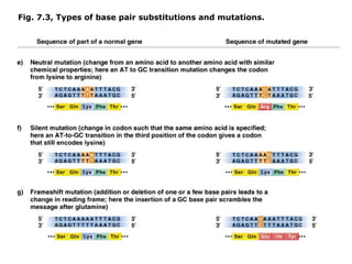
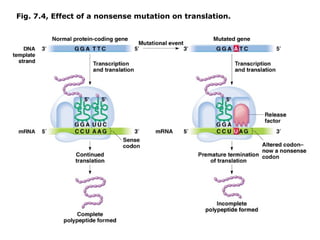
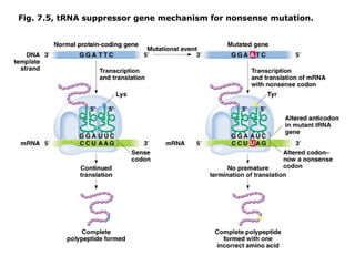
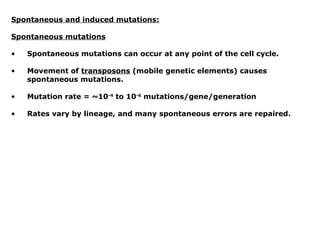
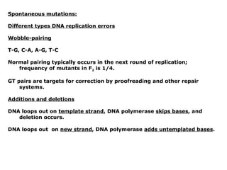
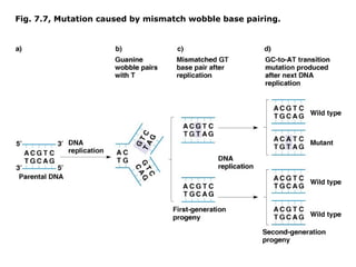
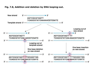
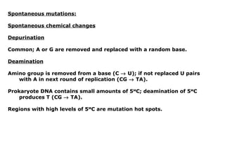
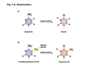
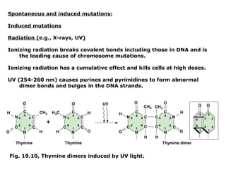
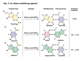
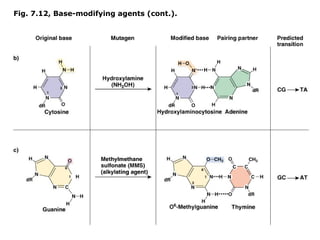
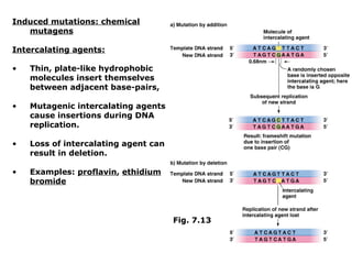
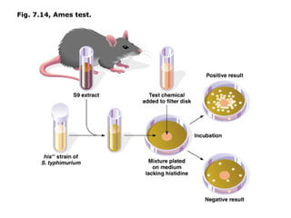
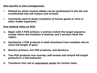
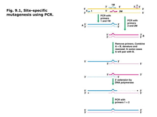
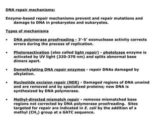
Chapter 7a - DNA mutation and repair:

•   Mutation and adaptation

•   Types of mutations

•   DNA repair mechanisms
Mutation and adaptation:

Jean-Baptiste Lamarck (1744-1829)

•   Proposed “inheritance of acquired traits” ~1801; induction by the
    environment; also known as transformism or transmutation.

•   Ideas largely ignored or attacked during his lifetime.

•   Never won the acceptance and esteem of his colleagues and died in
    poverty and obscurity.

•   Today Lamarck is associated with a discredited theory of heredity
    (but Lamarckism persisted until 1930s/1940s).
Charles Darwin (1809-1882)

•   Heritable adaptive variation results from random mutation and
    natural selection (1859, The Origin of Species).

•   Contrary to Larmarck, inheritance of adaptive traits does not result
    from induction by environmental influences.

•   But differential survival (selection) and heritable variation
    (originally arising from mutation).

•   Years following Darwin and rediscovery of Mendel resulted in
    controversy (until 1930s/1940s) about the relative importance of
    mutation and selection.

•   Largely resolved by theoretical and empirical work of Fisher,
    Haldane, and Wright (see chapter 21 lectures).
Experimental test of Lamarck’s “inheritance of acquired traits”

Salvador Luria and Max Delbrück (1943)

•    An E. coli population started from one cell should show different
     patterns of T1 resistance depending on which theory is correct.

1.   Adaptive theory states that cells are induced to become resistant
     when T1 is added; proportion of resistant cells should be the same
     for all cultures with the same genetic background.

2.   Mutation theory states that random events confer resistance to T1;
     duplicate cultures with the same genetic background should show
     different numbers of T1 resistant cells.
Fig. 7.2, Fluctuating populations of E. coli infected with T1 phage.
Luria and Delbrück (1943)

Adaptive theory prediction: proportions or resistant cells are the same


Mutation theory prediction: proportions are function of genotypes
Mutation

•    Substitution, deletion, or insertion of a base pair.

•    Chromosomal deletion, insertion, or rearrangement.

Somatic mutations occur in somatic cells and only affect the individual in
   which the mutation arises.

Germ-line mutations alter gametes and passed to the next generation.

Mutations are quantified in two ways:

1.   Mutation rate = probability of a particular type of mutation per unit
     time (or generation).

2.   Mutation frequency = number of times a particular mutation occurs
     in a population of cells or individuals.
Types of mutations:

Two types of point mutations:

1.   Base pair substitutions.

      1.       Transitions

           •     Convert a purine-pyrimidine to the other purine-pyrimidine.

           •     4 types of transitions; A ↔ G and T ↔ C

           •     Most transitions results in synonymous substitution.

      2.       Transversions

           •     Convert a purine-pyrimidine to a pyrimidine-purine.

           •     8 types of transversions; A ↔ T, G ↔ C, A ↔ C, and G ↔ T

           •     Transversion more likely to result in nonsyn substitution.

2.   Base pair deletions and insertions
Types of mutations in ORFs:

Nonsynonymous/missense mutation

Base pair substitution results in substitution of a different amino acid.

Nonsense mutation

Base pair substitution results in a stop codon (and shorter polypeptide).

Neutral nonsynonymous mutation

Base pair substitution results in substitution of an amino acid with
   similar chemical properties (protein function is not altered).

Synonymous/silent mutation

Base pair substitution results in the same amino acid.

Frameshift mutations:

Deletions or insertions (not divisible by 3) result in translation of
    incorrect amino acids, stops codons (shorter polypeptides), or read-
    through of stop codons (longer polypeptides).
Fig. 7.3, Types of base pair substitutions and mutations.
Fig. 7.4, Effect of a nonsense mutation on translation.
Reverse mutations and suppressor mutations:

Forward mutation

Mutation changes wild type to mutant.

Reverse mutation (back mutation)

Mutation changes mutant to wild type.

     •   Reversion to the wild type amino acid restores function.

     •   Reversion to another amino acid partly or fully restores
         function.

Suppressor mutation

Occur at sites different from the original mutation and mask or
   compensate for the initial mutation without reversing it.

     •   Intragenic suppressors occur on the same codon;
         e.g., nearby addition restores a deletion

     •   Intergenic suppressors occur on a different gene.
Intergenic suppressor genes:

•   Many function in mRNA translation.

•   Each suppressor gene works on only one type of nonsense,
    missense, of frameshift mutation.

•   Suppressor genes often encode tRNAs, which possess anti-codons
    that recognize stop codons and insert an amino acid.

•   Three classes of tRNA nonsense suppressors, one for each stop
    codon (UAG, UAA, UGA).

•   tRNA suppressor genes coexist with wild type tRNAS.

•   tRNA suppressors compete with release factors, which are important
    for proper amino acid chain termination.

•   Small number of read-through polypeptides are produced; tandem
    stop codons (UAGUAG) are required to result in correct translation
    termination.
Fig. 7.5, tRNA suppressor gene mechanism for nonsense mutation.
Spontaneous and induced mutations:

Spontaneous mutations

•   Spontaneous mutations can occur at any point of the cell cycle.

•   Movement of transposons (mobile genetic elements) causes
    spontaneous mutations.

•   Mutation rate = ~10-4 to 10-6 mutations/gene/generation

•   Rates vary by lineage, and many spontaneous errors are repaired.
Spontaneous mutations:

Different types DNA replication errors

Wobble-pairing

T-G, C-A, A-G, T-C

Normal pairing typically occurs in the next round of replication;
   frequency of mutants in F2 is 1/4.

GT pairs are targets for correction by proofreading and other repair
    systems.

Additions and deletions

DNA loops out on template strand, DNA polymerase skips bases, and
   deletion occurs.

DNA loops out on new strand, DNA polymerase adds untemplated bases.
Fig. 7.7, Mutation caused by mismatch wobble base pairing.
Fig. 7.8, Addition and deletion by DNA looping-out.
Spontaneous mutations:

Spontaneous chemical changes

Depurination

Common; A or G are removed and replaced with a random base.

Deamination

Amino group is removed from a base (C → U); if not replaced U pairs
   with A in next round of replication (CG → TA).

Prokaryote DNA contains small amounts of 5MC; deamination of 5MC
    produces T (CG → TA).

Regions with high levels of 5MC are mutation hot spots.
Fig. 7.9, Deamination.
Spontaneous and induced mutations:

Induced mutations

Radiation (e.g., X-rays, UV)

Ionizing radiation breaks covalent bonds including those in DNA and is
    the leading cause of chromosome mutations.

Ionizing radiation has a cumulative effect and kills cells at high doses.

UV (254-260 nm) causes purines and pyrimidines to form abnormal
    dimer bonds and bulges in the DNA strands.


Fig. 19.10, Thymine dimers induced by UV light.
Induced mutations: chemical mutagens

Base analogs

•   Similar to normal bases, incorporated into DNA during replication.

•   Some cause mis-pairing (e.g., 5-bromouracil).

•   Not all are mutagenic.
Fig. 7.11a, Mutagenic efffects
of 5-bromouracil
Fig. 7.11b, Mutagenic efffects
of 5-bromouracil
Induced mutations: Chemical mutagens

Base modifying agents, act at any stage of the cell cycle:

•   Deaminating agents

•   Hydroxylating agents

•   Alkylating agents
Fig. 7.12, Base-modifying agents.
Fig. 7.12, Base-modifying agents (cont.).
Induced mutations: chemical
   mutagens

Intercalating agents:

•   Thin, plate-like hydrophobic
    molecules insert themselves
    between adjacent base-pairs,

•   Mutagenic intercalating agents
    cause insertions during DNA
    replication.

•   Loss of intercalating agent can
    result in deletion.

•   Examples: proflavin, ethidium
    bromide


                                      Fig. 7.13
Detecting environmental mutations: Ames Test (after Bruce Ames)

Ames Test is an inexpensive method used to screen possible carcinogens
   and mutagens.

•   Histidine auxotroph Salmonella typhimurium (requires Histidine to
    grow) are mixed with rat liver enzymes and plated on media lacking
    histidine.

•   Liver enzymes are required to detect mutagens that are converted to
    carcinogenic forms by the liver (e.g., procarcinogens).

•   Test chemical is then added to medium.

•   Control plates show only a small # of revertants (bacteria cells
    growing without histidine).

•   Plates innoculated with mutagens or procarcinogens show a larger #
    of revertants.
Fig. 7.14, Ames test.
Site-specific in vitro mutagenesis:

•    Method by which mutant alleles can be synthesized in the lab and
     transformed into cell culture and animals.

•    Commonly used to study mutations of human genes in mice or
     other model organisms.

One method relies on PCR:

1.   Begin with 4 PCR primers; 2 primers match the target sequence
     except where the mutation is desired, and 2 primers flank the
     region.

2.   Synthesize 2 PCR products in both directions from mutation site to
     cover full length of gene

3.   Remove primers, mix PCR products, and denature.

4.   Two PCR products now overlap; self-anneal and extend full length
     products in a thermalcycler.

5.   Transform into cell or expression vector for further tests.
Fig. 9.1, Site-specific
mutagenesis using PCR.
DNA repair mechanisms:

Enzyme-based repair mechanisms prevent and repair mutations and
   damage to DNA in prokaryotes and eukaryotes.

Types of mechanisms

•   DNA polymerase proofreading - 3’-5’ exonuclease activity corrects
    errors during the process of replication.

•   Photoreactivation (also called light repair) - photolyase enzyme is
    activated by UV light (320-370 nm) and splits abnormal base
    dimers apart.

•   Demethylating DNA repair enzymes - repair DNAs damaged by
    alkylation.

•   Nucleotide excision repair (NER) - Damaged regions of DNA unwind
    and are removed and by specialized proteins; new DNA is
    synthesized by DNA polymerase.

•   Methyl-directed mismatch repair - removes mismatched base
    regions not corrected by DNA polymerase proofreading. Sites
    targeted for repair are indicated in E. coli by the addition of a
    methyl (CH3) group at a GATC sequence.
Fig. 7.16 Nucleotide excision repair (NER) of pyrimidine dimmer
and other damage-induced distortions of DNA
Fig. 7.17 Mechanism of mismatch correction repair

mutation ppt

  • 1.
    Chapter 7a -DNA mutation and repair: • Mutation and adaptation • Types of mutations • DNA repair mechanisms
  • 2.
    Mutation and adaptation: Jean-BaptisteLamarck (1744-1829) • Proposed “inheritance of acquired traits” ~1801; induction by the environment; also known as transformism or transmutation. • Ideas largely ignored or attacked during his lifetime. • Never won the acceptance and esteem of his colleagues and died in poverty and obscurity. • Today Lamarck is associated with a discredited theory of heredity (but Lamarckism persisted until 1930s/1940s).
  • 3.
    Charles Darwin (1809-1882) • Heritable adaptive variation results from random mutation and natural selection (1859, The Origin of Species). • Contrary to Larmarck, inheritance of adaptive traits does not result from induction by environmental influences. • But differential survival (selection) and heritable variation (originally arising from mutation). • Years following Darwin and rediscovery of Mendel resulted in controversy (until 1930s/1940s) about the relative importance of mutation and selection. • Largely resolved by theoretical and empirical work of Fisher, Haldane, and Wright (see chapter 21 lectures).
  • 4.
    Experimental test ofLamarck’s “inheritance of acquired traits” Salvador Luria and Max Delbrück (1943) • An E. coli population started from one cell should show different patterns of T1 resistance depending on which theory is correct. 1. Adaptive theory states that cells are induced to become resistant when T1 is added; proportion of resistant cells should be the same for all cultures with the same genetic background. 2. Mutation theory states that random events confer resistance to T1; duplicate cultures with the same genetic background should show different numbers of T1 resistant cells.
  • 5.
    Fig. 7.2, Fluctuatingpopulations of E. coli infected with T1 phage. Luria and Delbrück (1943) Adaptive theory prediction: proportions or resistant cells are the same Mutation theory prediction: proportions are function of genotypes
  • 6.
    Mutation • Substitution, deletion, or insertion of a base pair. • Chromosomal deletion, insertion, or rearrangement. Somatic mutations occur in somatic cells and only affect the individual in which the mutation arises. Germ-line mutations alter gametes and passed to the next generation. Mutations are quantified in two ways: 1. Mutation rate = probability of a particular type of mutation per unit time (or generation). 2. Mutation frequency = number of times a particular mutation occurs in a population of cells or individuals.
  • 7.
    Types of mutations: Twotypes of point mutations: 1. Base pair substitutions. 1. Transitions • Convert a purine-pyrimidine to the other purine-pyrimidine. • 4 types of transitions; A ↔ G and T ↔ C • Most transitions results in synonymous substitution. 2. Transversions • Convert a purine-pyrimidine to a pyrimidine-purine. • 8 types of transversions; A ↔ T, G ↔ C, A ↔ C, and G ↔ T • Transversion more likely to result in nonsyn substitution. 2. Base pair deletions and insertions
  • 8.
    Types of mutationsin ORFs: Nonsynonymous/missense mutation Base pair substitution results in substitution of a different amino acid. Nonsense mutation Base pair substitution results in a stop codon (and shorter polypeptide). Neutral nonsynonymous mutation Base pair substitution results in substitution of an amino acid with similar chemical properties (protein function is not altered). Synonymous/silent mutation Base pair substitution results in the same amino acid. Frameshift mutations: Deletions or insertions (not divisible by 3) result in translation of incorrect amino acids, stops codons (shorter polypeptides), or read- through of stop codons (longer polypeptides).
  • 9.
    Fig. 7.3, Typesof base pair substitutions and mutations.
  • 10.
    Fig. 7.3, Typesof base pair substitutions and mutations.
  • 11.
    Fig. 7.4, Effectof a nonsense mutation on translation.
  • 12.
    Reverse mutations andsuppressor mutations: Forward mutation Mutation changes wild type to mutant. Reverse mutation (back mutation) Mutation changes mutant to wild type. • Reversion to the wild type amino acid restores function. • Reversion to another amino acid partly or fully restores function. Suppressor mutation Occur at sites different from the original mutation and mask or compensate for the initial mutation without reversing it. • Intragenic suppressors occur on the same codon; e.g., nearby addition restores a deletion • Intergenic suppressors occur on a different gene.
  • 13.
    Intergenic suppressor genes: • Many function in mRNA translation. • Each suppressor gene works on only one type of nonsense, missense, of frameshift mutation. • Suppressor genes often encode tRNAs, which possess anti-codons that recognize stop codons and insert an amino acid. • Three classes of tRNA nonsense suppressors, one for each stop codon (UAG, UAA, UGA). • tRNA suppressor genes coexist with wild type tRNAS. • tRNA suppressors compete with release factors, which are important for proper amino acid chain termination. • Small number of read-through polypeptides are produced; tandem stop codons (UAGUAG) are required to result in correct translation termination.
  • 14.
    Fig. 7.5, tRNAsuppressor gene mechanism for nonsense mutation.
  • 15.
    Spontaneous and inducedmutations: Spontaneous mutations • Spontaneous mutations can occur at any point of the cell cycle. • Movement of transposons (mobile genetic elements) causes spontaneous mutations. • Mutation rate = ~10-4 to 10-6 mutations/gene/generation • Rates vary by lineage, and many spontaneous errors are repaired.
  • 16.
    Spontaneous mutations: Different typesDNA replication errors Wobble-pairing T-G, C-A, A-G, T-C Normal pairing typically occurs in the next round of replication; frequency of mutants in F2 is 1/4. GT pairs are targets for correction by proofreading and other repair systems. Additions and deletions DNA loops out on template strand, DNA polymerase skips bases, and deletion occurs. DNA loops out on new strand, DNA polymerase adds untemplated bases.
  • 17.
    Fig. 7.7, Mutationcaused by mismatch wobble base pairing.
  • 18.
    Fig. 7.8, Additionand deletion by DNA looping-out.
  • 19.
    Spontaneous mutations: Spontaneous chemicalchanges Depurination Common; A or G are removed and replaced with a random base. Deamination Amino group is removed from a base (C → U); if not replaced U pairs with A in next round of replication (CG → TA). Prokaryote DNA contains small amounts of 5MC; deamination of 5MC produces T (CG → TA). Regions with high levels of 5MC are mutation hot spots.
  • 20.
  • 21.
    Spontaneous and inducedmutations: Induced mutations Radiation (e.g., X-rays, UV) Ionizing radiation breaks covalent bonds including those in DNA and is the leading cause of chromosome mutations. Ionizing radiation has a cumulative effect and kills cells at high doses. UV (254-260 nm) causes purines and pyrimidines to form abnormal dimer bonds and bulges in the DNA strands. Fig. 19.10, Thymine dimers induced by UV light.
  • 22.
    Induced mutations: chemicalmutagens Base analogs • Similar to normal bases, incorporated into DNA during replication. • Some cause mis-pairing (e.g., 5-bromouracil). • Not all are mutagenic.
  • 23.
    Fig. 7.11a, Mutagenicefffects of 5-bromouracil
  • 24.
    Fig. 7.11b, Mutagenicefffects of 5-bromouracil
  • 25.
    Induced mutations: Chemicalmutagens Base modifying agents, act at any stage of the cell cycle: • Deaminating agents • Hydroxylating agents • Alkylating agents
  • 26.
  • 27.
  • 28.
    Induced mutations: chemical mutagens Intercalating agents: • Thin, plate-like hydrophobic molecules insert themselves between adjacent base-pairs, • Mutagenic intercalating agents cause insertions during DNA replication. • Loss of intercalating agent can result in deletion. • Examples: proflavin, ethidium bromide Fig. 7.13
  • 29.
    Detecting environmental mutations:Ames Test (after Bruce Ames) Ames Test is an inexpensive method used to screen possible carcinogens and mutagens. • Histidine auxotroph Salmonella typhimurium (requires Histidine to grow) are mixed with rat liver enzymes and plated on media lacking histidine. • Liver enzymes are required to detect mutagens that are converted to carcinogenic forms by the liver (e.g., procarcinogens). • Test chemical is then added to medium. • Control plates show only a small # of revertants (bacteria cells growing without histidine). • Plates innoculated with mutagens or procarcinogens show a larger # of revertants.
  • 30.
  • 31.
    Site-specific in vitromutagenesis: • Method by which mutant alleles can be synthesized in the lab and transformed into cell culture and animals. • Commonly used to study mutations of human genes in mice or other model organisms. One method relies on PCR: 1. Begin with 4 PCR primers; 2 primers match the target sequence except where the mutation is desired, and 2 primers flank the region. 2. Synthesize 2 PCR products in both directions from mutation site to cover full length of gene 3. Remove primers, mix PCR products, and denature. 4. Two PCR products now overlap; self-anneal and extend full length products in a thermalcycler. 5. Transform into cell or expression vector for further tests.
  • 32.
  • 33.
    DNA repair mechanisms: Enzyme-basedrepair mechanisms prevent and repair mutations and damage to DNA in prokaryotes and eukaryotes. Types of mechanisms • DNA polymerase proofreading - 3’-5’ exonuclease activity corrects errors during the process of replication. • Photoreactivation (also called light repair) - photolyase enzyme is activated by UV light (320-370 nm) and splits abnormal base dimers apart. • Demethylating DNA repair enzymes - repair DNAs damaged by alkylation. • Nucleotide excision repair (NER) - Damaged regions of DNA unwind and are removed and by specialized proteins; new DNA is synthesized by DNA polymerase. • Methyl-directed mismatch repair - removes mismatched base regions not corrected by DNA polymerase proofreading. Sites targeted for repair are indicated in E. coli by the addition of a methyl (CH3) group at a GATC sequence.
  • 34.
    Fig. 7.16 Nucleotideexcision repair (NER) of pyrimidine dimmer and other damage-induced distortions of DNA
  • 35.
    Fig. 7.17 Mechanismof mismatch correction repair