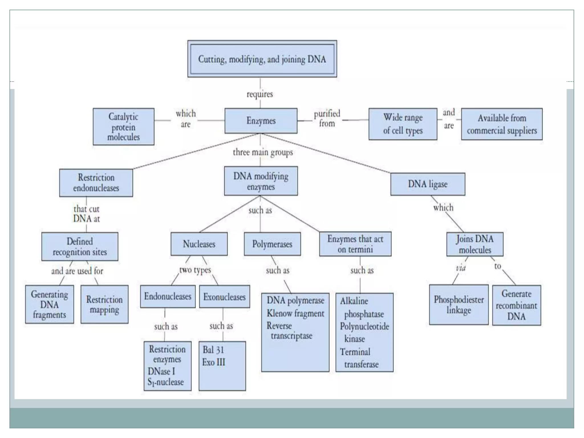
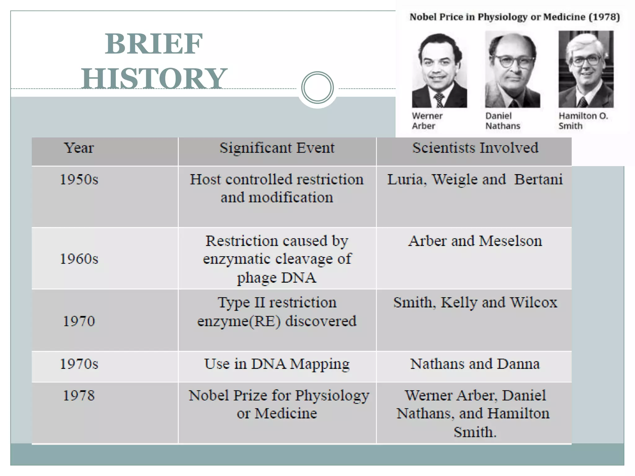
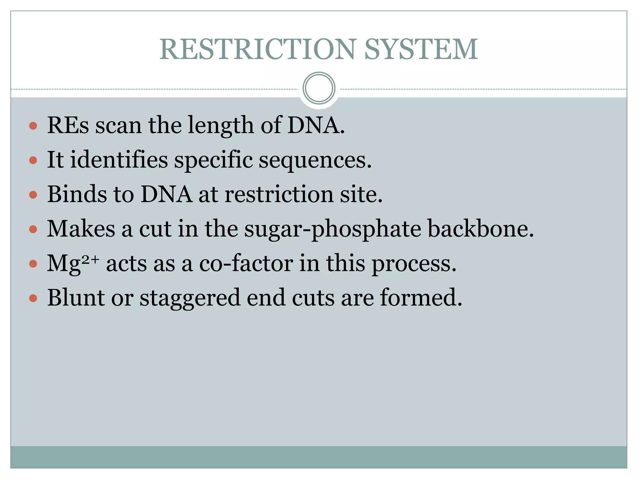
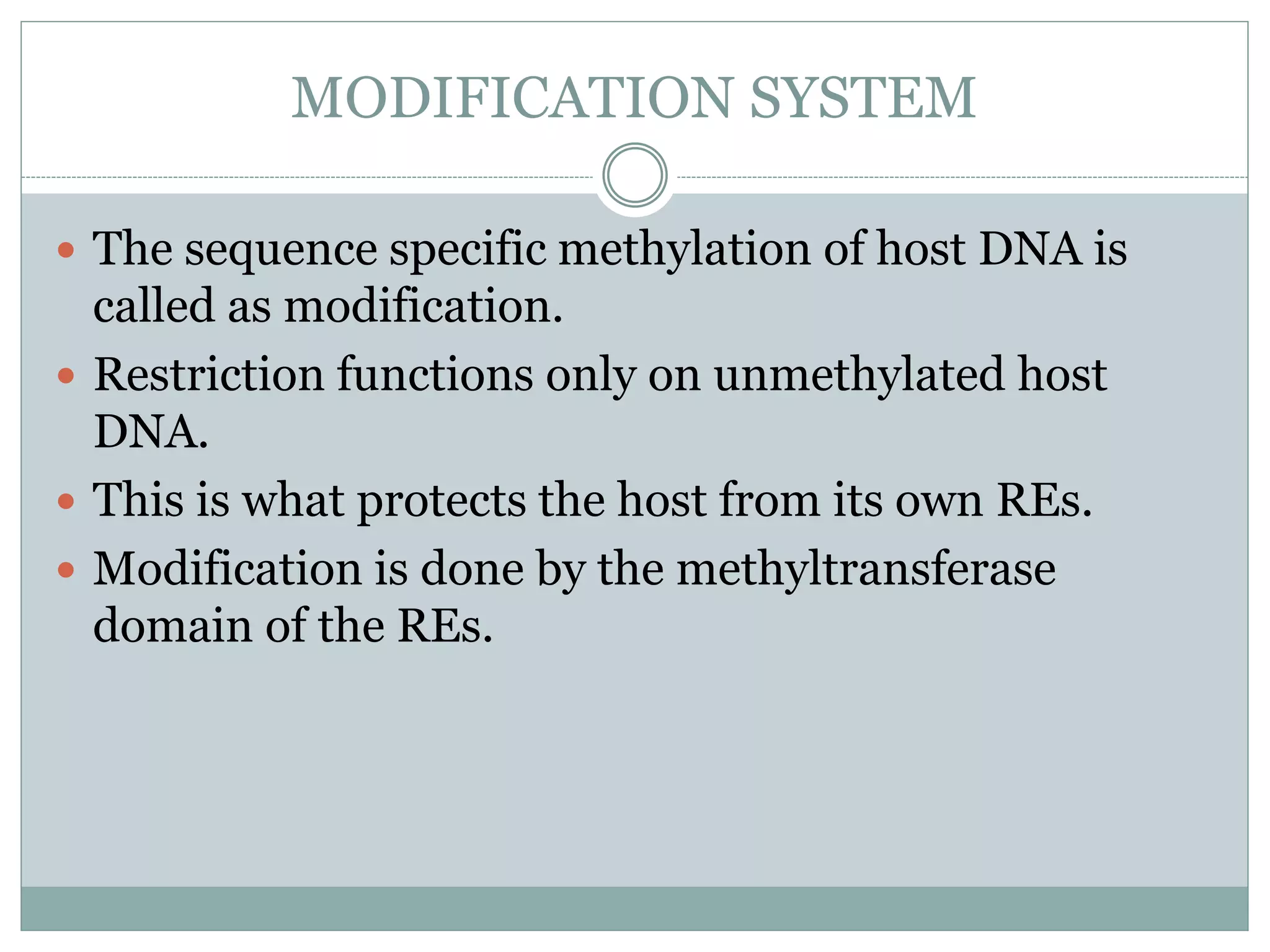
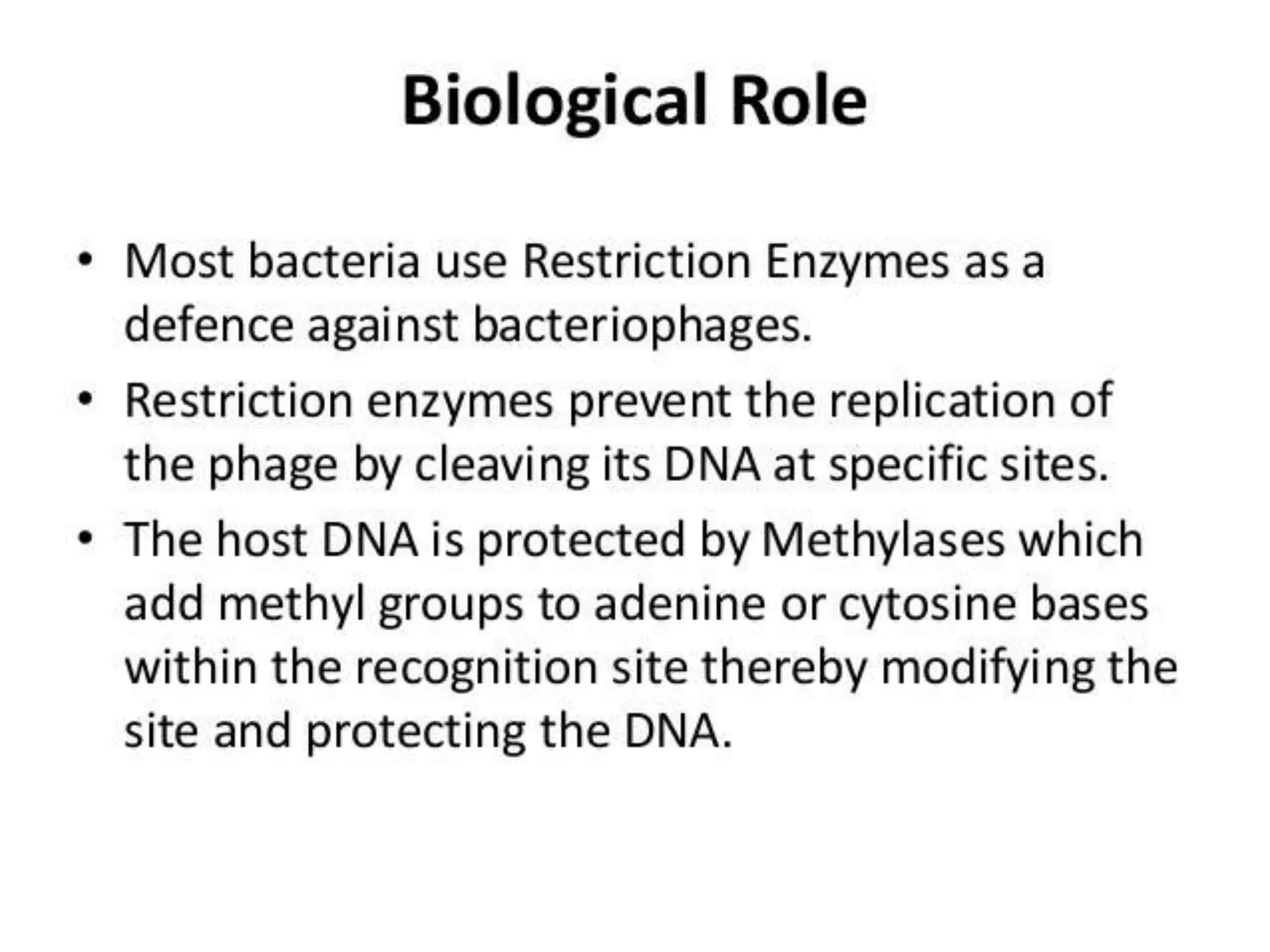
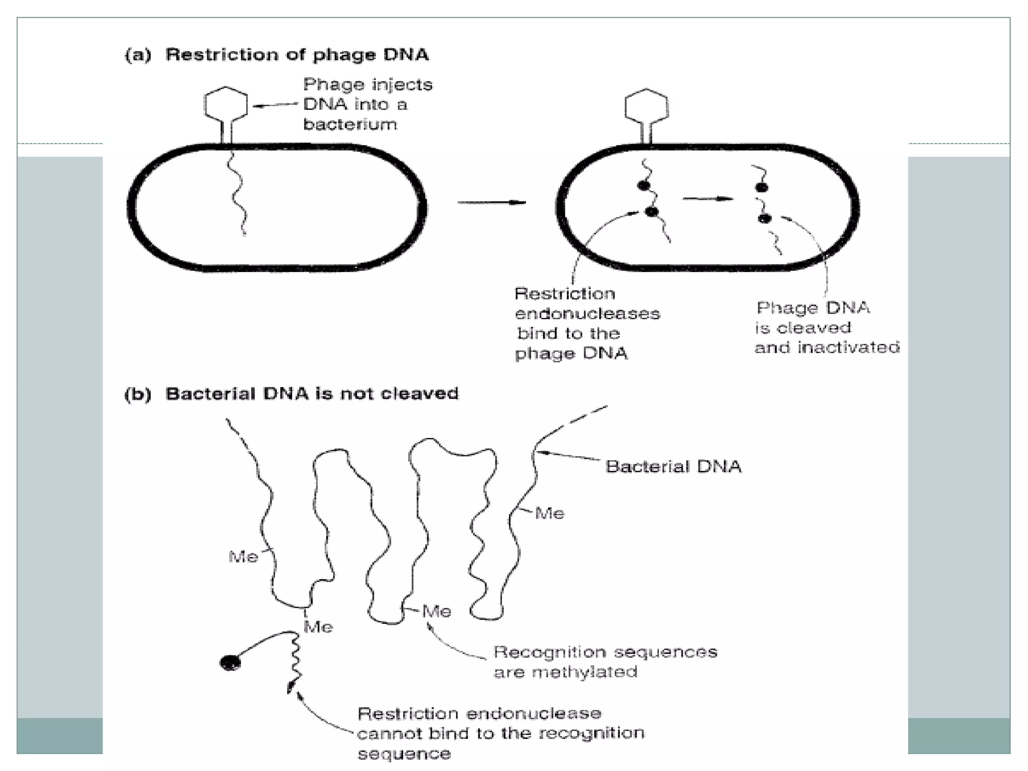
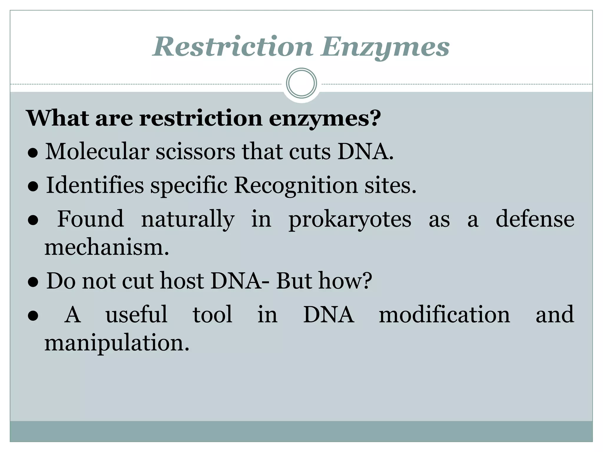
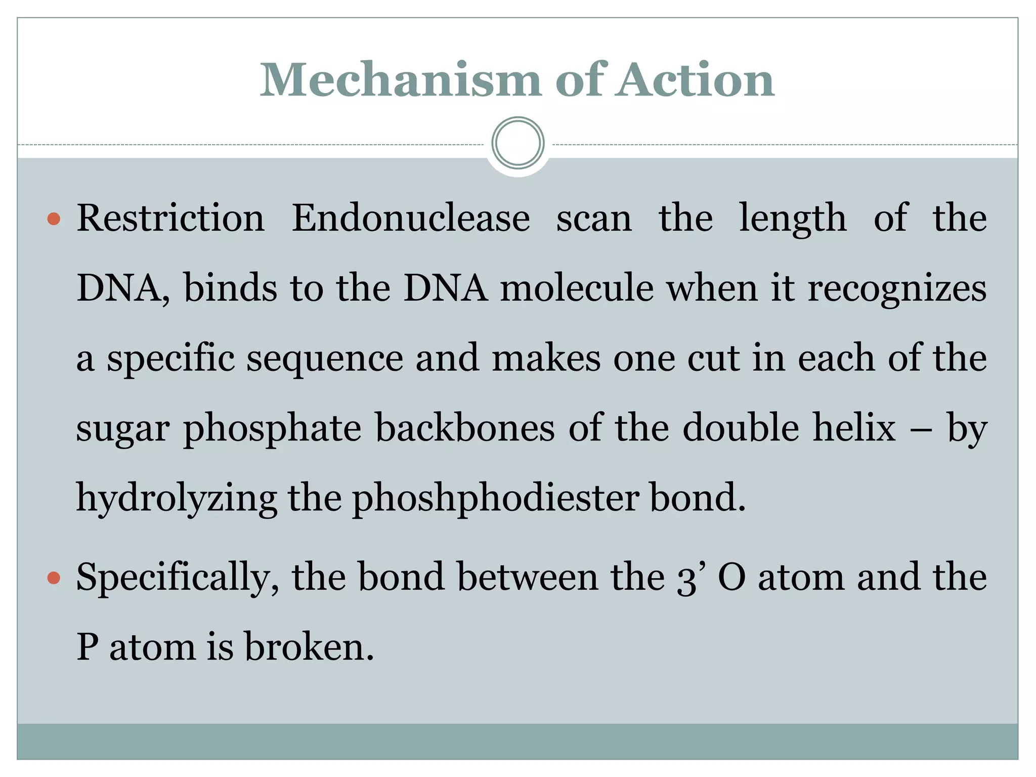
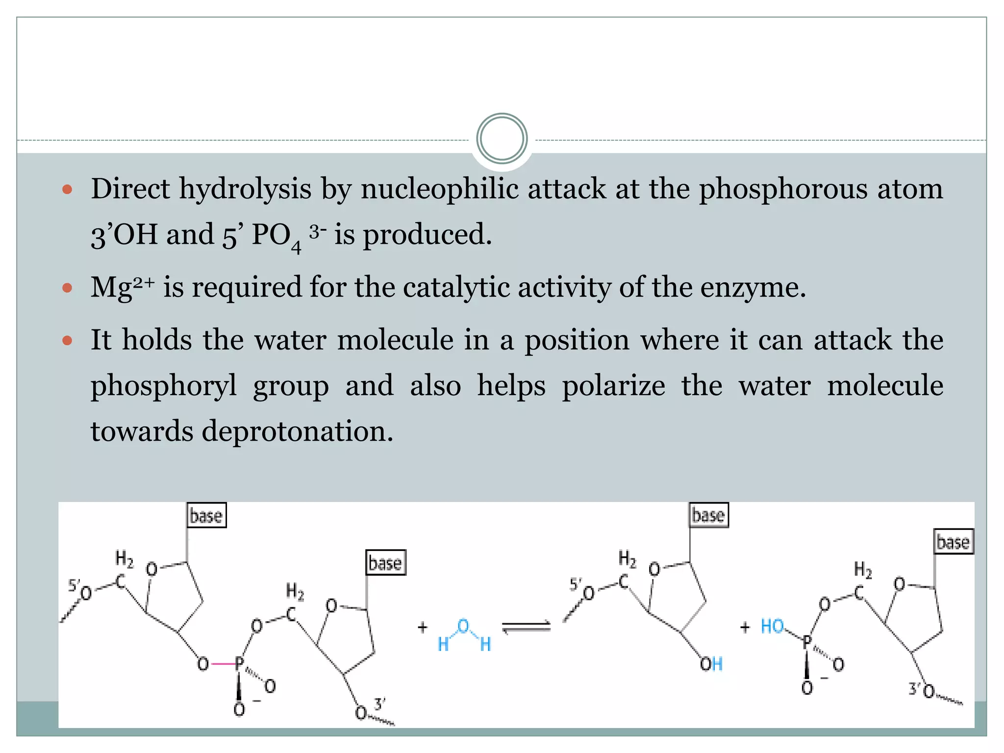
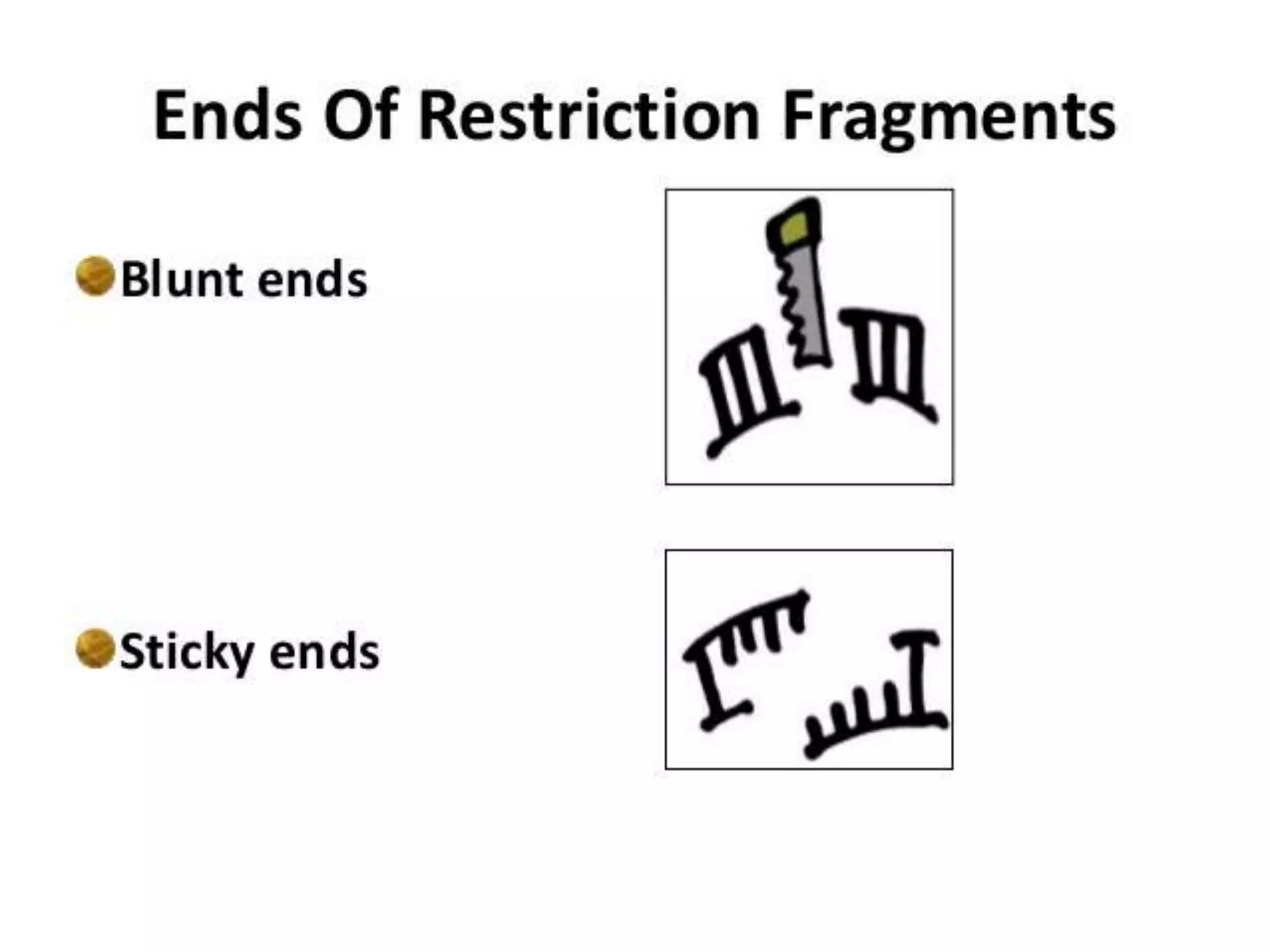
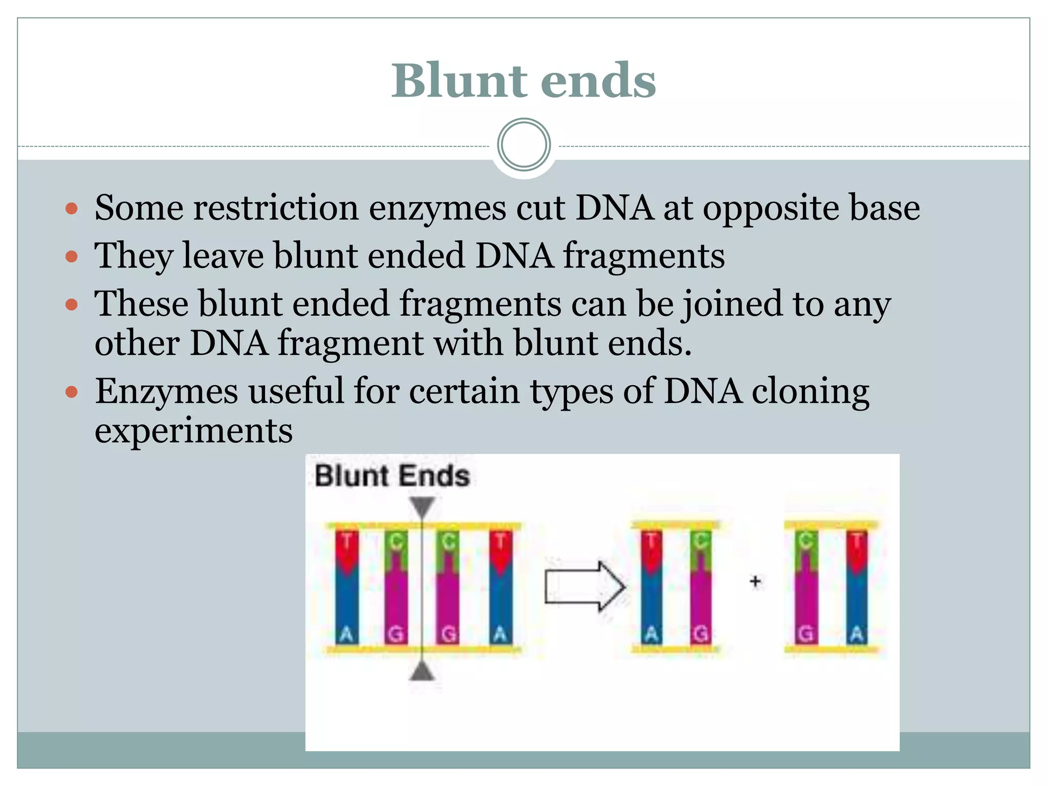
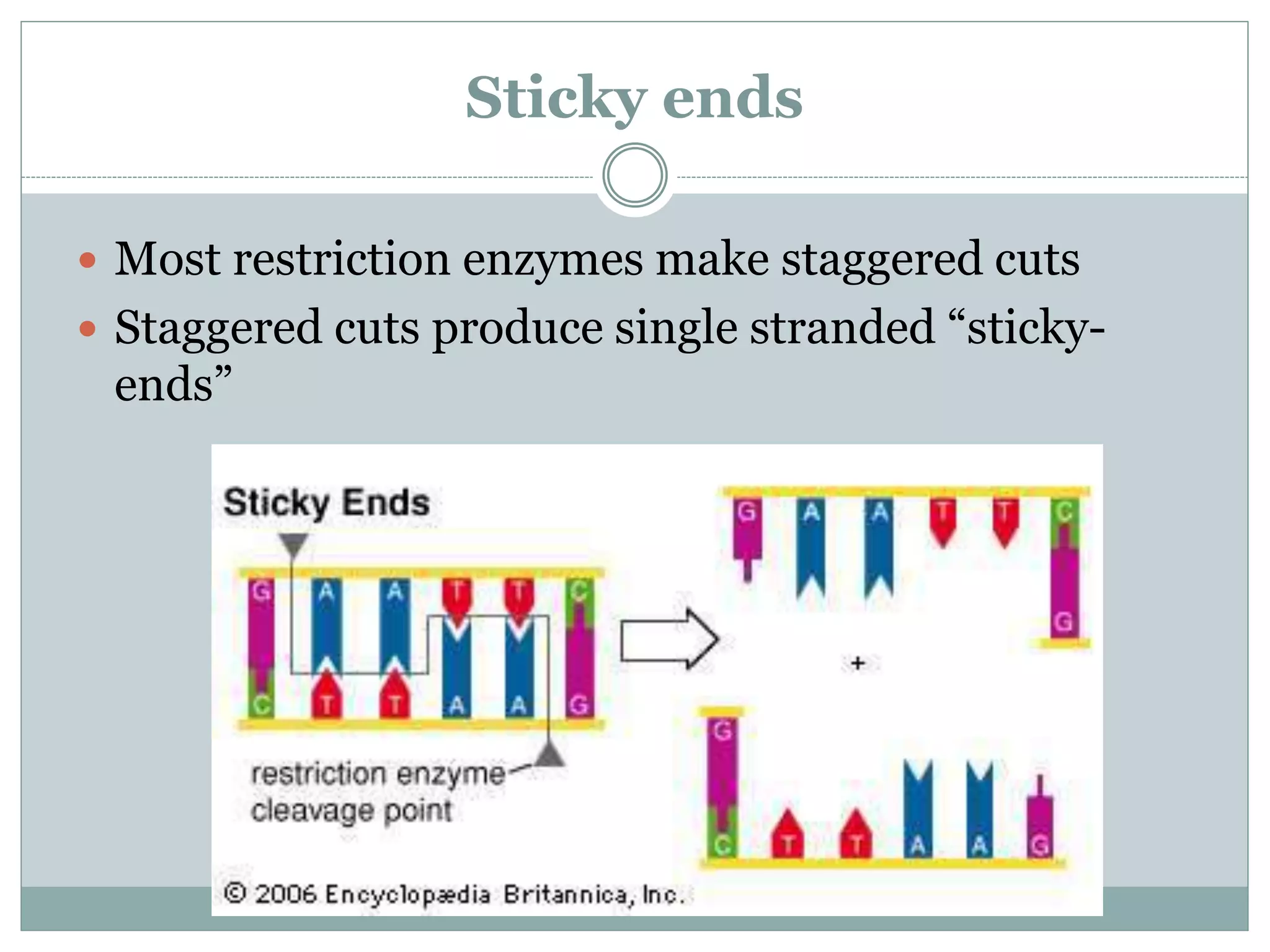
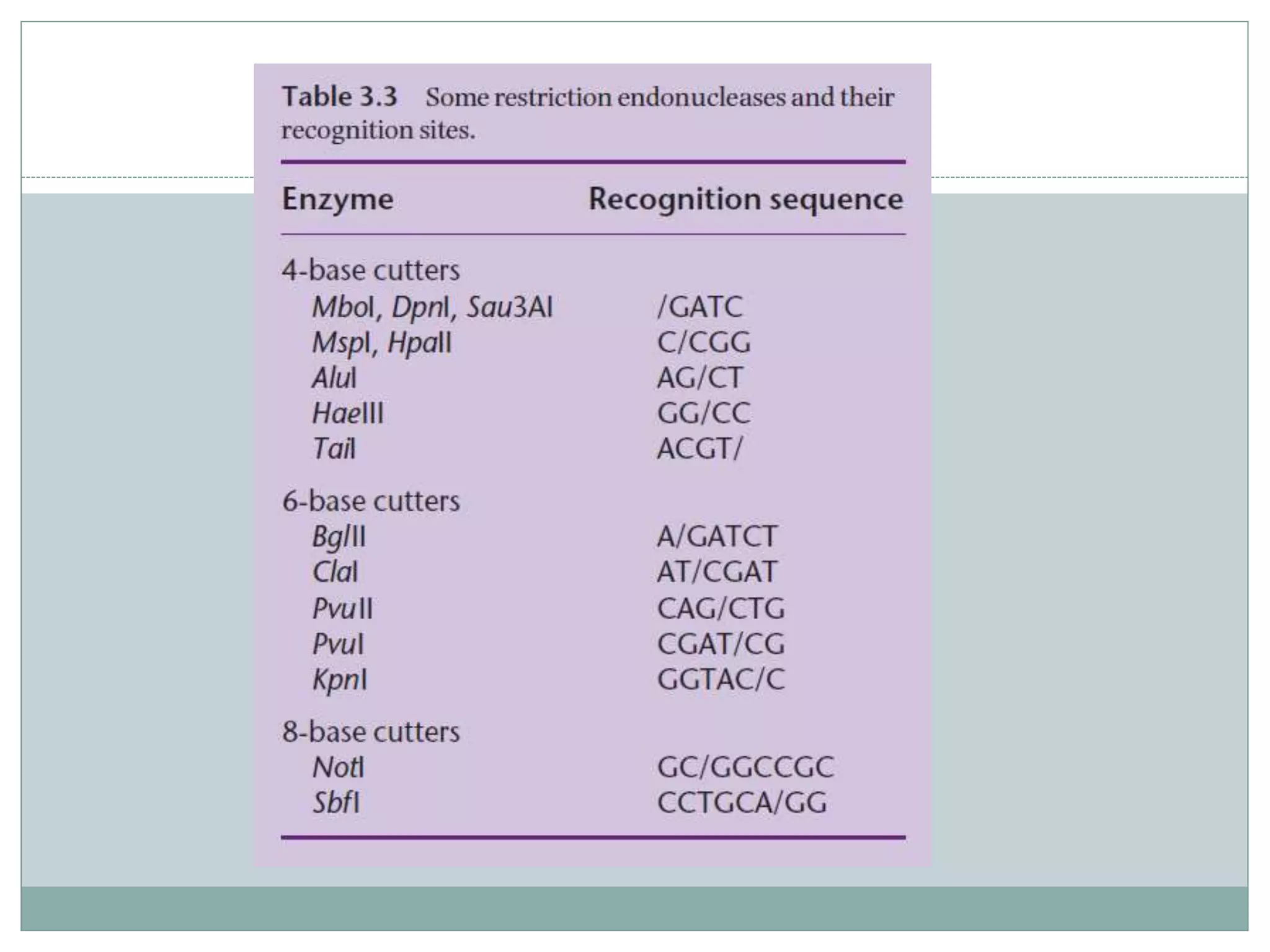
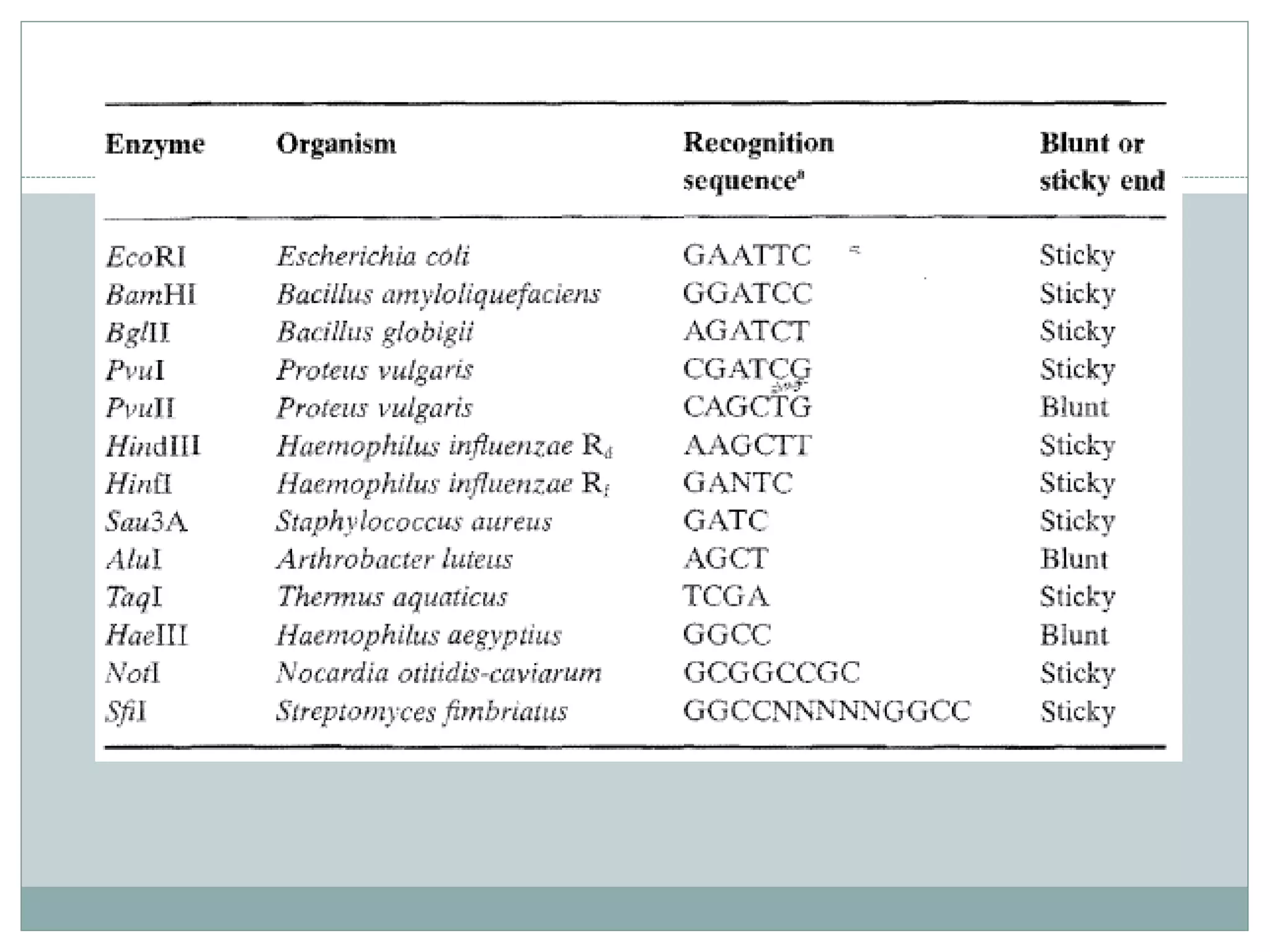
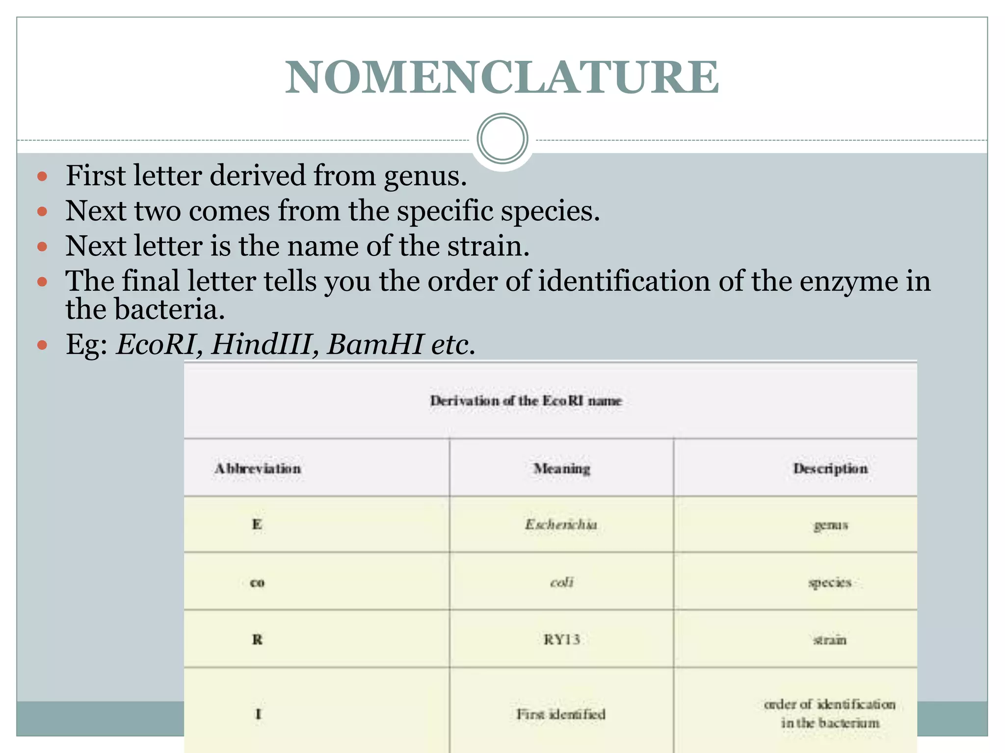
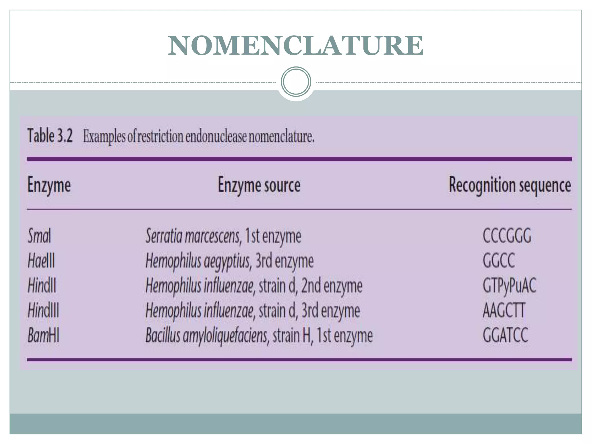
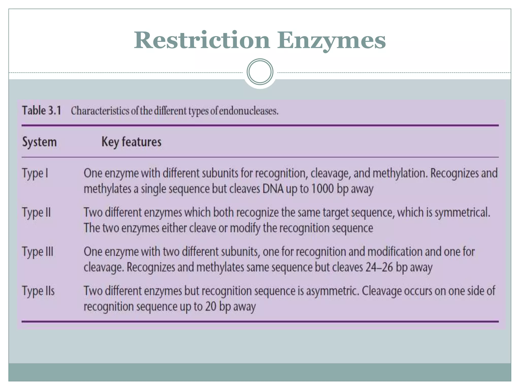
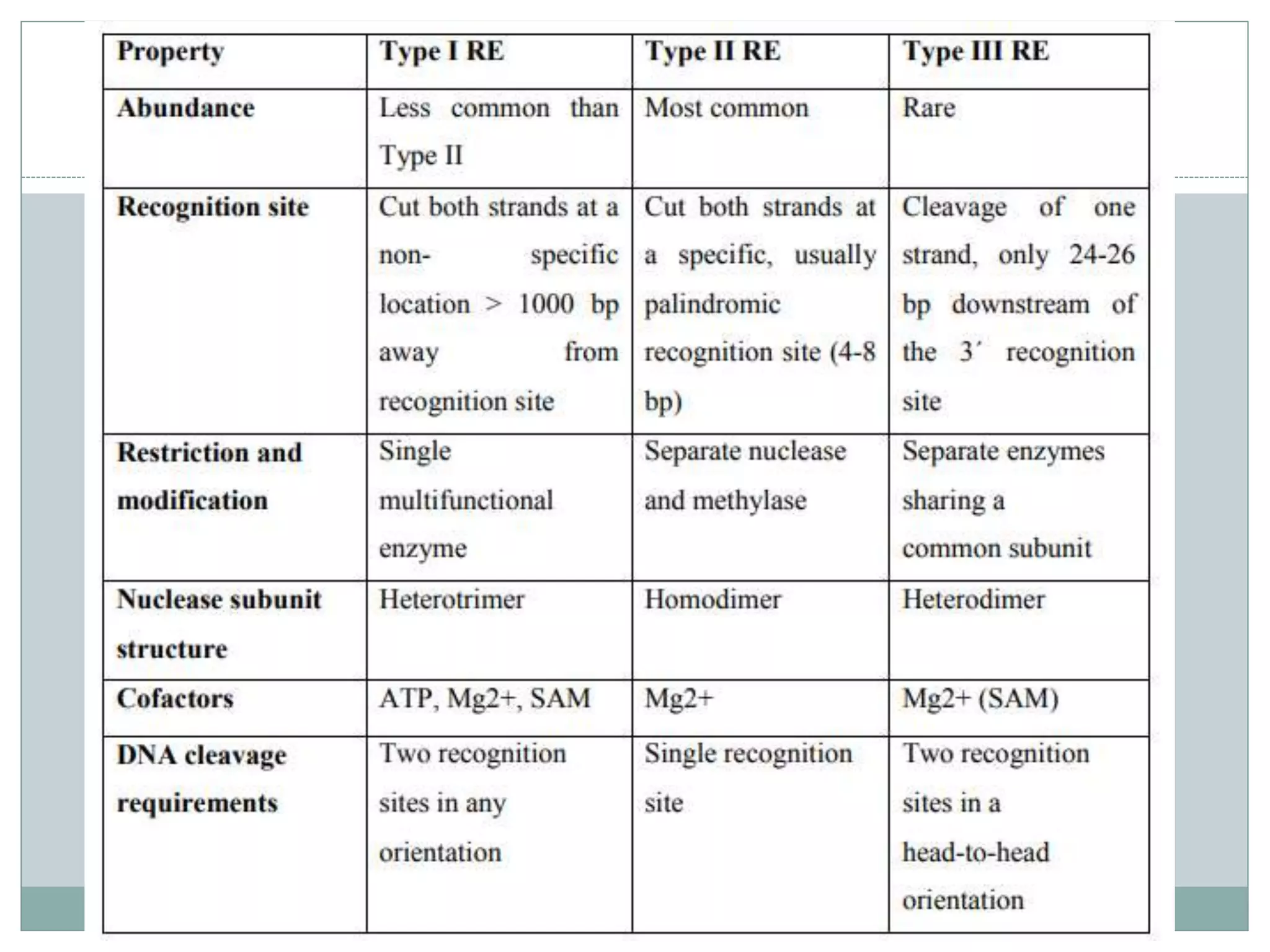
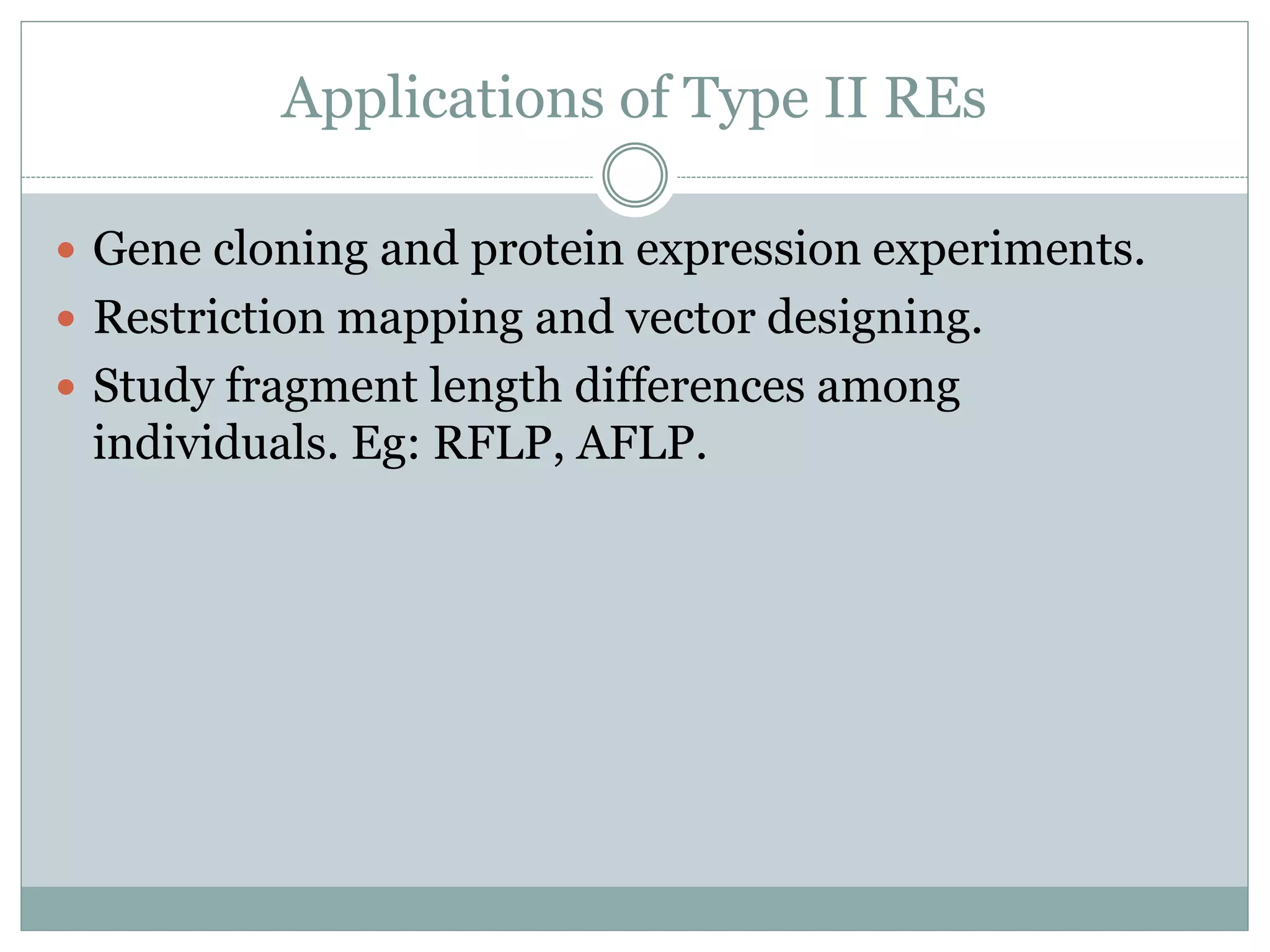
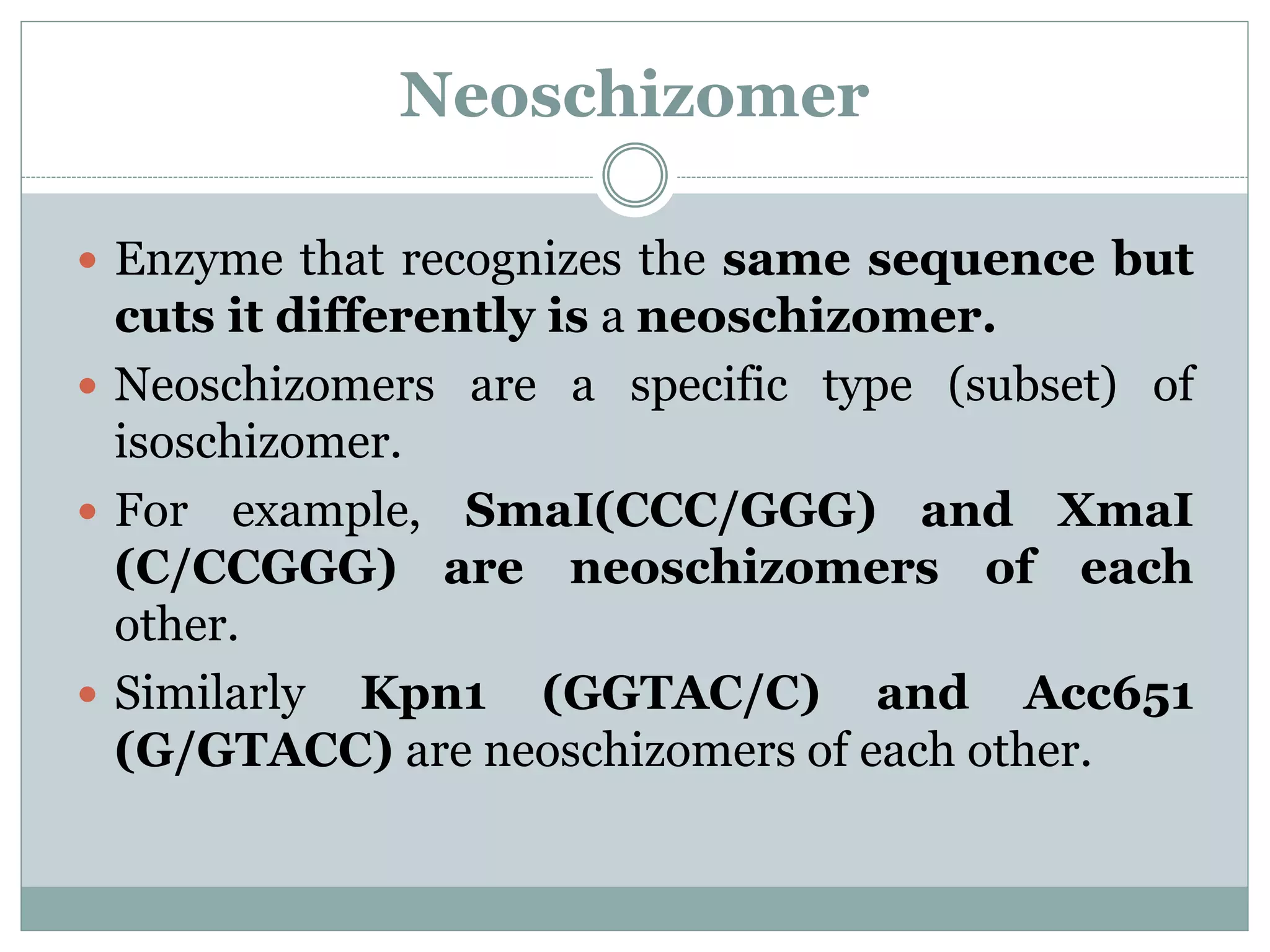
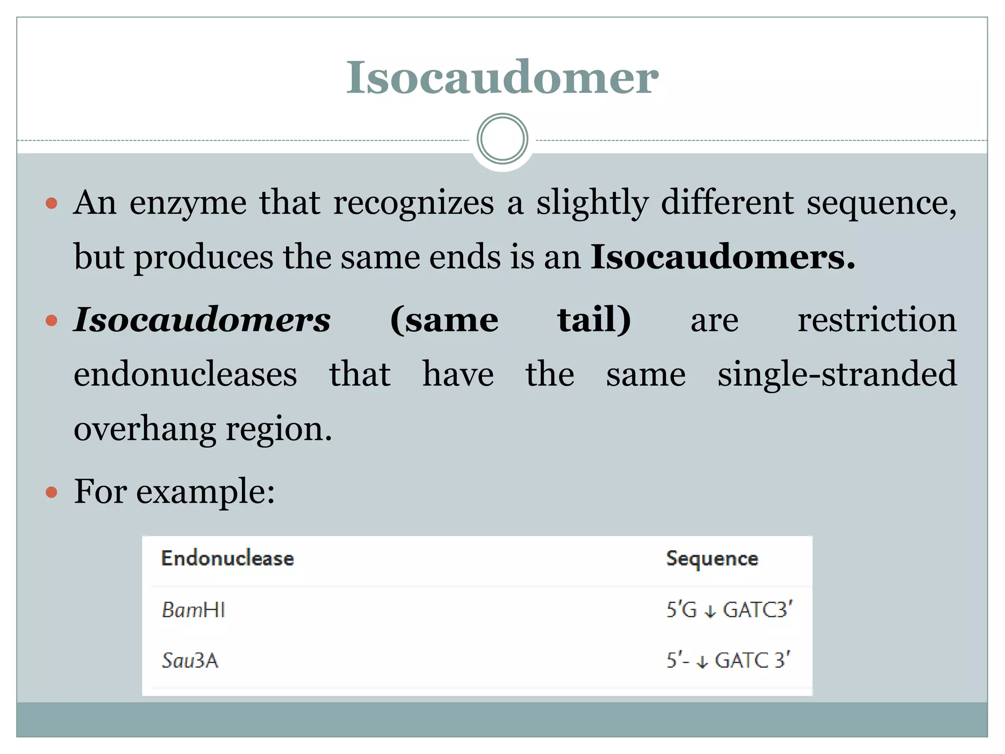
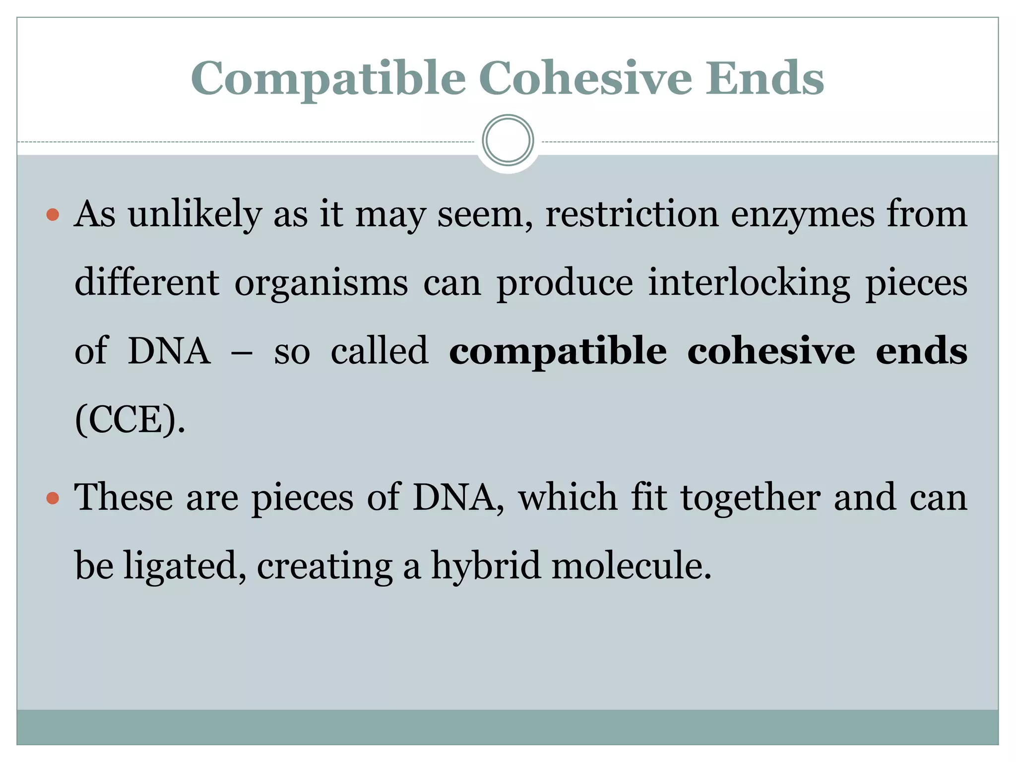
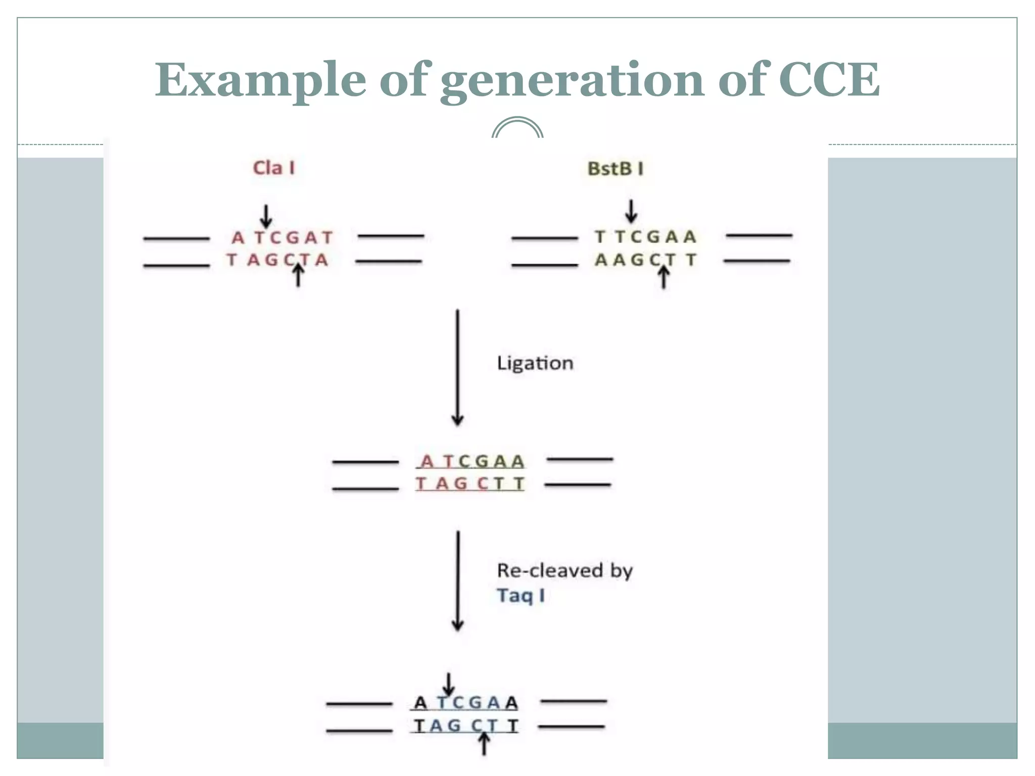
BT1502
UNIT I
Restriction and Modification
systems (RM system) – Biological
importance, Restriction enzymes
BRIEF
HISTORY
Restriction Modification system (RM system)
 The restriction modification system (RM
system) is found in bacteria and other prokaryotic
organisms, and provides a defense against foreign
DNA, such as that borne by bacteriophages.
 The RM system was first discovered by Salvatore
Luria and Mary Human in 1952 and 1953.
RESTRICTION SYSTEM
 REs scan the length of DNA.
 It identifies specific sequences.
 Binds to DNA at restriction site.
 Makes a cut in the sugar-phosphate backbone.
 Mg2+ acts as a co-factor in this process.
 Blunt or staggered end cuts are formed.
MODIFICATION SYSTEM
 The sequence specific methylation of host DNA is
called as modification.
 Restriction functions only on unmethylated host
DNA.
 This is what protects the host from its own REs.
 Modification is done by the methyltransferase
domain of the REs.
Restriction Enzymes
What are restriction enzymes?
● Molecular scissors that cuts DNA.
● Identifies specific Recognition sites.
● Found naturally in prokaryotes as a defense
mechanism.
● Do not cut host DNA- But how?
● A useful tool in DNA modification and
manipulation.
Mechanism of Action
 Restriction Endonuclease scan the length of the
DNA, binds to the DNA molecule when it recognizes
a specific sequence and makes one cut in each of the
sugar phosphate backbones of the double helix – by
hydrolyzing the phoshphodiester bond.
 Specifically, the bond between the 3’ O atom and the
P atom is broken.
 Direct hydrolysis by nucleophilic attack at the phosphorous atom
3’OH and 5’ PO4
3- is produced.
 Mg2+ is required for the catalytic activity of the enzyme.
 It holds the water molecule in a position where it can attack the
phosphoryl group and also helps polarize the water molecule
towards deprotonation.
Blunt ends
 Some restriction enzymes cut DNA at opposite base
 They leave blunt ended DNA fragments
 These blunt ended fragments can be joined to any
other DNA fragment with blunt ends.
 Enzymes useful for certain types of DNA cloning
experiments
Sticky ends
 Most restriction enzymes make staggered cuts
 Staggered cuts produce single stranded “sticky-
ends”
NOMENCLATURE
 First letter derived from genus.
 Next two comes from the specific species.
 Next letter is the name of the strain.
 The final letter tells you the order of identification of the enzyme in
the bacteria.
 Eg: EcoRI, HindIII, BamHI etc.
NOMENCLATURE
Types of Restriction Enzymes
FUNCTIONAL ROLES OF R-M SYSTEM
S-adenosyl-L-methionine
TYPE I ENDONUCLEASES
 First to be identified by Arber and Meselson.
 Asymmetric recognition sequence.
 Requires various co-factors including SAM, ATP and
Mg2+.
 Single enzyme that performs restriction and
modification functions.
 Contains 3 subunits HsdS, HsdM and HsdR.
 two R(restriction) subunits
 two M(methylation) subunits
 one S(specifity) subunits
TYPE I ENDONUCLEASES
 Are the most complex, consisting of three polypeptides: R
(restriction), M (modification), and S (specificity).
 The resulting complex can both cleave and methylate DNA.
 Both reactions require ATP, and cleavage often occurs a
considerable distance from the recognition site.
 The S subunit determines the specificity of both restriction
and methylation.
 Cleavage occurs at variable distances from the recognition
sequence, so discrete bands are not easily visualized by gel
electrophoresis.
TYPE II ENDONUCLEASES
 First identified in 1970 (HindII).
 Most commonly used in genetic manipulation
experiments.
 Recognizes 4-8 bp long pallindromic sequences.
 Cleaves within (mostly) the recognition sequence.
 Only Mg2+ required as cofactor, doesn’t require SAM
or ATP for function.
TYPE II ENDONUCLEASES
 Are the simplest and the most prevalent.
 Instead of working as a complex, the methyltransferase and endonuclease are
encoded as two separate proteins and act independently (there is no specificity
protein).
 Both proteins recognize the same recognition site, and therefore compete for
activity.
 The methyltransferase acts as a monomer, methylating the duplex one strand at a
time.
 The endonuclease acts as a homodimer, which facilitates the cleavage of both
strands.
 Cleavage occurs at a defined position close to or within the recognition sequence,
thus producing discrete fragments during gel electrophoresis.
 For this reason, Type II systems are used in labs for DNA analysis and gene cloning.
TYPE III ENDONUCLEASES
 Cleave DNA at immediate vicinity, about 20-30 base
pairs away from recognition sequence.
 Recognizes two separate non-palindromic sequences
that are inversely oriented.
 ATP, SAM (not essential) and Mg2+ acts as co-factor.
 Separate enzymes for restriction and modification,
but share a common subunit.
TYPE III ENDONUCLEASES
 Have R (res) and M (mod) proteins that form a
complex of modification and cleavage.
 The M protein, however, can methylate on its own.
Methylation also only occurs on one strand of the DNA
unlike most other known mechanisms.
 The heterodimer formed by the R and M proteins
competes with itself by modifying and restricting the
same reaction.
 This results in incomplete digestion
TYPE IV ENDONUCLEASES
 Cleave only modified DNA (methylated,
hydroxymethylated and glucosylhydroxymethylated
bases).
 Recognition sequences have not been well defined
 Cleavage takes place ~30 bp away from one of the
sites
TYPE IV ENDONUCLEASES
 Are not true RM systems because they only contain a
restriction enzyme and not a methylase.
 Unlike the other types, type IV restriction enzymes
recognize and cut only modified DNA.
Restriction Enzymes
Applications of Type II REs
 Gene cloning and protein expression experiments.
 Restriction mapping and vector designing.
 Study fragment length differences among
individuals. Eg: RFLP, AFLP.
Learning Check
 What are restriction endonucleases?
 How are these enzymes named?
 What are the types of RE?
 What is the mechanism of action of RE?
 What are the main functions of RE?
 What is the difference between Sticky ends and blunt
ends?
PALINDROME SEQUENCES
 The mirror like palindrome in which the same forward
and backwards are on a single strand of DNA strand, as in
GTAATG
 The Inverted repeat palindromes is also a sequence
that reads the same forward and backwards, but the forward
and backward sequences are found in complementary DNA
strands (GTATAC being complementary to CATATG)
 Inverted repeat palindromes are more common and
have greater biological importance than mirror like
palindromes.
Star effect
 Under extreme conditions such as elevated pH or low
ionic strength, RE are capable of cleaving sequences which
are similar but not identical to their recognition sequence
 Example: The rate difference for EcoR I at its cognate site (5'-
GAATTC-3') and next-best site (5'-TAATTC-3') is of the order
of 105. Similarly, for EcoR V, cleavage at its cognate site (5'-
GATATC-3') is 106 times faster than at the next-best site (5'-
GTTATC-3').
 The term star activity was introduced by Mayer who
characterized the modified activity in EcoRI.
Star Activity
 Star activity is the relaxation or alteration of the specificity of
restriction enzyme mediated cleavage of DNA that can occur under
reaction conditions that differ significantly from those optimal for
the enzyme.
 Differences which can lead to star include low ionic strength,
high pH, and high (> 5% v/v) glycerol concentrations.
 Star activity can happen because of presence of Mg2+, as is seen in
HindIII, for example.
 In order to suppress star activity, it is recommend performing
reactions at lower glycerol concentrations, neutral pH, and
higher salt concentrations.
Isoschizomer
 Restriction enzymes specific to the same recognition sequence.
 Isoschizomers are pairs of restriction enzymes specific to the
same recognition sequence.
 For example, SphI (CGTAC/G) and BbuI (CGTAC/G) are
isoschizomers of each other.
 Another example: Restriction enzymes HpaII and MspI are
isoschizomers, as they both recognize the sequence 5'-CCGG-3'
when it is unmethylated.
 But when the second C of the sequence is methylated, only
MspI can recognize it while HpaII cannot.
Neoschizomer
 Enzyme that recognizes the same sequence but
cuts it differently is a neoschizomer.
 Neoschizomers are a specific type (subset) of
isoschizomer.
 For example, SmaI(CCC/GGG) and XmaI
(C/CCGGG) are neoschizomers of each
other.
 Similarly Kpn1 (GGTAC/C) and Acc651
(G/GTACC) are neoschizomers of each other.
Isocaudomer
 An enzyme that recognizes a slightly different sequence,
but produces the same ends is an Isocaudomers.
 Isocaudomers (same tail) are restriction
endonucleases that have the same single-stranded
overhang region.
 For example:
Compatible Cohesive Ends
 As unlikely as it may seem, restriction enzymes from
different organisms can produce interlocking pieces
of DNA – so called compatible cohesive ends
(CCE).
 These are pieces of DNA, which fit together and can
be ligated, creating a hybrid molecule.
Example of generation of CCE
Learning Check
 What are the modification enzymes?
 Which enzymes are used for modification of ends of
DNA?
 What is Star activity?
 What is the difference between isoschizomers and
neoschizomer?
 What is the significance of CCE?
Thank you

Restriction-Modification system, Types of Restriction enzymes

  • 1.
    BT1502 UNIT I Restriction andModification systems (RM system) – Biological importance, Restriction enzymes
  • 3.
  • 4.
    Restriction Modification system(RM system)  The restriction modification system (RM system) is found in bacteria and other prokaryotic organisms, and provides a defense against foreign DNA, such as that borne by bacteriophages.  The RM system was first discovered by Salvatore Luria and Mary Human in 1952 and 1953.
  • 5.
    RESTRICTION SYSTEM  REsscan the length of DNA.  It identifies specific sequences.  Binds to DNA at restriction site.  Makes a cut in the sugar-phosphate backbone.  Mg2+ acts as a co-factor in this process.  Blunt or staggered end cuts are formed.
  • 6.
    MODIFICATION SYSTEM  Thesequence specific methylation of host DNA is called as modification.  Restriction functions only on unmethylated host DNA.  This is what protects the host from its own REs.  Modification is done by the methyltransferase domain of the REs.
  • 9.
    Restriction Enzymes What arerestriction enzymes? ● Molecular scissors that cuts DNA. ● Identifies specific Recognition sites. ● Found naturally in prokaryotes as a defense mechanism. ● Do not cut host DNA- But how? ● A useful tool in DNA modification and manipulation.
  • 10.
    Mechanism of Action Restriction Endonuclease scan the length of the DNA, binds to the DNA molecule when it recognizes a specific sequence and makes one cut in each of the sugar phosphate backbones of the double helix – by hydrolyzing the phoshphodiester bond.  Specifically, the bond between the 3’ O atom and the P atom is broken.
  • 11.
     Direct hydrolysisby nucleophilic attack at the phosphorous atom 3’OH and 5’ PO4 3- is produced.  Mg2+ is required for the catalytic activity of the enzyme.  It holds the water molecule in a position where it can attack the phosphoryl group and also helps polarize the water molecule towards deprotonation.
  • 13.
    Blunt ends  Somerestriction enzymes cut DNA at opposite base  They leave blunt ended DNA fragments  These blunt ended fragments can be joined to any other DNA fragment with blunt ends.  Enzymes useful for certain types of DNA cloning experiments
  • 14.
    Sticky ends  Mostrestriction enzymes make staggered cuts  Staggered cuts produce single stranded “sticky- ends”
  • 17.
    NOMENCLATURE  First letterderived from genus.  Next two comes from the specific species.  Next letter is the name of the strain.  The final letter tells you the order of identification of the enzyme in the bacteria.  Eg: EcoRI, HindIII, BamHI etc.
  • 18.
  • 20.
  • 21.
    FUNCTIONAL ROLES OFR-M SYSTEM S-adenosyl-L-methionine
  • 22.
    TYPE I ENDONUCLEASES First to be identified by Arber and Meselson.  Asymmetric recognition sequence.  Requires various co-factors including SAM, ATP and Mg2+.  Single enzyme that performs restriction and modification functions.  Contains 3 subunits HsdS, HsdM and HsdR.  two R(restriction) subunits  two M(methylation) subunits  one S(specifity) subunits
  • 23.
    TYPE I ENDONUCLEASES Are the most complex, consisting of three polypeptides: R (restriction), M (modification), and S (specificity).  The resulting complex can both cleave and methylate DNA.  Both reactions require ATP, and cleavage often occurs a considerable distance from the recognition site.  The S subunit determines the specificity of both restriction and methylation.  Cleavage occurs at variable distances from the recognition sequence, so discrete bands are not easily visualized by gel electrophoresis.
  • 24.
    TYPE II ENDONUCLEASES First identified in 1970 (HindII).  Most commonly used in genetic manipulation experiments.  Recognizes 4-8 bp long pallindromic sequences.  Cleaves within (mostly) the recognition sequence.  Only Mg2+ required as cofactor, doesn’t require SAM or ATP for function.
  • 25.
    TYPE II ENDONUCLEASES Are the simplest and the most prevalent.  Instead of working as a complex, the methyltransferase and endonuclease are encoded as two separate proteins and act independently (there is no specificity protein).  Both proteins recognize the same recognition site, and therefore compete for activity.  The methyltransferase acts as a monomer, methylating the duplex one strand at a time.  The endonuclease acts as a homodimer, which facilitates the cleavage of both strands.  Cleavage occurs at a defined position close to or within the recognition sequence, thus producing discrete fragments during gel electrophoresis.  For this reason, Type II systems are used in labs for DNA analysis and gene cloning.
  • 26.
    TYPE III ENDONUCLEASES Cleave DNA at immediate vicinity, about 20-30 base pairs away from recognition sequence.  Recognizes two separate non-palindromic sequences that are inversely oriented.  ATP, SAM (not essential) and Mg2+ acts as co-factor.  Separate enzymes for restriction and modification, but share a common subunit.
  • 27.
    TYPE III ENDONUCLEASES Have R (res) and M (mod) proteins that form a complex of modification and cleavage.  The M protein, however, can methylate on its own. Methylation also only occurs on one strand of the DNA unlike most other known mechanisms.  The heterodimer formed by the R and M proteins competes with itself by modifying and restricting the same reaction.  This results in incomplete digestion
  • 28.
    TYPE IV ENDONUCLEASES Cleave only modified DNA (methylated, hydroxymethylated and glucosylhydroxymethylated bases).  Recognition sequences have not been well defined  Cleavage takes place ~30 bp away from one of the sites
  • 29.
    TYPE IV ENDONUCLEASES Are not true RM systems because they only contain a restriction enzyme and not a methylase.  Unlike the other types, type IV restriction enzymes recognize and cut only modified DNA.
  • 30.
  • 32.
    Applications of TypeII REs  Gene cloning and protein expression experiments.  Restriction mapping and vector designing.  Study fragment length differences among individuals. Eg: RFLP, AFLP.
  • 33.
    Learning Check  Whatare restriction endonucleases?  How are these enzymes named?  What are the types of RE?  What is the mechanism of action of RE?  What are the main functions of RE?  What is the difference between Sticky ends and blunt ends?
  • 34.
    PALINDROME SEQUENCES  Themirror like palindrome in which the same forward and backwards are on a single strand of DNA strand, as in GTAATG  The Inverted repeat palindromes is also a sequence that reads the same forward and backwards, but the forward and backward sequences are found in complementary DNA strands (GTATAC being complementary to CATATG)  Inverted repeat palindromes are more common and have greater biological importance than mirror like palindromes.
  • 35.
    Star effect  Underextreme conditions such as elevated pH or low ionic strength, RE are capable of cleaving sequences which are similar but not identical to their recognition sequence  Example: The rate difference for EcoR I at its cognate site (5'- GAATTC-3') and next-best site (5'-TAATTC-3') is of the order of 105. Similarly, for EcoR V, cleavage at its cognate site (5'- GATATC-3') is 106 times faster than at the next-best site (5'- GTTATC-3').  The term star activity was introduced by Mayer who characterized the modified activity in EcoRI.
  • 36.
    Star Activity  Staractivity is the relaxation or alteration of the specificity of restriction enzyme mediated cleavage of DNA that can occur under reaction conditions that differ significantly from those optimal for the enzyme.  Differences which can lead to star include low ionic strength, high pH, and high (> 5% v/v) glycerol concentrations.  Star activity can happen because of presence of Mg2+, as is seen in HindIII, for example.  In order to suppress star activity, it is recommend performing reactions at lower glycerol concentrations, neutral pH, and higher salt concentrations.
  • 37.
    Isoschizomer  Restriction enzymesspecific to the same recognition sequence.  Isoschizomers are pairs of restriction enzymes specific to the same recognition sequence.  For example, SphI (CGTAC/G) and BbuI (CGTAC/G) are isoschizomers of each other.  Another example: Restriction enzymes HpaII and MspI are isoschizomers, as they both recognize the sequence 5'-CCGG-3' when it is unmethylated.  But when the second C of the sequence is methylated, only MspI can recognize it while HpaII cannot.
  • 38.
    Neoschizomer  Enzyme thatrecognizes the same sequence but cuts it differently is a neoschizomer.  Neoschizomers are a specific type (subset) of isoschizomer.  For example, SmaI(CCC/GGG) and XmaI (C/CCGGG) are neoschizomers of each other.  Similarly Kpn1 (GGTAC/C) and Acc651 (G/GTACC) are neoschizomers of each other.
  • 39.
    Isocaudomer  An enzymethat recognizes a slightly different sequence, but produces the same ends is an Isocaudomers.  Isocaudomers (same tail) are restriction endonucleases that have the same single-stranded overhang region.  For example:
  • 40.
    Compatible Cohesive Ends As unlikely as it may seem, restriction enzymes from different organisms can produce interlocking pieces of DNA – so called compatible cohesive ends (CCE).  These are pieces of DNA, which fit together and can be ligated, creating a hybrid molecule.
  • 41.
  • 42.
    Learning Check  Whatare the modification enzymes?  Which enzymes are used for modification of ends of DNA?  What is Star activity?  What is the difference between isoschizomers and neoschizomer?  What is the significance of CCE?
  • 43.