Cvs ie-csbrp
•Download as PPTX, PDF•
27 likes•8,811 views

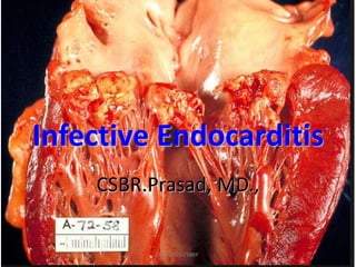
Infective endocarditis is an infection of the heart valves or endocardium that leads to the formation of vegetations composed of thrombotic debris and organisms. It can be acute or subacute in onset. Acute infective endocarditis is commonly caused by Staphylococcus aureus and has an abrupt onset, while subacute bacterial endocarditis is commonly caused by Streptococcus viridans and has a more insidious onset. Complications of infective endocarditis include infected emboli, which can lead to conditions such as Roth spots, Osler's nodes, and Janeway lesions. Non-infective causes of endocardial vegetations include non-bacterial thrombot

Recommended
More Related Content
What's hot
What's hot (20)
Viewers also liked
Viewers also liked (20)
Similar to Cvs ie-csbrp
Similar to Cvs ie-csbrp (20)
More from Prasad CSBR
More from Prasad CSBR (20)
Recently uploaded
Recently uploaded (20)
Cvs ie-csbrp
- 2. • What is THE requisite for inflammation? • Name some structures devoid of vasculature? • What are the vessel wall changes that incite thrombosis? APR-2015-CSBRP
- 3. Infective Endocarditis (IE) • IE is most common of common endocardial diseases • Should be kept in mind in the DDs in a case of PUO APR-2015-CSBRP
- 4. Infective Endocarditis (IE) Def: Infection of cardiac valves, endocardium or arteriovenous shunts (Boyd) Infective endocarditis (IE) is a microbial infection of the heart valves or the mural endocardium that leads to the formation of vegetations composed of thrombotic debris and organisms, often associated with destruction of the underlying cardiac tissues (Robbins) APR-2015-CSBRP
- 5. Infective Endocarditis (IE) Types: 1.Acute – Stormy onset 2.Subacute – Insidious onset APR-2015-CSBRP
- 6. Infective Endocarditis (IE) Organisms: 1. Acute – Staphylococcus aureus 2. Subacute – Streptococcus viridans APR-2015-CSBRP
- 7. Infective Endocarditis (IE) Feature Acute bacterial IE SABE Valve status Normal valve Abnormal valve Organism Highly virulent organism: Staph. aureus Lower virulence Strep. viridans Onset Abrupt fever, fatigue, weakness Slow less dramatic Prognosis Poor usually needs surgery; high mortality Relatively good cured with antibiotics APR-2015-CSBRP
- 8. Infective Endocarditis (IE) Organisms: 1. IV drug abuse: Staphylococcus aureus (50%) 2. In patients with cardiac prosthesis: – Early infections: • 50% Staph. Epidermidis • 20% Gram negative bacilli • 10% Fungi – Late infections: • Staphylococcus and Streptococcus together 60% • HACEK 20% 3. Most common fungal endocarditis: – Candida – Aspergillus APR-2015-CSBRP
- 9. ETIOLOGY & PATHOGENESIS Two Factors interact in the causation of IE: 1. Small Thrombotic masses 2. Bacteraemia APR-2015-CSBRP
- 10. ETIOLOGY & PATHOGENESIS SMALL THROMBOTIC MASSES: • Results from Haemodynamic Injury to endocardial surface by high pressure jets striking the Endocardium / turbulent flow • Examples: – Mitral Valve Prolapse – Congenital Bicuspid Aortic valve – Chronic RHD (AR) – Prosthetic valves – CHD: VSD, TOF, PDA APR-2015-CSBRP
- 11. ETIOLOGY & PATHOGENESIS BACTERAEMIA: Organisms - Mode of entry: 1. Organisms are carried to the heart in the blood – bacteremia – Tooth extraction, dental procedures – Instrumentation of urinary tract, GIT 2. Direct inoculation during cardiac surgery 3. Ascend in an indwelling intravascular catheter APR-2015-CSBRP
- 12. ETIOLOGY & PATHOGENESIS HOST FACTORS IN INFECTIVE ENDOCARDITIS – Neutropenia – Immunodeficiency – Malignancy – Immunosuppression – Diabetes mellitus – Alcohol – IV Drug abuse APR-2015-CSBRP
- 13. IE Lesions - VEGETATIONS • Large friable masses on the valves – Along the lines of closure – At the margin of septal defect – Arteriovenous fistulas • Encroach on to: – the adjacent endocardium – chordae tendinae – into sinus of Valsalva • Perforation of cusps • Rupture of chordae tendinae (result in sudden incompetence) • Abscess may form and surround the valve ring (ring abscess) • Perforation of interventricular septum APR-2015-CSBRP
- 14. The aortic valve shows three thin and delicate cusps. The coronary artery orifices can be seen just above. The endocardium is smooth, beneath which can be seen a red-brown myocardium. The aorta above the valve displays a smooth intima with no atherosclerosis APR-2015-CSBRP
- 15. This is infective endocarditis. The aortic valve demonstrates a large, irregular, reddish tan vegetation.Virulent organisms, such as Staphylococcus aureus, produce an "acute" bacterial endocarditis, while some organisms such as Streptococcus viridans produce a "subacute" bacterial endocarditis. APR-2015-CSBRP
- 16. A mitral valve vegetation caused by bacterial endocarditis APR-2015-CSBRP
- 17. Infective endocarditis spreading to myocardium, gross APR-2015-CSBRP
- 18. Aortic valve acute endocarditis. The probe extends through a perforated leaflet. APR-2015-CSBRP
- 19. A normal aortic valve is shown at the top to compare with an aortic valve at the bottom being destroyed by vegetations with infective endocarditis. APR-2015-CSBRP
- 23. IE Lesions - VEGETATIONS Microscopy: – Layers of fibrin and platelets – Colonies of causative organism – Little inflammation (if valve was normal) – Inflammatory response is seen • In previously damaged valves which are vascularized • Vegetations in the endocardium • Vegetations in the blood vessels APR-2015-CSBRP
- 26. IE Lesions - Pathogenesis “Organisms rarely attack the valves directly” APR-2015-CSBRP
- 27. IE Lesions - Complications • Infected thromboemboli – Myocardium – Spleen (pain on breathing) – Kidney (hematuria) – Brain (Transient neurological dysfunction) • Microinfarcts • Microabscesses • Transient neurological dysfunction • Glomerulonephritis • Jane way lesions (petechiae in the skin / mucus membranes) • Roth spots (painful nodules in pulp of fingers / toes) • Splinter hemorrhages • Osler’s nodes (vasculitis) APR-2015-CSBRP
- 28. Acute Infective Endocarditis with embolic manifestations APR-2015-CSBRP
- 37. APR-2015-CSBRP
- 38. APR-2015-CSBRP
- 39. Clinical presentations • Begins insidiously • Fatigue • Lassitude • Fever • Sweating at night • Anorexia • Loss of weight • Splenomegaly • New murmurs • Leucocytosis • High ESR NOTE: Repeated blood cultures usually, but not always demonstrate the organism APR-2015-CSBRP
- 41. Noninfected Vegetations • Nonbacterial thrombotic endocarditis NBTE • Libman-Sack’s endocarditis • Rheumatic fever APR-2015-CSBRP
- 42. Nonbacterial thrombotic endocarditis NBTE • Synonyms: Merantic or Terminal Endocarditis • Conditions associated with: – Malignancies • Lung, pancreas, stomach – Wasting diseases • Vegetations: – On Normal valves – Along the lines of closure – Small 1-5mm – Sterile – Composed of platelets and fibrin • Complications: – Emboli 15-60% APR-2015-CSBRP
- 43. Trousseau syndrome • The Trousseau's Syndrome is a sign involving episodes of vessel inflammation due to blood clot (thrombophlebitis) which are recurrent or appearing in different locations over time (migratory thrombophlebitis). The location of the clot is tender and the clot can be felt as a nodule under the skin APR-2015-CSBRP
- 44. Migratory thrombophlebitis APR-2015-CSBRP SEEN IN: Gliomas Pancreatic adenocarcinoma Pulmonary adenocarcinoma
- 49. Libman-Sacks endocarditis Endocarditis of Systemic Lupus Erythematosus • Seen in SLE • Vegetations: • Small (1 to 4 mm in diameter) • Single or multiple • Pink warty (verrucous) vegetations • Sterile • May be located on the undersurfaces of the atrioventricular valves, the chords, mural endocardium APR-2015-CSBRP
- 51. Libman-Sacks endocarditis, Micro Histologically the vegetations consist of: • Finely granular, fibrinous eosinophilic material • Composed of cellular debris including nuclear remnants (hematoxyphilic bodies) • Vegetations are often associated with an intense valvulitis, characterized by fibrinoid necrosis of the valve substance APR-2015-CSBRP
- 52. Dr. Emanuel Libman Dr.Benjamin Sacks Libman-Sacks endocarditis APR-2015-CSBRP
- 53. APR-2015-CSBRP
- 54. Comparison of the four major forms of vegetative endocarditis Rheumatic fever IE NBTE LSE Small, Warty Firm, friable Large Bulky Irregular Small, Warty Friable Medium sized Flat, verrucous Irregular Along the lines of closure Valve cusps & Mural endocardium Along the lines of closure Both surfaces of the valves may be involved In pockets of valves Sterile (no organism) Non-sterile (full of bacteria) Sterile (no organism) Sterile (no organism) Embolization is uncommon Embolization is very common Embolization is common Embolization is uncommon RHD Infective endocarditis Cancers (AML- M3, Stomach, Pancreas) SLE APR-2015-CSBRP
- 55. Comparison of the four major forms of vegetative endocarditis SLE APR-2015-CSBRP
- 57. APR-2015-CSBRP
- 58. • ►M.c cause of community acquired native valve endocarditis in neonate is Staphylococcus aureus ►M.c cause of community acquired native valve endocarditis (overall and age >2 months) is streptococci ►M.c cause of native valve endocarditis in health care associated patients : Staphylococcus aureus ►M.c cause of infective endocarditis in prosthetic valve endocarditis <2 months and 2-12 months: coagulase negative staphylococcus > 12 months: Streptococcus (They say it is similar to native valve endocarditis) ►M.c cause of infective endocarditis in IV drug users: Over all: Staph. Aureus Right side: Staph. Aureus Left side: Enterococcus > Staph. Aureus (Marginally) APR-2015-CSBRP
- 59. APR-2015-CSBRP
- 60. Comparison of the four major forms of vegetative endocarditis APR-2015-CSBRP
- 62. Non-infective Endocarditis Non-bacterial thrombotic endocarditis, gross APR-2015-CSBRP
- 64. This is the tricuspid valve. The leaflets and thin and delicate. Just like the mitral valve, the leaflets have thin chordae tendineae that attach the leaflet margins to the papillary muscles of the ventricular wall below. APR-2015-CSBRP
- 65. The small verrucous vegetations seen along the closure line of this mitral valve are associated with acute rheumatic fever. These warty vegetations average only a few millimeters and form along the line of valve closure over areas of endocardial inflammation. Such verrucae are too small to cause serious cardiac problems. APR-2015-CSBRP
Editor's Notes
- Acute bacterial IE: Usually occurs on previously-normal heart valves Infecting bug is highly virulent (e.g., Staph aureus) and produces necrotizing, destructive lesions Onset is stormy (abrupt fever, fatigue, weakness) Prognosis is poor (hard to cure with antibiotics; usually needs surgery; high mortality) Subacute bacterial IE Usually occurs on an abnormal heart valve Infecting bug is of lower virulence (e.g., Strep viridans) and causes less destructive lesions Onset is less dramatic Prognosis is relatively good (most patients are cured with antibiotics after a period of weeks/months)
- HACEK = Hemophilus, Actinobacillus, Cardiobacterium, Eiknella and Kingella.
- HACEK = Hemophilus, Actinobacillus, Cardiobacterium, Eiknella and Kingella.
- The aortic valve shows three thin and delicate cusps. The coronary artery orifices can be seen just above.The endocardium is smooth, beneath which can be seen a red-brown myocardium. The aorta above the valve displays a smooth intima with no atherosclerosis.
- This is infective endocarditis. The aortic valve demonstrates a large, irregular, reddish tan vegetation.Virulent organisms, such as Staphylococcus aureus, produce an "acute" bacterial endocarditis, while some organisms such as Streptococcus viridans produce a "subacute" bacterial endocarditis.
- In this case, the infective endocarditis demonstrates how the infection tends to spread from the valve surface. Here, vegetations can be seen on the endocardial surfaces, and the infection is extending into to underlying myocardium.
- bacterial endocarditis – aortic valve vegetationsed, destruction of valves
- Little inflammation (if valve was normal): As the normal valves are not vascularized, there is little or no inflammatory reaction.
- Microscopically, the valve in infective endocarditis demonstrates friable vegetations of fibrin and platelets (pink) mixed with inflammatory cells and bacterial colonies (blue). The friability explains how portions of the vegetation can break off and embolize.
- Flourish as there is no immune attack = as there is no inflammation, the organisms are protected from the attack by PMNs and macrophages.
- Glomerulonephritis: Before the advent of antibiotics, acute proliferative GN caused by immune complexes developed in 75% of patients. GN is now uncommon seen only in <10% of cases of SABE.
- Osler nodes are red-purple, slightly raised, tender lumps, often with a pale centre. Pain often precedes the development of the visible lesion by up to 24 hours. They are typically found on the fingers and/or toes. They can occur at any time during the course of endocarditis (usually subacute) and last from hours to several days. Janeway lesions are non-tender, often haemorrhagic (bleeding into the skin), and occur mostly on the palms and soles including the thenar and hypothenar eminences (at the base of the thumb and little finger respectively). They tend to last days to weeks before healing totally. They are more commonly seen in acute endocarditis, when bacteria such as Staphylococcus aureus may be cultured from them. The histology is usually consistent with septic micro-embolism (i.e. bacteria may be found within the blood vessels).
- Diagnosis by these guidelines, often called the Duke Criteria, requires either pathologic or clinical criteria; if clinical criteria are used, 2 major, 1 major + 3 minor, or 5 minor criteria are required for diagnosis. † Janeway lesions are small erythematous or hemorrhagic, macular, nontender lesions on the palms and soles and are the consequence of septic embolic events. ‡ Osler nodes are small, tender subcutaneous nodules that develop in the pulp of the digits or occasionally more proximally in the fingers and persist for hours to several days. § Roth spots are oval retinal hemorrhages with pale centers. Modified from Durack DT, et al: New criteria for diagnosis of infective endocarditis: utilization of specific echocardiographic findings. Am J Med, 96:200, 1994, and Karchmer AW: Infective Endocarditis. In Braunwald E, Zipes DP, Libby P (eds): Heart Disease. A Textbook of Cardiovascular Medicine, 6th ed. Philadelphia, WB Saunders, 2001, p 1723.
- NBTE is often encountered in debilitated patients, such as those with cancer or sepsis—hence the previous term marantic endocarditis (root word marasmus, relating to malnutrition). It frequently occurs concomitantly with deep venous thromboses, pulmonary emboli, or other findings suggesting an underlying systemic hypercoagulable state. Indeed, there is a striking association with mucinous adenocarcinomas.
- Figure 12-26 Nonbacterial thrombotic endocarditis (NBTE). A, Nearly complete row of thrombotic vegetations along the line of closure of the mitral(arrows). B, Photomicrograph of NBTE, showing bland thrombus, with virtually no inflammation in the valve cusp (c) or the thrombotic deposit (t).is only loosely attached to the cusp (arrow).
- Figure 12-26 Nonbacterial thrombotic endocarditis (NBTE). A, Nearly complete row of thrombotic vegetations along the line of closure of the mitral(arrows). B, Photomicrograph of NBTE, showing bland thrombus, with virtually no inflammation in the valve cusp (c) or the thrombotic deposit (t).is only loosely attached to the cusp (arrow).
- The valve is seen on the left, and a bland vegetation is seen on the right. It appears pink because it is composed of fibrin and platelets. It displays about as much morphologic variation as a brown paper bag. Such bland vegetations are typical of the non-infective forms of endocarditis.
- The valve is seen on the Right, and a bland vegetation is seen on the Left. It appears pink because it is composed of fibrin and platelets. It displays about as much morphologic variation as a brown paper bag. Such bland vegetations are typical of the non-infective forms of endocarditis.
- Here are flat, pale tan, spreading vegetations over the mitral valve surface and even on the chordae tendineae. This patient has systemic lupus erythematosus. Thus, these vegetations that can be on any valve or even on endocardial surfaces are consistent with Libman-Sacks endocarditis. These vegetations appear in about 4% of SLE patients and rarely cause problems because they are not large and rarely embolize. Note also the thickened, shortened, and fused chordae tendineae that represent remote rheumatic heart disease.
- Here are flat, pale tan, spreading vegetations over the mitral valve surface and even on the chordae tendineae. This patient has systemic lupus erythematosus. Thus, these vegetations that can be on any valve or even on endocardial surfaces are consistent with Libman-Sacks endocarditis. These vegetations appear in about 4% of SLE patients and rarely cause problems because they are not large and rarely embolize. Note also the thickened, shortened, and fused chordae tendineae that represent remote rheumatic heart disease.
- Benjamin Sacks was born on August 14th, 1896. He graduated from high school in three years, and began his collegiate career at Johns Hopkins University in Maryland, earning recognition as a Phi Beta Kappa for devotion to learning. Sacks advanced his education at Johns Hopkins in the School of Medicine. After he earned his M.D., he became licensed to practice medicine in 1922 in the State of New York. His career began at The Mount Sinai Hospital in Manhattan. In 1923, Sacks, along with partner Dr. Emanuel Libman, discovered an uncommon heart ailment called the Libman-Sacks Syndrome.(1) This particular type of heart ailment is reported postmortem in approximately 50% of fatal lupus cases. Sacks’ work at The Mount Sinai Hospital earned him respect and praise from the medical community. Louis Gross, M.D., the Director of Laboratories for Mount Sinai, had this to say about Dr. Benjamin Sacks in a letter of recommendation to United States Surgeon General Hugh S. Cumming: Dr. Sacks is a man of striking personality. He is what is called a “natural born leader of men.” He is an extraordinarily able teacher and is a constant stimulus to those about him. He is exceptionally well trained for investigative work.(2) In the early 1950's Dr. Sacks served as a technical advisor on for the movie industry. By 1959, he had moved to Los Angeles. Sacks worked with actors including Cary Grant, Bing Crosby, and Barry Fitzgerald. Dr. Benjamin Sacks was known for his attention to detail, creating sets that were identical to a real hospital or doctor’s office. [Sacks] was flattered when a Los Angeles medico visited the set [of 'People Will Talk', a medical drama starring Cary Grant], scrutinized everything with a critical eye, and then said, “All right, wheel the patient in. I am ready.” (3) Sacks was an essential part of any production team he served. When asked what his job entailed, Sacks said, “I start with the story and make certain the dialogue shapes up correctly from a physician’s point of view. I talk over the backgrounds with the art department, look at costumes for both men and women, and work closely with the property department to see that every detail matches real life.” In the mid-1950’s, Sacks fell victim to failing health, and was forced to stop practicing medicine. He continued his role as a technical advisor to the film studios. His interests shifted towards the study of history, and Sacks found himself engaged in researching the Southwest, specifically Arizona. He was captivated by the years 1850 to 1875, when Arizona became a Territory after ceding from Mexico as a result of the Mexican-American War. For close to 20 years, he traveled the country and searched in repositories great and small. Dr. Sacks went about his research much like a detective would go about solving a crime, piecing together new hypotheses and theories while collecting data and pieces of the puzzle. He thought about his work while driving in his car, and even when lying in bed.(4) In 1959, Dr. Sacks met with Arizona Senator Barry Goldwater and Phoenix historian Bert Fireman in Washington, D.C. Out of this meeting was born the Arizona Historical Foundation, an institution “dedicated to preserving Arizona’s rich political, business and legal history.”(5) Goldwater was founder and the original president, Fireman executive director, and Sacks historical consultant. Sacks’ desire was to see the historical societies in Arizona take a more scholarly approach to analyzing their state’s rich history, and to focus on answering new questions that had yet to be considered. In 1964, Sacks published his first book about the history of Arizona. Entitled “Be It Enacted: the Creation of the Territory of Arizona,” Sacks delved into the Library of Congress archives and spent countless hours mining the country for information on the pioneers of the Arizona Territory. His work “will be long remembered in Arizona as a masterpiece of historical investigation and exposition,” believed John Alexander Carroll, then a Professor of History at the University of Arizona.(6) The exhaustive research received an Award of Merit from the American Association for State and Loral History. In 1970, Sacks released his second book, “Arizona’s Angry Man: United States Marshal Milton B. Duffield.” In the foreword, Senator Goldwater praised Sacks for his exceptional research. The study was the result of relentless research and astute scholarship of B. Sacks, retired physician and Historical Consultant of the [Arizona Historical] Foundation, who for two decades has devoted his entire energies to research into a multitude of problems in the early Territorial history of Arizona. At the same time he has been of willing and erudite assistance to many scholars, both within and outside our universities and state, offering advice and in many instances providing access to the remarkable research material he has accumulated.(7) This wealth of knowledge, now known as The Sacks Collection of the American West, is located at the Arizona Historical Foundation in the Hayden Library on the Tempe campus of Arizona State University. A perfectionist in every sense of the word, Sacks compiled note cards containing bits of information, and filed them by subject in an exceptionally large card catalog. Many of the cards have a stamped number on them, which can be traced to the Manuscript Collection. Sacks often copied primary documents by hand, and filed them in boxes to keep his research in order. The attention to detail is an unusual, remarkable quality that is often not seen in collections of this size. Dr. Benjamin Sacks died on May 2nd, 1971. His achievements will continue to enrich the lives of those who seek his knowledge, and the body of research he left behind will support generations of avid historians of Arizona.. <http://www.ahfweb.org/collections_sacksbio.html>
- LSE sounds like SLE, easy to remember the condition associated with Libman-Sacks endocarditis.
- Figure 12-24 Comparison of the four major forms of vegetative endocarditis. The rheumatic fever phase of rheumatic heart disease (RHD) is marked by small, warty vegetations along the lines of closure of the valve leaflets. Infective endocarditis (IE) is characterized by large, irregular masses on the valve cusps that can extend onto the chordae (see Fig. 12-25A). Nonbacterial thrombotic endocarditis (NBTE) typically exhibits small, bland vegetations, usually attached at the line of closure. One or many may be present (see Figure 12-26). Libman-Sacks endocarditis (LSE) has small or medium-sized vegetations on either or both sides of the valve leaflets.
- The small pink vegetation on the rightmost cusp margin represents the typical finding with non-bacterial thrombotic endocarditis (or so-called "marantic endocarditis"). This is non-infective. It tends to occur in persons with a hypercoagulable state (Trousseau's syndrome, a paraneoplastic syndrome associated with malignancies) and in very ill persons.
- Libman-Sacks endocarditis
- The small verrucous vegetations seen along the closure line of this mitral valve are associated with acute rheumatic fever. These warty vegetations average only a few millimeters and form along the line of valve closure over areas of endocardial inflammation. Such verrucae are too small to cause serious cardiac problems.