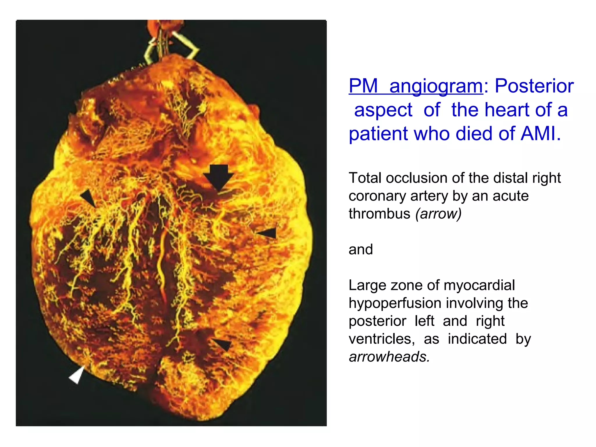
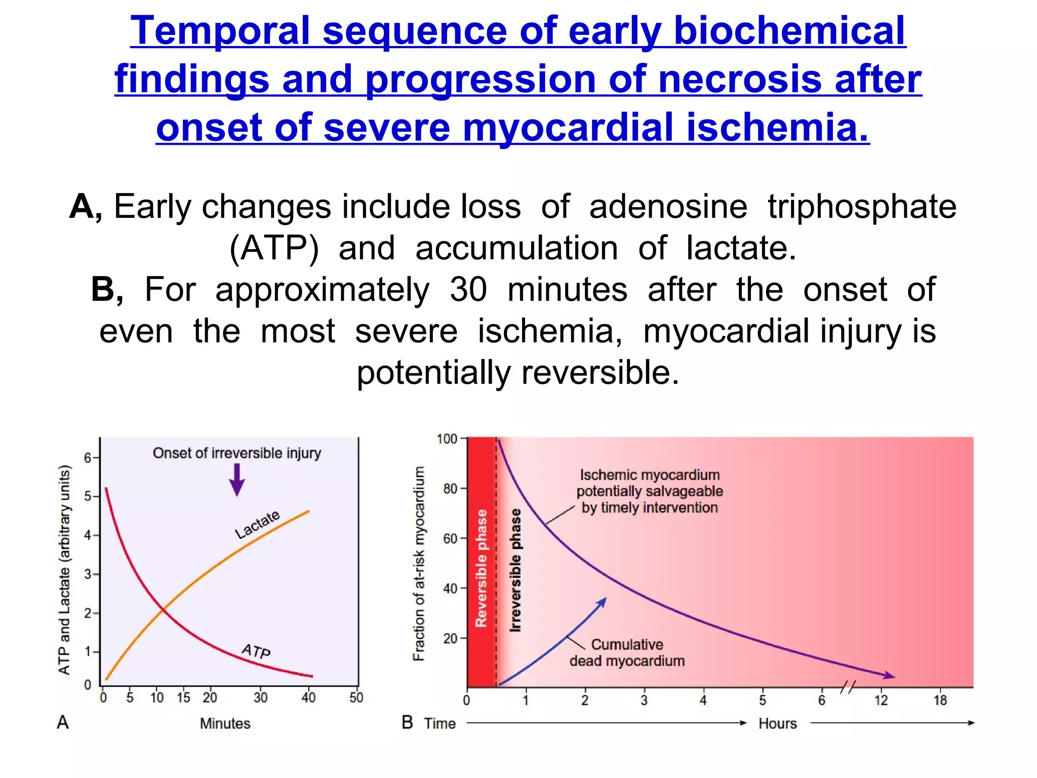
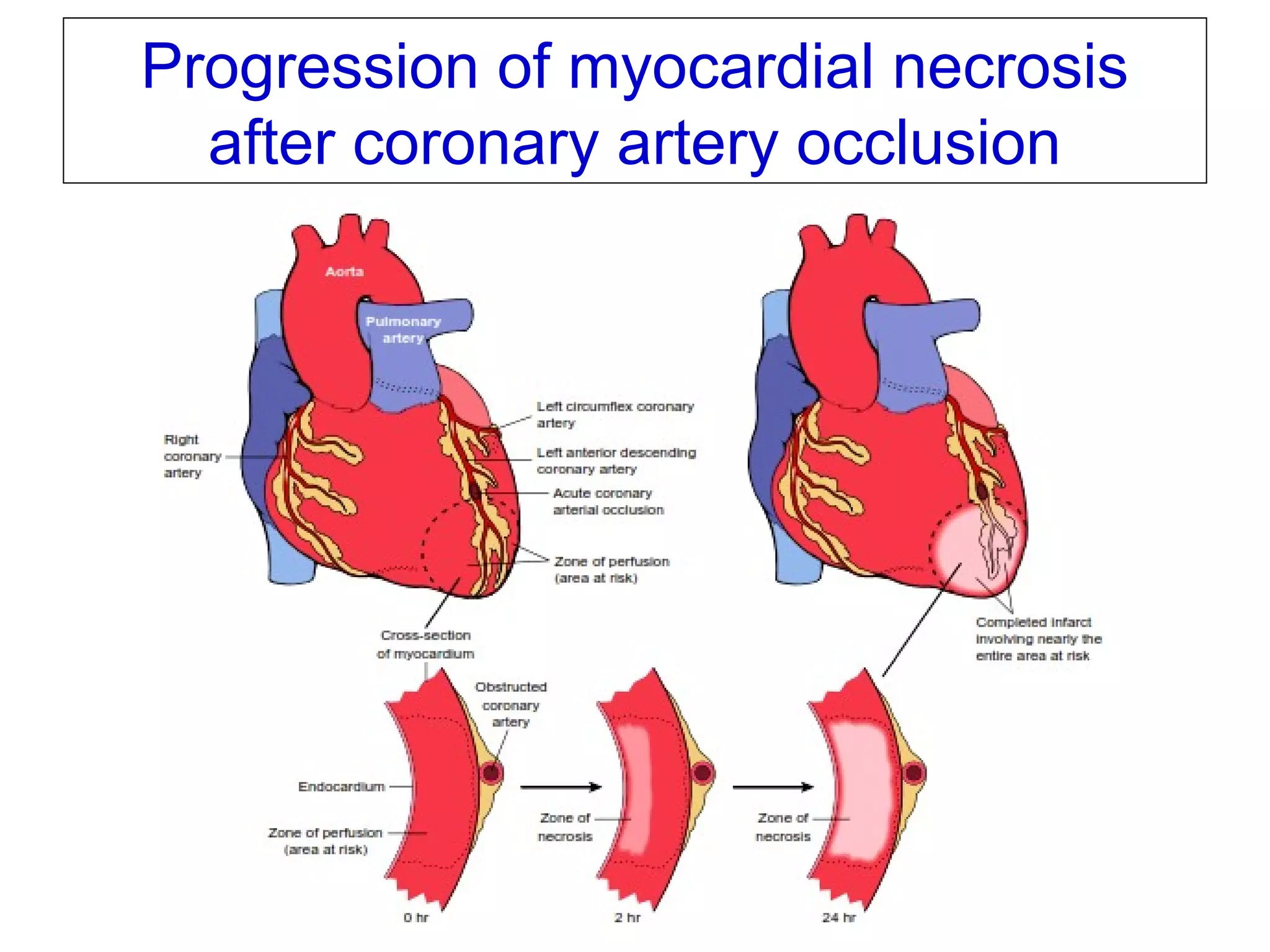
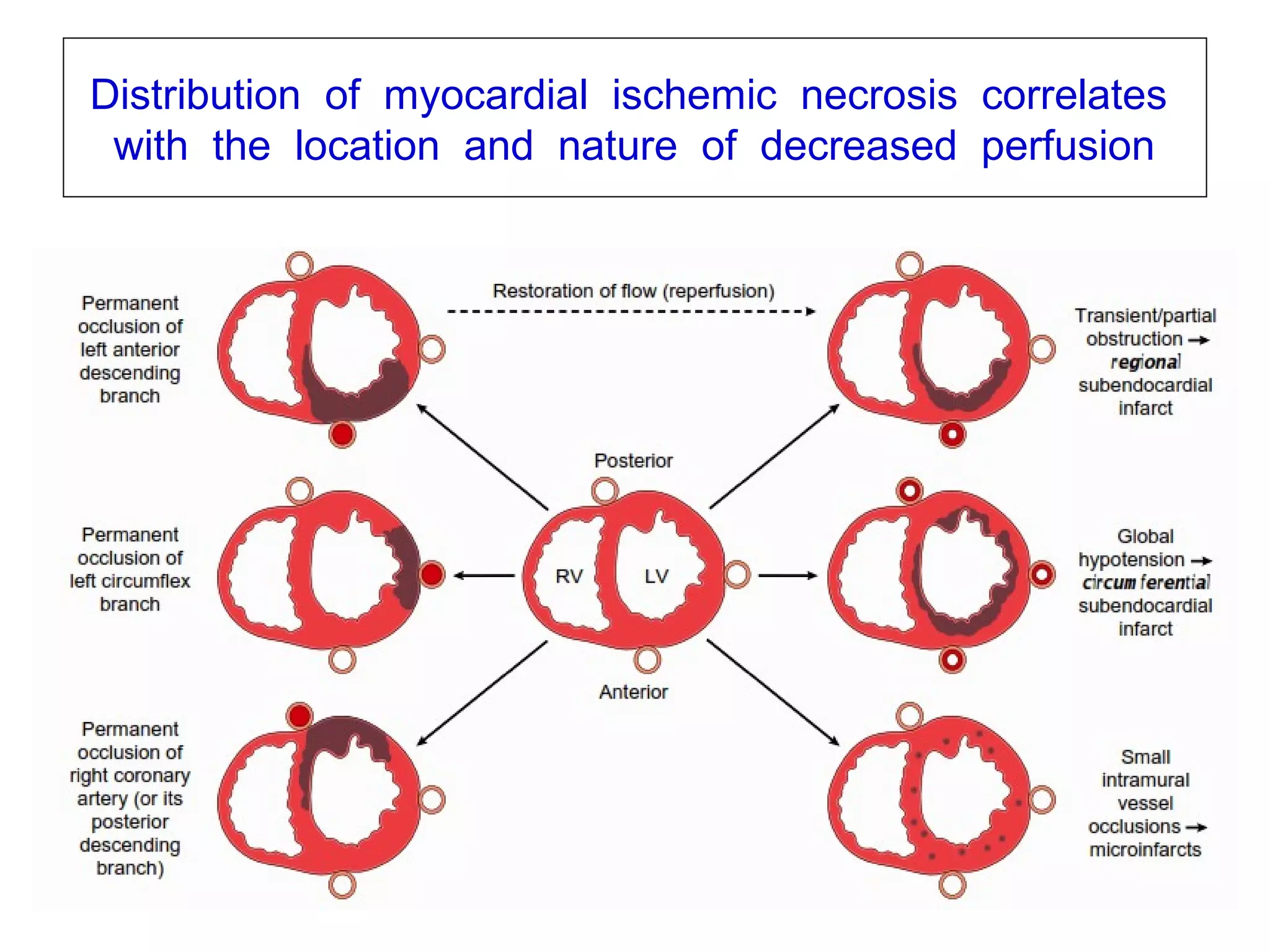
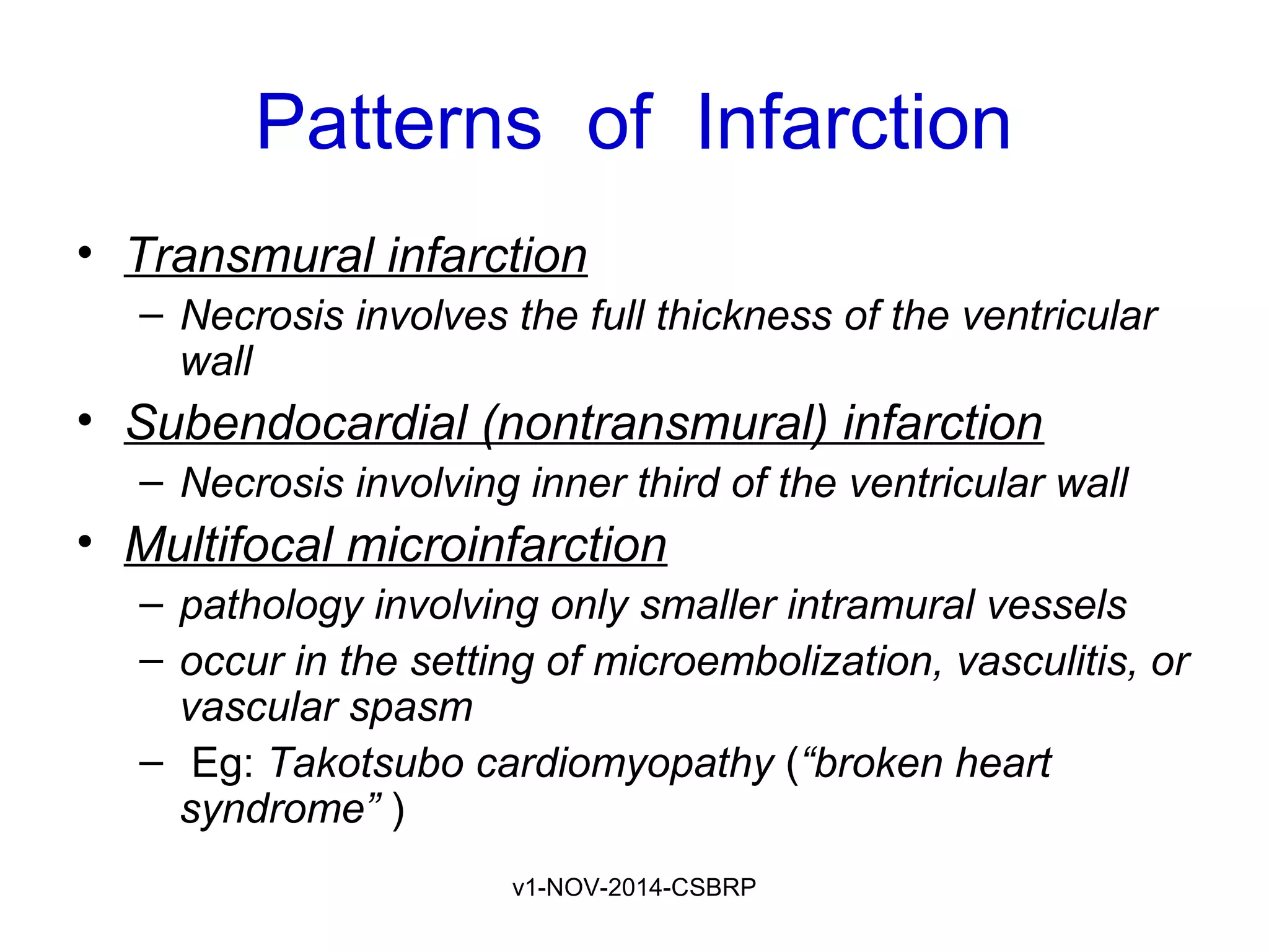
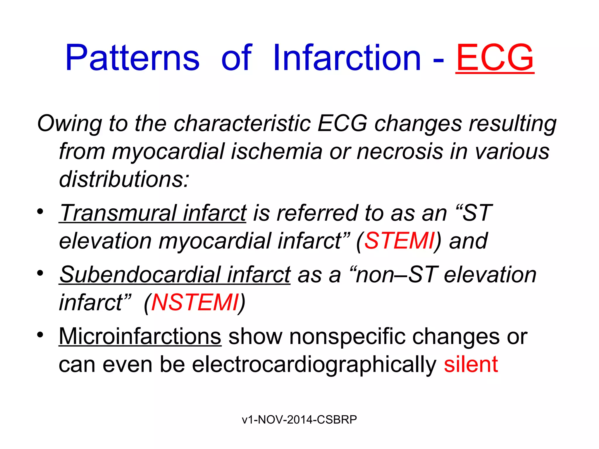
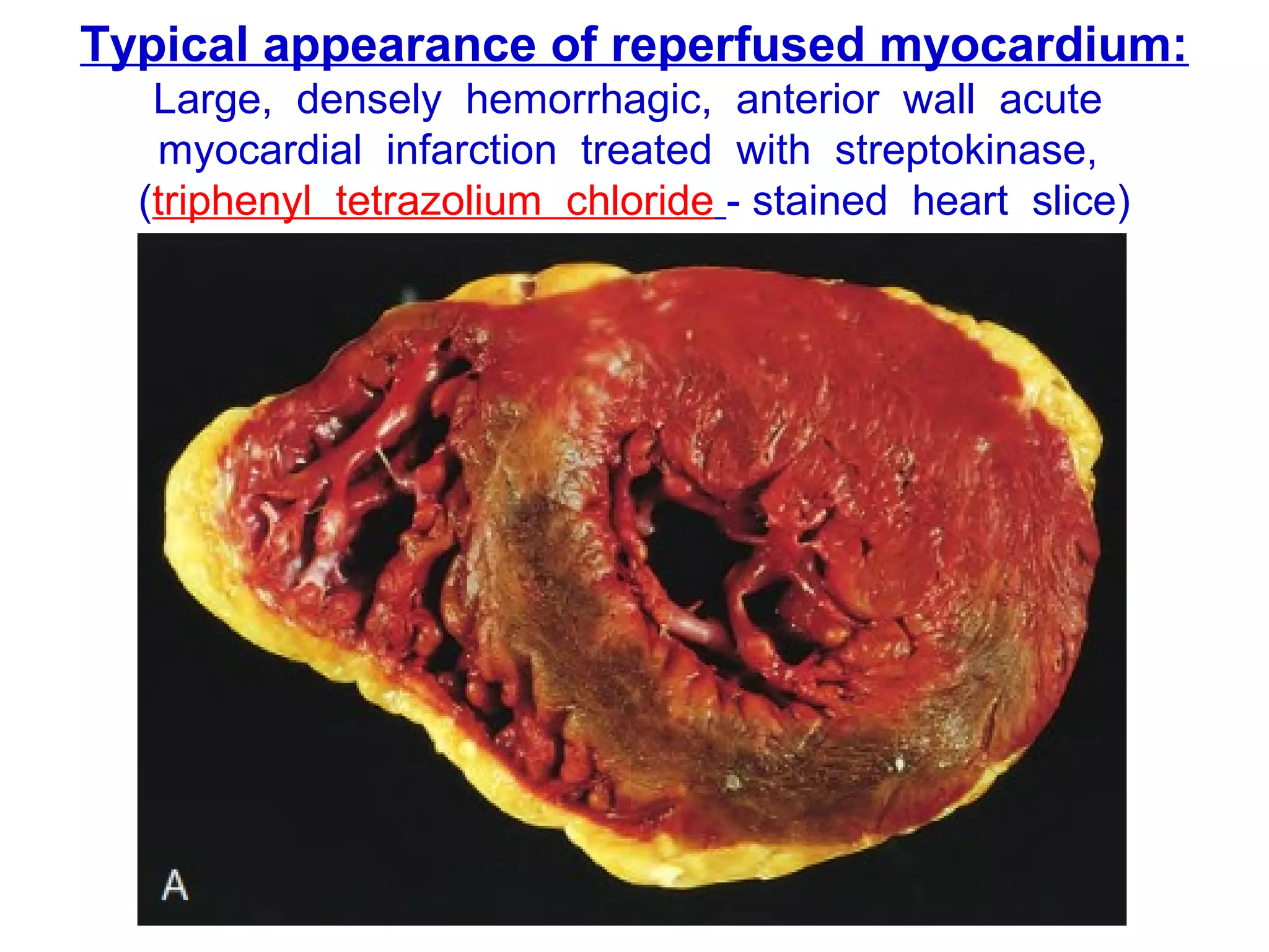
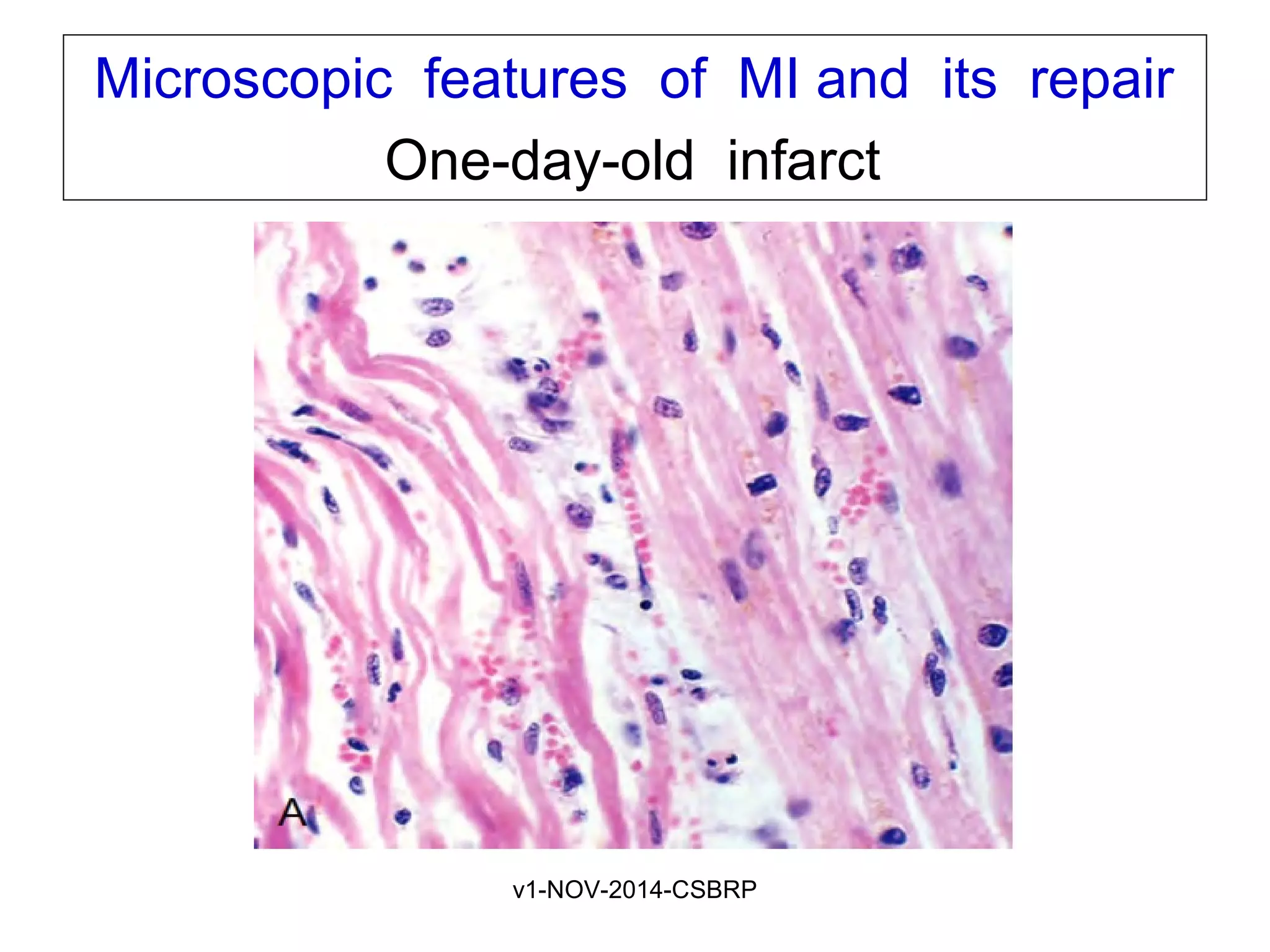
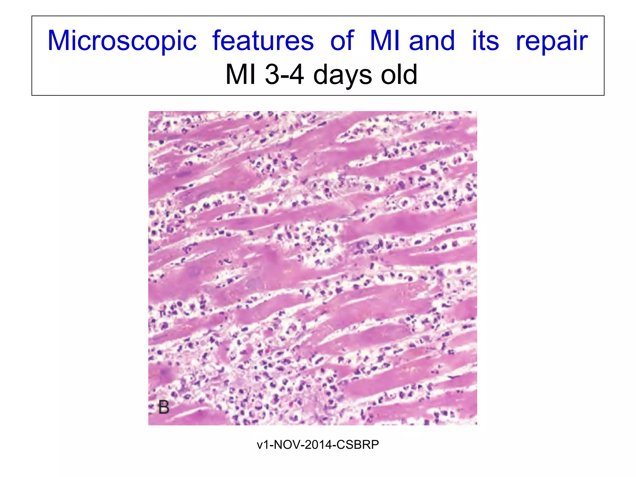
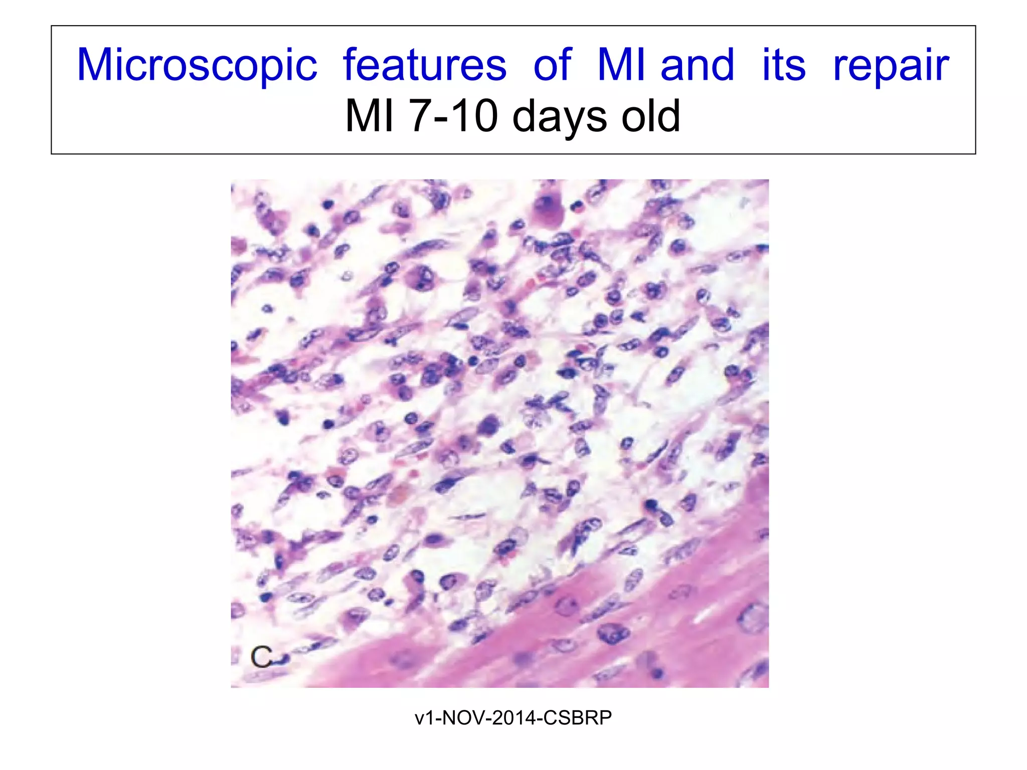
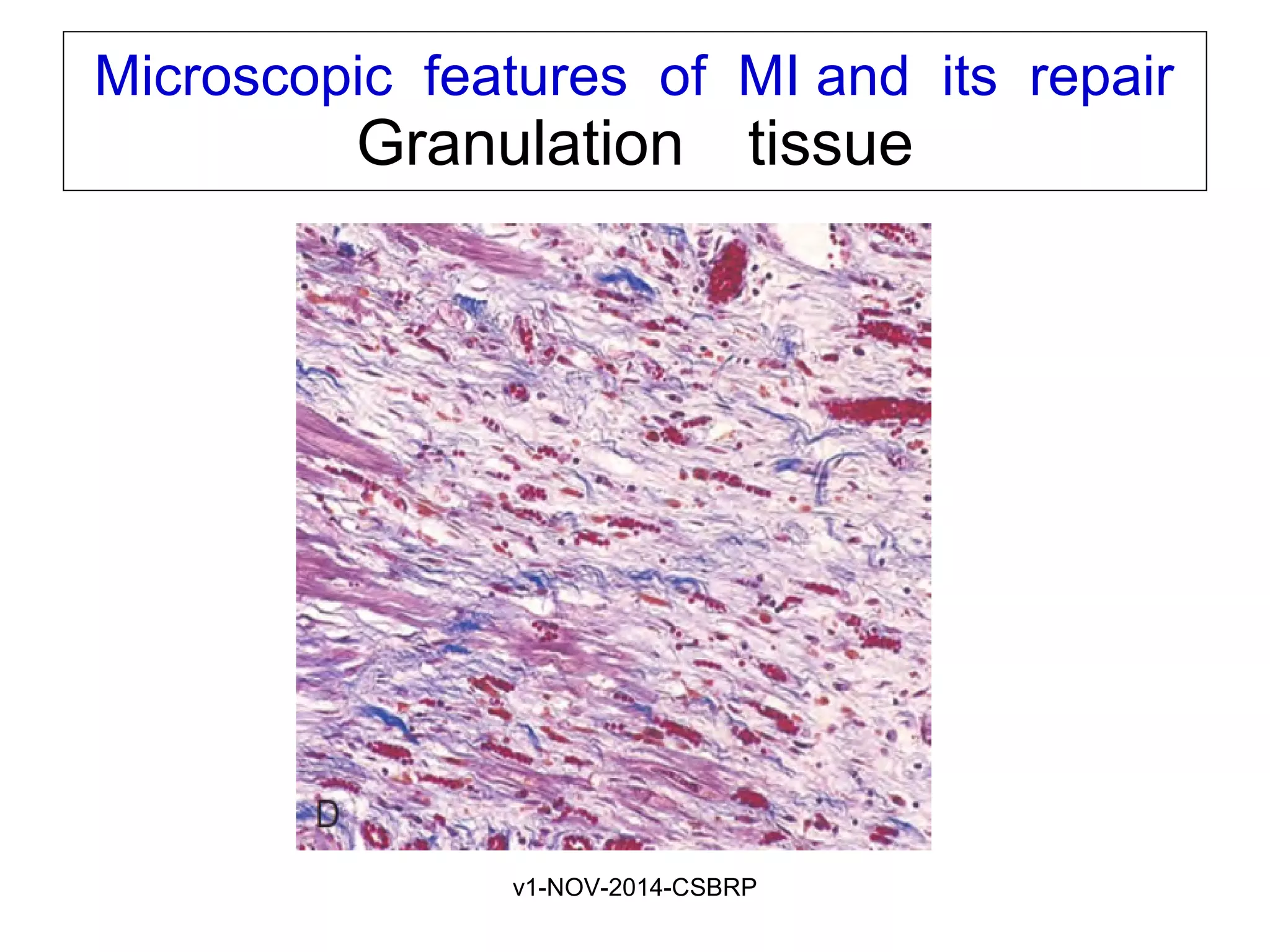
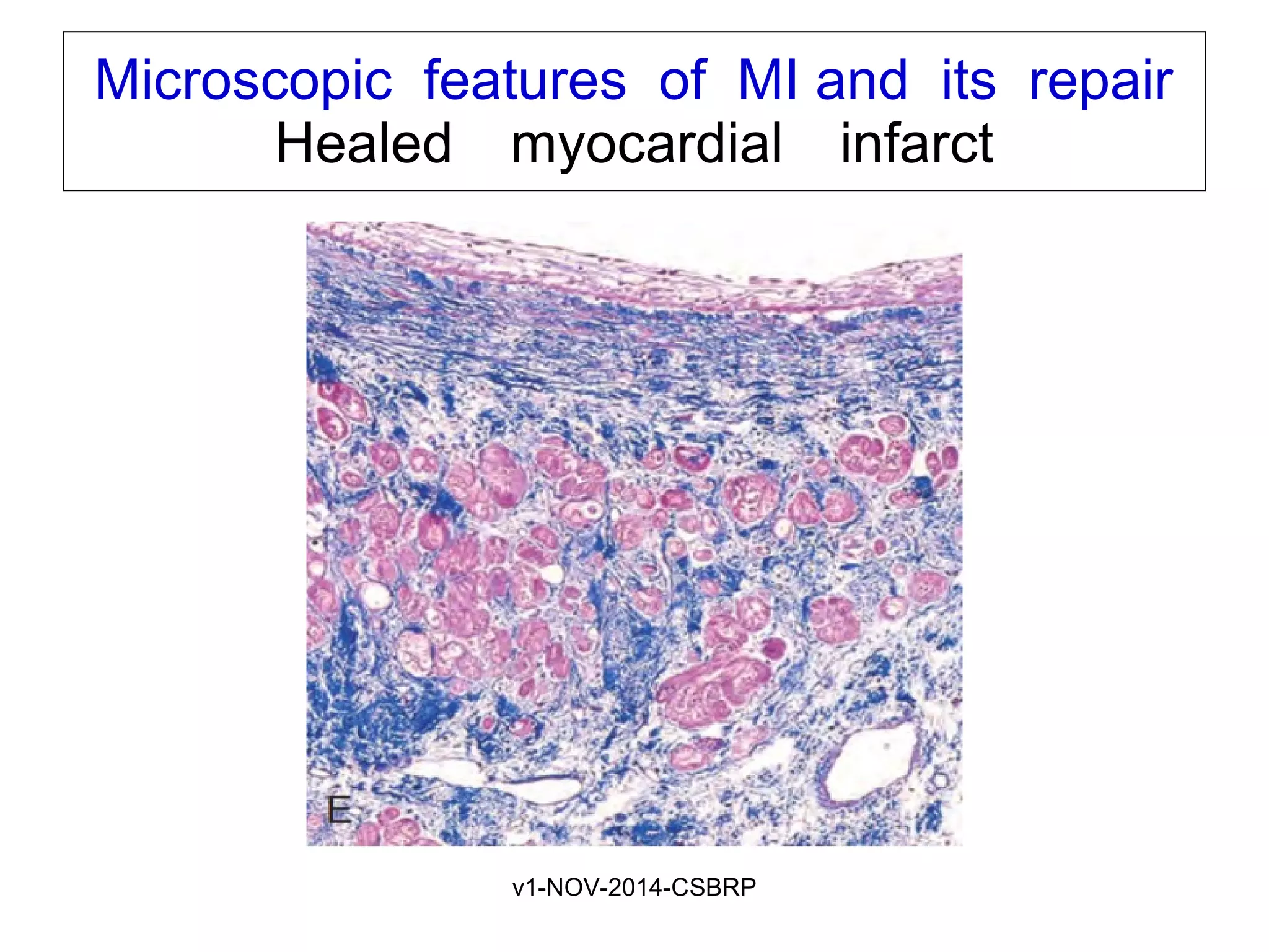
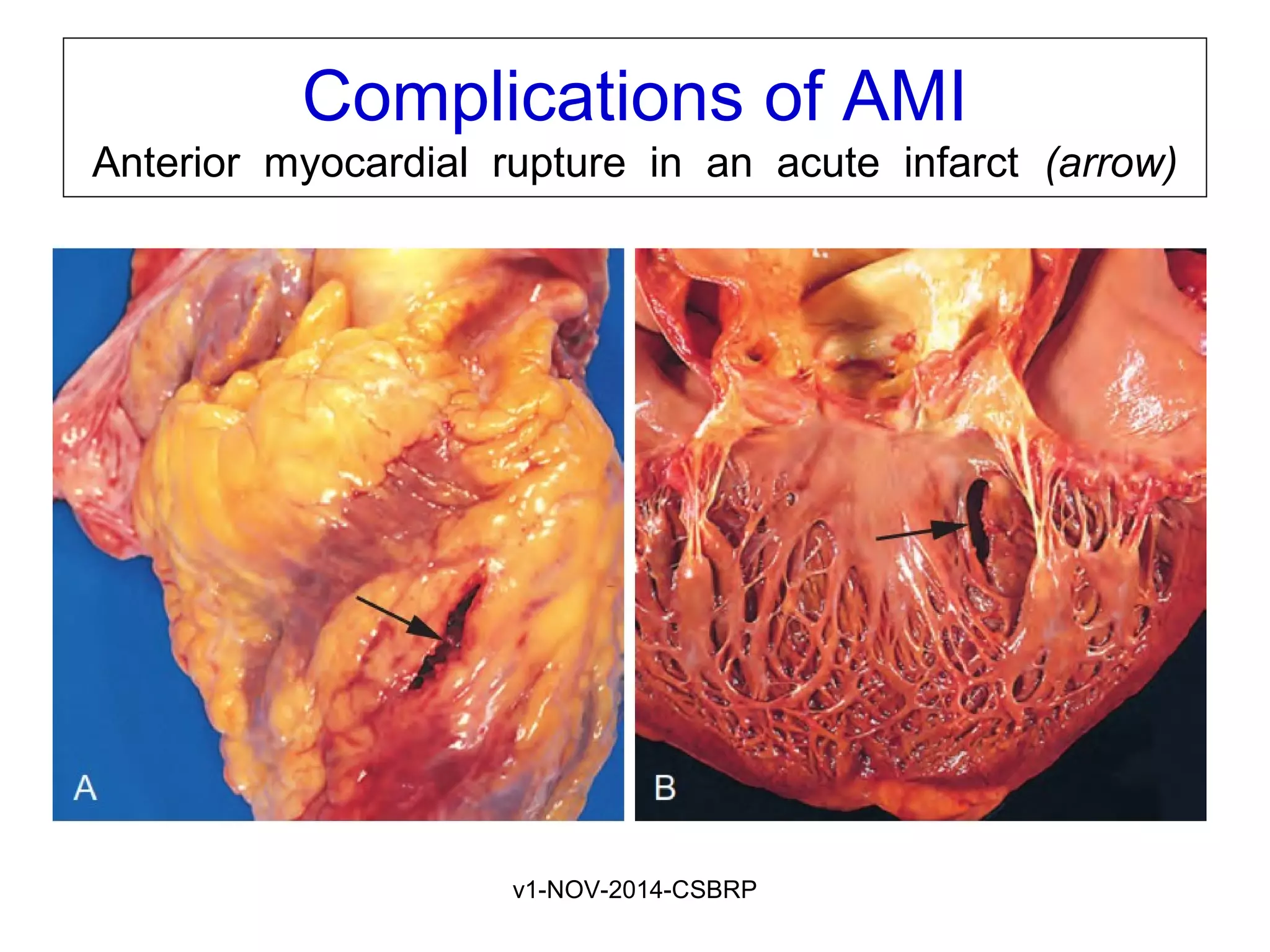
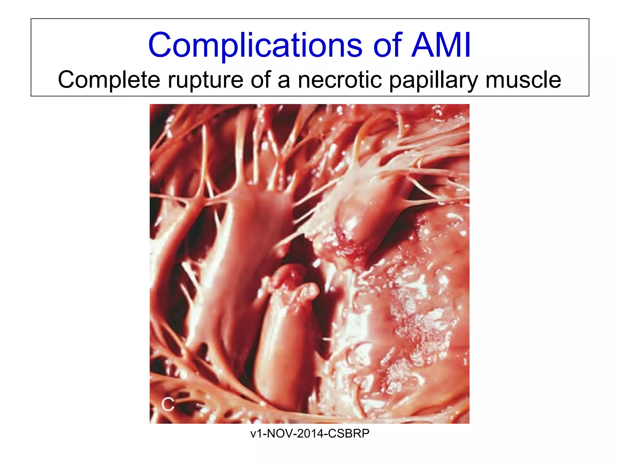
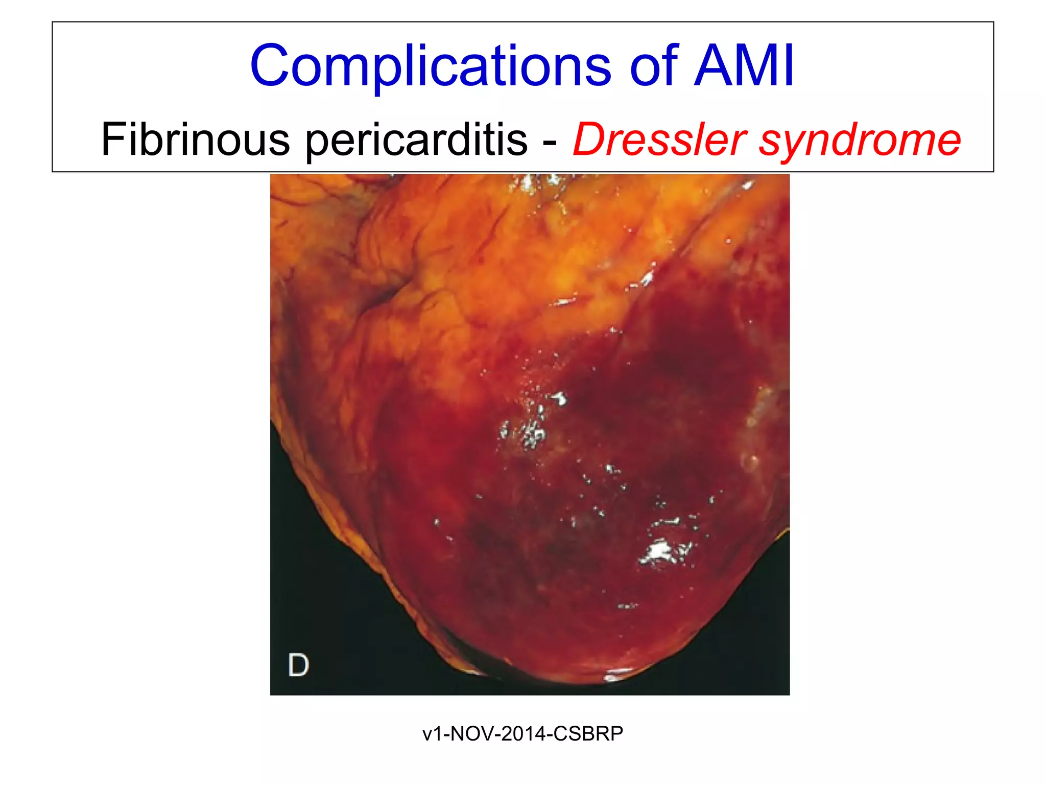
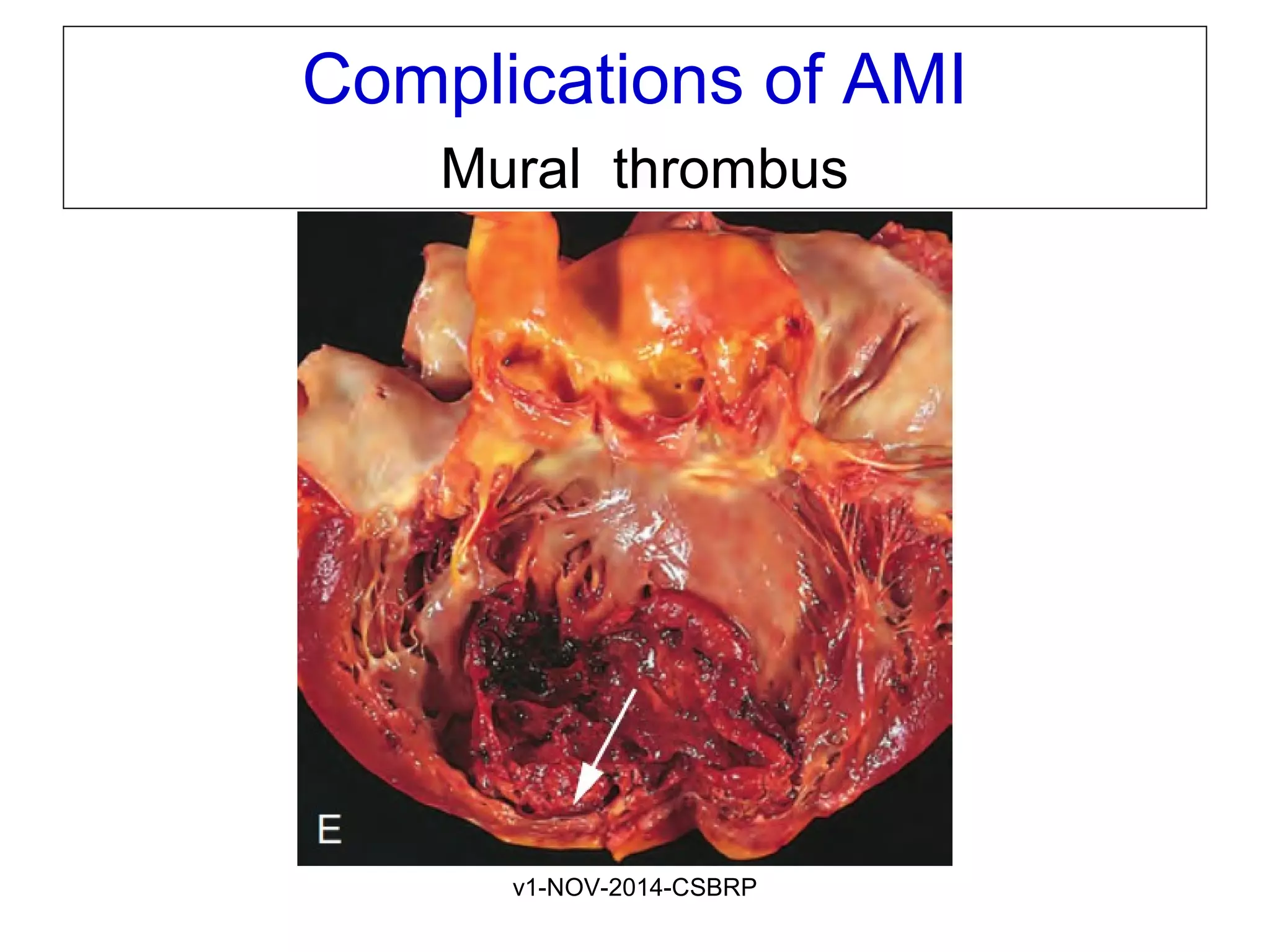
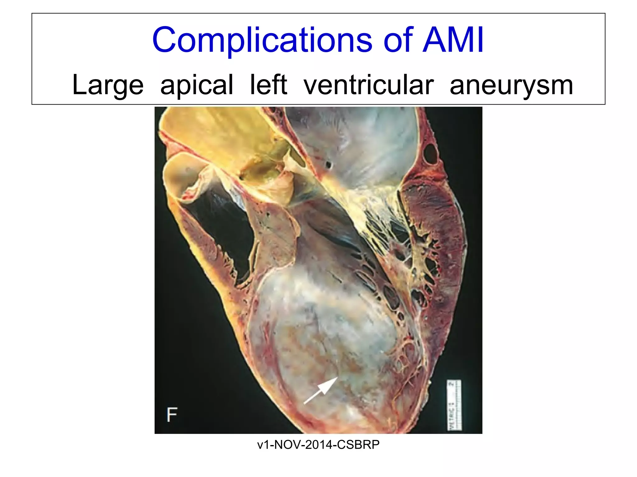
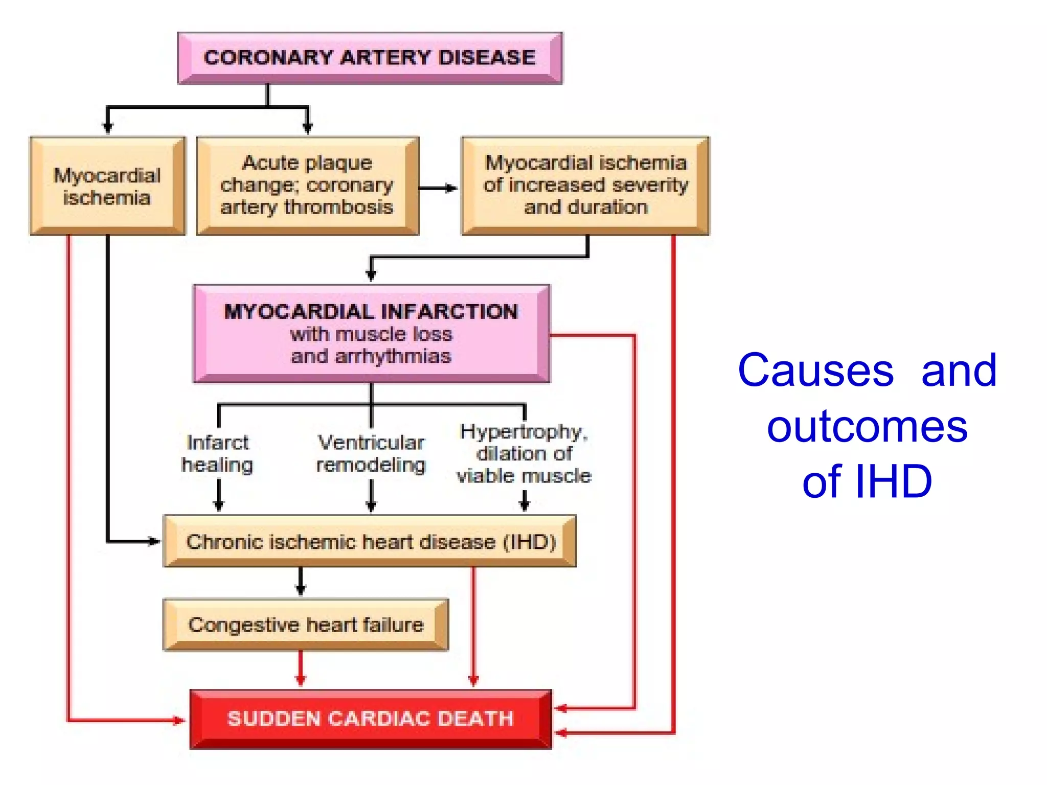
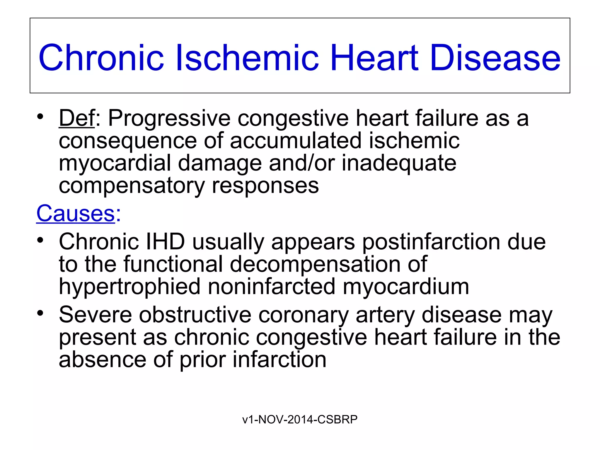
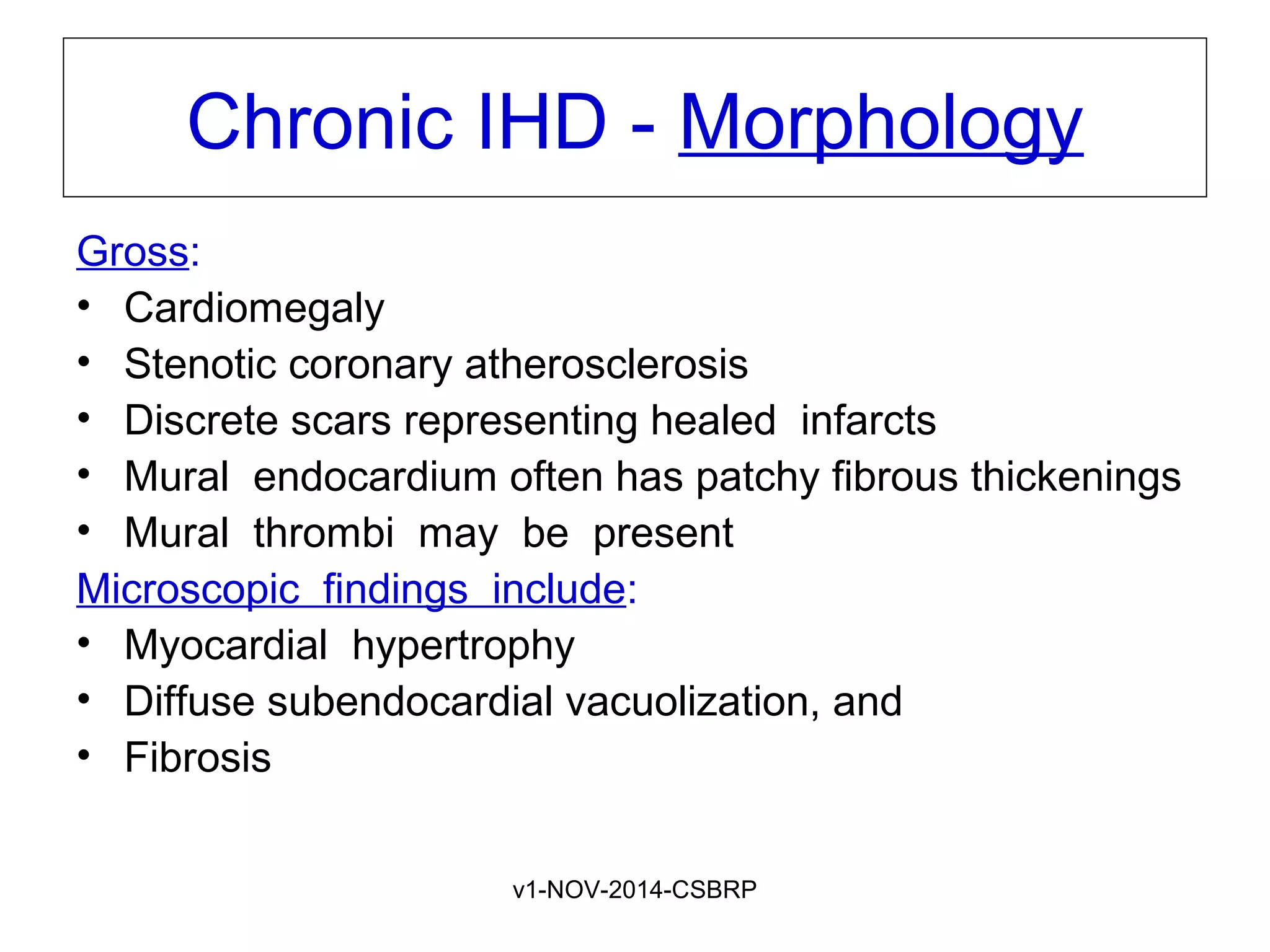
v1-NOV-2014-CSBRP
Lab Diagnosis of AMI
CSBR.Prasad, MD.,
PM angiogram: Posterior
aspect of the heart of a
patient who died of AMI.
Total occlusion of the distal right
coronary artery by an acute
thrombus (arrow)
and
Large zone of myocardial
hypoperfusion involving the
posterior left and right
ventricles, as indicated by
arrowheads.
v1-NOV-2014-CSBRP
Temporal sequence of early biochemical
findings and progression of necrosis after
onset of severe myocardial ischemia.
A, Early changes include loss of adenosine triphosphate
(ATP) and accumulation of lactate.
B, For approximately 30 minutes after the onset of
even the most severe ischemia, myocardial injury is
potentially reversible.
v1-NOV-2014-CSBRP
Progression of myocardial necrosis
after coronary artery occlusion
v1-NOV-2014-CSBRP
Distribution of myocardial ischemic necrosis correlates
with the location and nature of decreased perfusion
v1-NOV-2014-CSBRP
Patterns of Infarction
• Transmural infarction
– Necrosis involves the full thickness of the ventricular
wall
• Subendocardial (nontransmural) infarction
– Necrosis involving inner third of the ventricular wall
• Multifocal microinfarction
– pathology involving only smaller intramural vessels
– occur in the setting of microembolization, vasculitis, or
vascular spasm
– Eg: Takotsubo cardiomyopathy (“broken heart
syndrome” )
v1-NOV-2014-CSBRP
Patterns of Infarction - ECG
Owing to the characteristic ECG changes resulting
from myocardial ischemia or necrosis in various
distributions:
• Transmural infarct is referred to as an “ST
elevation myocardial infarct” (STEMI) and
• Subendocardial infarct as a “non–ST elevation
infarct” (NSTEMI)
• Microinfarctions show nonspecific changes or
can even be electrocardiographically silent
v1-NOV-2014-CSBRP
Infarct Modification by Reperfusion
“time is myocardium”
• Reperfusion: is the restoration of blood flow to ischemic
myocardium threatened by infarction
• The goal: is to salvage cardiac muscle at risk and limit
infarct size
– Prompt reperfusion is the preeminent objective for treatment of
patients with AMI
– This can be accomplished by a host of coronary interventions:
• Thrombolysis
• Angioplasty
• Stent placement or
• CABG
The first 3 to 4 hours following obstruction are critical
v1-NOV-2014-CSBRP
Typical appearance of reperfused myocardium:
Large, densely hemorrhagic, anterior wall acute
myocardial infarction treated with streptokinase,
(triphenyl tetrazolium chloride - stained heart slice)
v1-NOV-2014-CSBRP
Microscopic features of MI and its repair
One-day-old infarct
v1-NOV-2014-CSBRP
Microscopic features of MI and its repair
MI 3-4 days old
v1-NOV-2014-CSBRP
Microscopic features of MI and its repair
MI 7-10 days old
v1-NOV-2014-CSBRP
Microscopic features of MI and its repair
Granulation tissue 
v1-NOV-2014-CSBRP
Microscopic features of MI and its repair
Healed myocardial infarct   
v1-NOV-2014-CSBRP
Effects of reperfusion on
myocardial viability and function
v1-NOV-2014-CSBRP
Reperfusion
• Reperfusion not only salvages reversibly injured cells but
also alters the morphology of lethally injured cells
• The effects of reperfusion on myocardial viability and
function:
• Clearly beneficial
• Can trigger deleterious complications:
– Arrhythmias
– Reperfusion injury
– Endothelial swelling that occludes capillaries (no-reflow)
– Biochemical abnormalities may also persist for days to weeks in
reperfused myocytes
• Stunned myocardium
• Hibernation
v1-NOV-2014-CSBRP
Clinical Features of AMI
• Prolonged chest pain
– > 30 minutes
– Crushing, stabbing, or squeezing
– Retrosternal
– Radiating to left arm along ulnar border
– Associated with profuse sweating
– Nausea and vomiting (involvement of posterior-
inferior ventricle with secondary vagal stimulation)
• No chest pain
– Diabetic neuropathy
– Cardiac transplants
• Dyspnea
v1-NOV-2014-CSBRP
Laboratory diagnosis of AMI
• The laboratory evaluation of MI is based on
measuring the blood levels of proteins that leak
out of irreversibly damaged myocytes
– Cardiacspecific troponins T and I (cTnT and cTnI)
– Creatine kinase (CK-MB)
– LDH
– AST
– Myoglobin
v1-NOV-2014-CSBRP
Laboratory diagnosis of AMI
• Time to elevation of CK-MB, cTnT and cTnI
is 3 to 12 hrs
• CK-MB and cTnI peak at 24 hours
• CK-MB returns to normal in 48-72 hrs, cTnI
in 5-10 days, and cTnT in 5 to 14 days
v1-NOV-2014-CSBRP
Laboratory diagnosis of AMI
v1-NOV-2014-CSBRP
Basic principles of management
• Half of the deaths associated with acute MI occur within 1 hour of
onset, most commonly due to a fatal arrhythmia
• AMI therapeutic interventions include:
– Morphine to relieve pain and improve dyspneic symptoms
– Prompt reperfusion to salvage myocardium
– Antiplatelet agents such as aspirin, P2Y12 receptor inhibitors, and
GPIIb/IIIa inhibitors
– Anticoagulant therapy with unfractionated heparin, low-molecular-weight
heparin, direct thrombin inhibitors, and/or factor Xa inhibitors to prevent
coronary artery clot propagation
– Nitrates to induce vasodilation and reverse vasospasm
– Beta blockers to decrease myocardial oxygen demand and to reduce
risk of arrhythmias
– Antiarrhythmics to manage arrhythmias
– Angiotensin-converting enzyme (ACE) inhibitors to limit ventricular
dilation
– Oxygen supplementation to improve blood oxygen saturation
v1-NOV-2014-CSBRP
Prognostic factors in AMI
Factors associated with a poorer prognosis
include:
• Advanced age
• Female gender
• Diabetes mellitus, and
• Previous MI (cumulative effect)
v1-NOV-2014-CSBRP
Complications of AMI
1. Contractile dysfunction
– Cardiogenic shock
1. Arrhythmias
2. Myocardial rupture
3. Ventricular aneurysm
4. Pericarditis
5. Infarct expansion
6. Mural thrombus
7. Papillary muscle dysfunction
8. Progressive late heart failure (Chronic IHD)
v1-NOV-2014-CSBRP
Complications of AMI
Anterior myocardial rupture in an acute infarct (arrow)
v1-NOV-2014-CSBRP
Complications of AMI
Complete rupture of a necrotic papillary muscle
v1-NOV-2014-CSBRP
Complications of AMI
Fibrinous pericarditis - Dressler syndrome
v1-NOV-2014-CSBRP
Complications of AMI
Mural thrombus
v1-NOV-2014-CSBRP
Complications of AMI
Large apical left ventricular aneurysm
v1-NOV-2014-CSBRP
Causes and
outcomes
of IHD
v1-NOV-2014-CSBRP
Chronic Ischemic Heart Disease
• Def: Progressive congestive heart failure as a
consequence of accumulated ischemic
myocardial damage and/or inadequate
compensatory responses
Causes:
• Chronic IHD usually appears postinfarction due
to the functional decompensation of
hypertrophied noninfarcted myocardium
• Severe obstructive coronary artery disease may
present as chronic congestive heart failure in the
absence of prior infarction
v1-NOV-2014-CSBRP
Chronic IHD - Morphology
Gross:
• Cardiomegaly
• Stenotic coronary atherosclerosis
• Discrete scars representing healed infarcts
• Mural endocardium often has patchy fibrous thickenings
• Mural thrombi may be present
Microscopic findings include:
• Myocardial hypertrophy
• Diffuse subendocardial vacuolization, and
• Fibrosis

Cvs lab dxami-csbrp

  • 1.
  • 2.
    PM angiogram: Posterior aspectof the heart of a patient who died of AMI. Total occlusion of the distal right coronary artery by an acute thrombus (arrow) and Large zone of myocardial hypoperfusion involving the posterior left and right ventricles, as indicated by arrowheads.
  • 3.
    v1-NOV-2014-CSBRP Temporal sequence ofearly biochemical findings and progression of necrosis after onset of severe myocardial ischemia. A, Early changes include loss of adenosine triphosphate (ATP) and accumulation of lactate. B, For approximately 30 minutes after the onset of even the most severe ischemia, myocardial injury is potentially reversible.
  • 4.
    v1-NOV-2014-CSBRP Progression of myocardialnecrosis after coronary artery occlusion
  • 5.
    v1-NOV-2014-CSBRP Distribution of myocardialischemic necrosis correlates with the location and nature of decreased perfusion
  • 6.
    v1-NOV-2014-CSBRP Patterns of Infarction •Transmural infarction – Necrosis involves the full thickness of the ventricular wall • Subendocardial (nontransmural) infarction – Necrosis involving inner third of the ventricular wall • Multifocal microinfarction – pathology involving only smaller intramural vessels – occur in the setting of microembolization, vasculitis, or vascular spasm – Eg: Takotsubo cardiomyopathy (“broken heart syndrome” )
  • 7.
    v1-NOV-2014-CSBRP Patterns of Infarction- ECG Owing to the characteristic ECG changes resulting from myocardial ischemia or necrosis in various distributions: • Transmural infarct is referred to as an “ST elevation myocardial infarct” (STEMI) and • Subendocardial infarct as a “non–ST elevation infarct” (NSTEMI) • Microinfarctions show nonspecific changes or can even be electrocardiographically silent
  • 8.
    v1-NOV-2014-CSBRP Infarct Modification byReperfusion “time is myocardium” • Reperfusion: is the restoration of blood flow to ischemic myocardium threatened by infarction • The goal: is to salvage cardiac muscle at risk and limit infarct size – Prompt reperfusion is the preeminent objective for treatment of patients with AMI – This can be accomplished by a host of coronary interventions: • Thrombolysis • Angioplasty • Stent placement or • CABG The first 3 to 4 hours following obstruction are critical
  • 9.
    v1-NOV-2014-CSBRP Typical appearance ofreperfused myocardium: Large, densely hemorrhagic, anterior wall acute myocardial infarction treated with streptokinase, (triphenyl tetrazolium chloride - stained heart slice)
  • 10.
    v1-NOV-2014-CSBRP Microscopic features ofMI and its repair One-day-old infarct
  • 11.
    v1-NOV-2014-CSBRP Microscopic features ofMI and its repair MI 3-4 days old
  • 12.
    v1-NOV-2014-CSBRP Microscopic features ofMI and its repair MI 7-10 days old
  • 13.
    v1-NOV-2014-CSBRP Microscopic features ofMI and its repair Granulation tissue 
  • 14.
    v1-NOV-2014-CSBRP Microscopic features ofMI and its repair Healed myocardial infarct   
  • 15.
    v1-NOV-2014-CSBRP Effects of reperfusionon myocardial viability and function
  • 16.
    v1-NOV-2014-CSBRP Reperfusion • Reperfusion notonly salvages reversibly injured cells but also alters the morphology of lethally injured cells • The effects of reperfusion on myocardial viability and function: • Clearly beneficial • Can trigger deleterious complications: – Arrhythmias – Reperfusion injury – Endothelial swelling that occludes capillaries (no-reflow) – Biochemical abnormalities may also persist for days to weeks in reperfused myocytes • Stunned myocardium • Hibernation
  • 17.
    v1-NOV-2014-CSBRP Clinical Features ofAMI • Prolonged chest pain – > 30 minutes – Crushing, stabbing, or squeezing – Retrosternal – Radiating to left arm along ulnar border – Associated with profuse sweating – Nausea and vomiting (involvement of posterior- inferior ventricle with secondary vagal stimulation) • No chest pain – Diabetic neuropathy – Cardiac transplants • Dyspnea
  • 18.
    v1-NOV-2014-CSBRP Laboratory diagnosis ofAMI • The laboratory evaluation of MI is based on measuring the blood levels of proteins that leak out of irreversibly damaged myocytes – Cardiacspecific troponins T and I (cTnT and cTnI) – Creatine kinase (CK-MB) – LDH – AST – Myoglobin
  • 19.
    v1-NOV-2014-CSBRP Laboratory diagnosis ofAMI • Time to elevation of CK-MB, cTnT and cTnI is 3 to 12 hrs • CK-MB and cTnI peak at 24 hours • CK-MB returns to normal in 48-72 hrs, cTnI in 5-10 days, and cTnT in 5 to 14 days
  • 20.
  • 21.
    v1-NOV-2014-CSBRP Basic principles ofmanagement • Half of the deaths associated with acute MI occur within 1 hour of onset, most commonly due to a fatal arrhythmia • AMI therapeutic interventions include: – Morphine to relieve pain and improve dyspneic symptoms – Prompt reperfusion to salvage myocardium – Antiplatelet agents such as aspirin, P2Y12 receptor inhibitors, and GPIIb/IIIa inhibitors – Anticoagulant therapy with unfractionated heparin, low-molecular-weight heparin, direct thrombin inhibitors, and/or factor Xa inhibitors to prevent coronary artery clot propagation – Nitrates to induce vasodilation and reverse vasospasm – Beta blockers to decrease myocardial oxygen demand and to reduce risk of arrhythmias – Antiarrhythmics to manage arrhythmias – Angiotensin-converting enzyme (ACE) inhibitors to limit ventricular dilation – Oxygen supplementation to improve blood oxygen saturation
  • 22.
    v1-NOV-2014-CSBRP Prognostic factors inAMI Factors associated with a poorer prognosis include: • Advanced age • Female gender • Diabetes mellitus, and • Previous MI (cumulative effect)
  • 23.
    v1-NOV-2014-CSBRP Complications of AMI 1.Contractile dysfunction – Cardiogenic shock 1. Arrhythmias 2. Myocardial rupture 3. Ventricular aneurysm 4. Pericarditis 5. Infarct expansion 6. Mural thrombus 7. Papillary muscle dysfunction 8. Progressive late heart failure (Chronic IHD)
  • 24.
    v1-NOV-2014-CSBRP Complications of AMI Anteriormyocardial rupture in an acute infarct (arrow)
  • 25.
    v1-NOV-2014-CSBRP Complications of AMI Completerupture of a necrotic papillary muscle
  • 26.
    v1-NOV-2014-CSBRP Complications of AMI Fibrinouspericarditis - Dressler syndrome
  • 27.
  • 28.
    v1-NOV-2014-CSBRP Complications of AMI Largeapical left ventricular aneurysm
  • 29.
  • 30.
    v1-NOV-2014-CSBRP Chronic Ischemic HeartDisease • Def: Progressive congestive heart failure as a consequence of accumulated ischemic myocardial damage and/or inadequate compensatory responses Causes: • Chronic IHD usually appears postinfarction due to the functional decompensation of hypertrophied noninfarcted myocardium • Severe obstructive coronary artery disease may present as chronic congestive heart failure in the absence of prior infarction
  • 31.
    v1-NOV-2014-CSBRP Chronic IHD -Morphology Gross: • Cardiomegaly • Stenotic coronary atherosclerosis • Discrete scars representing healed infarcts • Mural endocardium often has patchy fibrous thickenings • Mural thrombi may be present Microscopic findings include: • Myocardial hypertrophy • Diffuse subendocardial vacuolization, and • Fibrosis

Editor's Notes

  • #3 Figure 12-9 Postmortem angiogram showing the posterior aspect of the heart of a patient who died during the evolution of acute myocardial infarction, demonstrating total occlusion of the distal right coronary artery by an acute thrombus (arrow) and a large zone of myocardial hypoperfusion involving the posterior left and right ventricles, as indicated by arrowheads, and having almost absent filling of capillaries. The heart has been fixed by coronary arterial perfusion with glutaraldehyde and cleared with methyl salicylate, followed by intracoronary injection of silicone polymer (yellow). (Photograph courtesy Lewis L. Lainey. Reproduced with permission from Schoen FJ: Interventional and Surgical Cardiovascular Pathology: Clinical Correlations and Basic Principles. Philadelphia, WB Saunders, 1989, p. 60.)
  • #4 Figure 12-10 Temporal sequence of early biochemical findings and progression of necrosis after onset of severe myocardial ischemia. A, Early changes include loss of adenosine triphosphate (ATP) and accumulation of lactate. B, For approximately 30 minutes after the onset of even the most severe ischemia, myocardial injury is potentially reversible. Thereafter, progressive loss of viability occurs that is complete by 6 to 12 hours. The benefits of reperfusion are greatest when it is achieved early, and are progressively lost when reperfusion is delayed. (Modified with permission from Antman E: Acute myocardial infarction. In Braunwald E, et al [eds]: Heart Disease: A Textbook of Cardiovascular Medicine, 6th ed. Philadelphia, WB Saunders, 2001, pp 1114-1231.)
  • #5 Figure 12-11 Progression of myocardial necrosis after coronary artery occlusion. Necrosis begins in a small zone of the myocardium beneath the endocardial surface in the center of the ischemic zone. The area that depends on the occluded vessel for perfusion is the “at risk” myocardium (shaded). Note that a very narrow zone of myocardium immediately beneath the endocardium is spared from necrosis because it can be oxygenated by diffusion from the ventricle.
  • #6 Figure 12-12 Distribution of myocardial ischemic necrosis correlates with the location and nature of decreased perfusion. Left, The positions of transmural acute infarcts resulting from occlusions of the major coronary arteries; top to bottom, left anterior descending, left circumflex, and right coronary arteries. Right, The types of infarcts that result from a partial or transient occlusion, global hypotension, or intramural small vessel occlusions.
  • #7 Transmural infarction: Necrosis involves the full thickness of the ventricular wall in the distribution of the affected coronary. Subendocardial (nontransmural) infarction. As the suben-docardial zone is normally the least perfused region of myocardium, this area is most vulnerable to any reduction in coronary flow. A subendocardial infarct—typically involving roughly the inner third of the ventricular wall—can Occur as a result of a plaque disruption followed by a coronary thrombus that becomes lysed (therapeutically or spontaneously) before myocardial necrosis extends across the full thickness of the wall. Subendocardial infarcts can also result from prolonged, severe reduction in systemic blood pressure, as in shock superimposed on chronic, otherwise noncritical, coronary stenoses. In the subendocardial infarcts That occur as a result of global hypotension, myocardial damage is usually circumferential, rather than being limited to the distribution of a single major coronary artery. Multifocal microinfarction. This pattern is seen when there is pathology involving only smaller intramural vessels. This may occur in the setting of microembolization, vasculitis, or vascular spasm, for example, due to endogenous catechols (epinephrine) or drugs (cocaine or ephedrine). Elevated levels of catechols also increase heart rate and myocardial contractility, exacerbating ischemia caused by the vasospasm. The outcome of such vasospasm can be sudden cardiac death (usually caused by a fatal arrhythmia) or an ischemic dilated cardiomyopathy, so-called takotsubo cardiomyopathy (also called “broken heart syndrome” because of the association with emotional duress)
  • #8 Owing to the characteristic electrocardiographic changes resulting from myocardial ischemia or necrosis in various distributions, a transmural infarct is sometimes referred to as an “ST elevation myocardial infarct” (STEMI) and a subendocardial infarct as a “non–ST elevation infarct” (NSTEMI).
  • #10 Figure 12-15 Consequences of myocardial ischemia followed by reperfusion. Gross (A) and microscopic (B) appearance of myocardium modified by reperfusion. A, Large, densely hemorrhagic, anterior wall acute myocardial infarction in a patient with left anterior descending artery thrombus treated with streptokinase, a fibrinolytic agent (triphenyl tetrazolium chloride-stained heart slice).
  • #11 One-day-old infarct showing coagulative necrosis and wavy fibers (elongated and narrow, as compared with adjacent normal fibers at right). Widened spaces between the dead fibers contain edema fluid and scattered neutrophils.
  • #12 B,  Dense  polymorphonuclear  leukocytic infiltrate in an acute myocardial infarction that is 3  to  4  days  old.
  • #13 C,  Removal  of  necrotic  myocytes by  phagocytosis  (approximately  7  to  10 days).
  • #14 D, Granulation tissue characterized by loose collagen and abundant capillaries. 
  • #15 E, Healed myocardial infarct,  in  which  the  necrotic  tissue  has been  replaced  by  a  dense  collagenous  scar.  The residual  cardiac  muscle  cells  show  evidence  of compensatory hypertrophy.
  • #16 Figure 12-16 Effects of reperfusion on myocardial viability and function. Following coronary occlusion, contractile function is lost within 2 minutes and viability begins to diminish after approximately 20 minutes. If perfusion is not restored (A), then nearly all myocardium in the affected region suffers death. B, If flow is restored, then some necrosis is prevented, myocardium is salvaged, and at least some function can return. The earlier reperfusion occurs, the greater the degree of salvage. However, the process of reperfusion itself may induce some damage (reperfusion injury), and return of function of salvaged myocardium may be delayed for hours to days (postischemic ventricular dysfunction or stunning).
  • #17 Thus, reperfusion not only salvages reversibly injured cells but also alters the morphology of lethally injured cells. The effects of reperfusion on myocardial viability and function are discussed later and summarized in Figure 12-16. Although clearly beneficial, reperfusion can trigger deleterious complications, including arrhythmias as well as damage superimposed on the original ischemia, so-called reperfusion injury. This term encompasses various forms of damage that can occur after restoration of flow to “vulnerable” myocardium that is ischemic but not yet irreversibly damaged (Fig. 12-16B). As discussed in Chapter 2, reperfusion injury may be mediated by oxidative stress, calcium overload, and inflammatory cells recruited after tissue reperfusion. Reperfusion-induced microvascular injury not only results in hemorrhage but can also cause endothelial swelling that occludes capillaries and may limit the reperfusion of critically injured myocardium (called no-reflow). Although the clinical significance of myocardial reperfusion injury is debated, it has been estimated that up to 50% (or more) of the ultimate infarct size can be attributed to its effects. Biochemical abnormalities (and their functional consequences) may also persist for days to weeks in reperfused myocytes. Such changes are thought to underlie a phenomenon referred to as stunned myocardium, a state of prolonged cardiac failure induced by short-term ischemia that usually recovers after several days. Myocardium that is subjected to chronic, sublethal ischemia may also enter into a state of lowered metabolism and function called hibernation. Subsequent revascularization (e.g., by CABG surgery, angioplasty, or stenting) often restores normal function to such hibernating myocardium.
  • #19 The laboratory evaluation of MI is based on measuring the blood levels of proteins that leak out of irreversibly damaged myocytes; the most useful of these molecules are cardiacspecific troponins T and I (cTnT and cTnI), and the MB fraction of creatine kinase (CK-MB) (Fig. 12-17). The diagnosis of myocardial injury is established when Blood levels of these cardiac biomarkers are elevated. The rate of appearance of these markers in the peripheral circulation depends on several factors, including their intracellular location and molecular weight, the blood flow and lymphatic drainage in the area of the infarct, and the rate of elimination of the marker from the blood. The most sensitive and specific biomarkers of myocardial damage are cardiac-specific proteins, particularly cTnT and cTnI (proteins that regulate calcium-mediated contraction of cardiac and skeletal muscle). Troponins I and T are not normally detectable in the circulation. Following an MI, levels of both begin to rise at 3-12 hours; cTnT levels peak somewhere between 12-48 hours while cTnI levels are maximal at 24 hours. Creatine kinase is an enzyme expressed in brain, myocardium, and skeletal muscle; it is a dimer composed of two isoforms designated “M” and “B.” While MM homodimers are found predominantly in cardiac and skeletal muscle, and BB homodimers in brain, lung, and many other tissues, MB heterodimers are principally localized to cardiac muscle (with considerably lesser amounts found in skeletal muscle). Thus, the MB form of creatine kinase (CK-MB) is sensitive but not specific, since it can also be elevated after skeletal muscle injury. CK-MB begins to rise within 3 to 12 hours of the onset of MI, peaks at about 24 hours, and returns to normal within approximately 48 to 72 hours.
  • #20 The laboratory evaluation of MI is based on measuring the blood levels of proteins that leak out of irreversibly damaged myocytes; the most useful of these molecules are cardiacspecific troponins T and I (cTnT and cTnI), and the MB fraction of creatine kinase (CK-MB) (Fig. 12-17). The diagnosis of myocardial injury is established when blood levels of these cardiac biomarkers are elevated. The rate of appearance of these markers in the peripheral circulation depends on several factors, including their intracellular location and molecular weight, the blood flow and lymphatic drainage in the area of the infarct, and the rate of elimination of the marker from the blood. The most sensitive and specific biomarkers of myocardial damage are cardiac-specific proteins, particularly cTnT and cTnI (proteins that regulate calcium-mediated contraction of cardiac and skeletal muscle). Troponins I and T are not normally detectable in the circulation. Following an MI, levels of both begin to rise at 3-12 hours; cTnT levels peak somewhere between 12-48 hours while cTnI levels are maximal at 24 hours. Creatine kinase is an enzyme expressed in brain, myocardium, and skeletal muscle; it is a dimer composed of two isoforms designated “M” and “B.” While MM homodimers are found predominantly in cardiac and skeletal muscle, and BB homodimers in brain, lung, and many other tissues, MB heterodimers are principally localized to cardiac muscle (with considerably lesser amounts found in skeletal muscle). Thus, the MB form of creatine kinase (CK-MB) is sensitive but not specific, since it can also be elevated after skeletal muscle injury. CK-MB begins to rise within 3 to 12 hours of the onset of MI, peaks at about 24 hours, and returns to normal within approximately 48 to 72 hours.
  • #24 Contractile dysfunction. Myocardial infarcts produce abnormalities in left ventricular function roughly proportional to their size. There is usually some degree of left ventricular failure with hypotension, pulmonary vascular congestion, and interstitial pulmonary transudates, which can progress to pulmonary edema and respiratory impairment. Severe “pump failure” (cardiogenic shock) occurs in 10% to 15% of patients following acute MI, generally with large infarcts involving more than 40% of the left ventricle. Cardiogenic shock has a nearly 70% mortality rate; it accounts for two thirds of in-hospital deaths in those patients admitted for MI. Right ventricular infarcts can cause right-sided heart failure associated with pooling of blood in the venous circulation and systemic hypotension. Arrhythmias. Many patients have myocardial irritability and/or conduction disturbances following MI that lead to potentially fatal arrhythmias. MI-associated arrhythmias include sinus bradycardia, atrial fibrillation, heart block, tachycardia, ventricular premature contractions, ventricular tachycardia, and ventricular fibrillation. Because of the location of portions of the atrioventricular conduction system (bundle of His) in the inferoseptal myocardium, infarcts involving this site can also be associated with heart block (see also the discussion concerning arrhythmias). Free-wall rupture occurs most frequently 2 to 4 days after MI, when coagulative necrosis, neutrophilic infiltration, and lysis of the myocardial connective tissue have appreciably weakened the infarcted myocardium; the anterolateral wall at the mid-ventricular level is the most common site. Pericarditis. A fibrinous or fibrinohemorrhagic pericarditis usually develops about the second or third day following a transmural infarct as a result of underlying myocardial inflammation (Dressler syndrome) Papillary muscle dysfunction. Although papillary muscle rupture after an MI may certainly result in precipitous onset of mitral (or tricuspid) valve incompetence, mostpost-infarct regurgitation results from ischemic dysfunction of a papillary muscle (and underlying myocardium), or later from ventricular dilation or from papillary muscle fibrosis and shortening.
  • #27 Fibrinous pericarditis, showing a dark, roughened epicardial surface overlying an acute infarct.
  • #28 Early expansion of anteroapical infarct with wall thinning (arrow) and mural thrombus.
  • #29 F, Large apical left ventricular aneurysm. The left ventricle is on the right in this apical four-chamber view of the heart.
  • #30 Figure 12-19 Schematic for the causes and outcomes of ischemic heart disease (IHD), showing the interrelationships among coronary artery disease, acute plaque change, myocardial ischemia, myocardial infarction, chronic IHD, congestive heart failure, and sudden cardiac death.