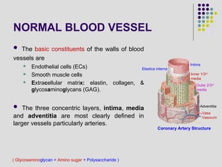
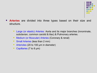
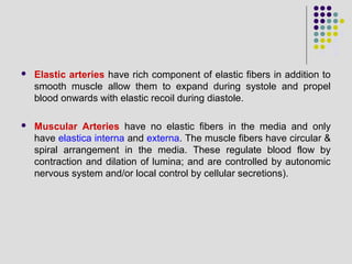
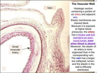
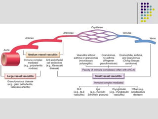
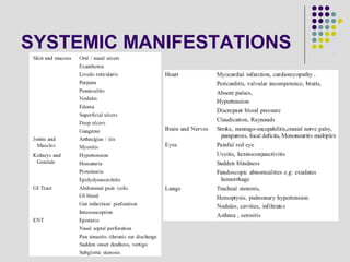
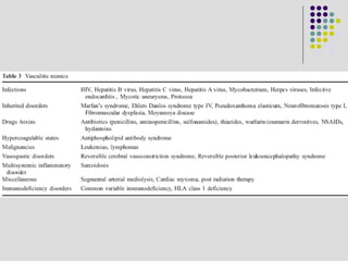
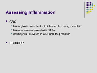
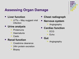
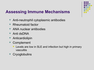
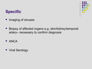
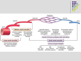
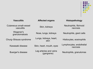
APPROACH TO
VASCULITIS
Dr. Muhammad Usman Shams
( Glycosaminoglycan = Amino sugar + Polysaccharide )
NORMAL BLOOD VESSEL
 The basic constituents of the walls of blood
vessels are
 Endothelial cells (ECs)
 Smooth muscle cells
 Extracellular matrix: elastin, collagen, &
glycosaminoglycans (GAG).
 The three concentric layers, intima, media
and adventitia are most clearly defined in
larger vessels particularly arteries.
Intima
Elastica interna
Inner 1/3rd
media
Outer 2/3rd
media
Adventitia
Coronary Artery Structure
Vasa
Vasorum
 Arteries are divided into three types based on their size and
structure.
 Large (or elastic) Arteries: Aorta and its major branches (innominate,
subclavian, common carotid & iliac) & Pulmonary arteries.
 Medium (or Muscular) Arteries (Coronary & renal)
 Small Arteries (less than 2 mm)
 Arterioles (20 to 100 µm in diameter)
 Capillaries (7 to 8 µm)
 Elastic arteries have rich component of elastic fibers in addition to
smooth muscle allow them to expand during systole and propel
blood onwards with elastic recoil during diastole.
 Muscular Arteries have no elastic fibers in the media and only
have elastica interna and externa. The muscle fibers have circular &
spiral arrangement in the media. These regulate blood flow by
contraction and dilation of lumina; and are controlled by autonomic
nervous system and/or local control by cellular secretions).
Small
muscular
Artery
Vein
The Vascular Wall
Histologic section
containing a portion of
an artery and adjacent
vein.
Elastic membranes are
stained black.
Because it is exposed
to higher blood
pressures, the artery
has a thicker wall that
maintains an open,
round lumen, even
when blood is absent.
Moreover, the elastin of
an artery is more
organized than in the
corresponding vein.
The vein has a larger,
but collapsed, lumen,
and the elastin in the
wall is diffusely
distributed.
VASCULITIS
A clinicopathologic process characterized by inflammation of and
damage to blood vessels, often resulting in complete or partial
occlusion of the involved vessels, with resulting ischemic damage to
the supplied organ/tissue.
 Fairly rare diseases
 Presentation: highly variable making delays in diagnosis common
 High morbidity and mortality
 Therapeutic challenge often requiring prolonged & intensive
immunosuppression.
CLASSIFICATION (Cause)
 Primary vasculitis – occurs in absence of recognized precipitating
cause/associated disease
 Secondary vasculitis
 secondary to established disease
 secondary to infection
 secondary to malignancy
 secondary to drugs
CLASSIFICATION (Vessel Size)
 Large vessel vasculitis
 Giant cell arteritis
 Takayasu’s arteritis
 Medium-sized vessel vasculitis
 Polyarteritis nodosa
 Kawasaki’s disease
 Primary granulomatous CNS vasculitis
 Small vessel vasculitis
 ANCA associated vasculitis
 Immune complex small vessel vasculitis
 Paraneoplastic small vessel vasculitis
 Inflammatory bowel disease vasculitis
Small Vessel Vasculitis
 ANCA associated
 Microscopic polyangiitis
 Wegeners’s granulomatosis
 Churg-Strauss syndrome
 Drug induced ANCA associated vasculitis – PTU and hydralazine
 Immune complex
 Cryoglobulinemic vasculitis
 Henoch-Schonlein purpura
 Lupus vasculitis
 Urticarial vasculitis
 Goodpasture’s syndrome
 Serum-sickness vasculitis
 Drug induced immune complex vasculitis
 Paraneoplastic
 Lymphoproliferative/Myeloproliferative/Carcinoma induced vasculitis
PATHOGENESIS
 Remains largely unknown
 Two possible mechanisms: (Most likely multifactorial)
 Infectious
 Non-infectious
INFECTIOUS VASCULITIS
 Caused by the direct invasion of infectious agents, usually bacteria
or fungi, and in particular Aspergillus and Mucor species.
 Vascular invasion can be
 part of a localized tissue infection (e.g., bacterial pneumonia or adjacent
to abscesses) OR
 from hematogenous seeding of bacteria during septicemia or
embolization from sepsis of infective endocarditis. (less common)
 Vascular infections can weaken arterial walls and culminate in
mycotic aneurysms, or can induce thrombosis and infarction.
NON-INFECTIOUS VASCULITIS
 The main immunological mechanisms that initiate
noninfectious vasculitis are
 Immune complex deposition
 Antineutrophil cytoplasmic antibodies (ANCA)
 Anti–endothelial cell antibodies.
Immune Complex Deposition
 Antibody and complement are typically detected in vasculitic lesions
or/and in circulation.
_________________
 Systemic immunological diseases, such as systemic lupus
erythematosus (SLE) and polyarteritis nodosa.
 Drug hypersensitivity: In some cases (e.g., penicillin) the drugs bind
to serum proteins; other agents, like streptokinase, are themselves
foreign proteins.
 Secondary to viral infections: Antibody to viral proteins forms
immune complexes that can be found in the serum and the vascular
lesions.
ANCA
 Anti-Neutrophil Cytoplasmic Antibodies
 Ab directed against proteins in the cytoplasmic granules of
PMN’s and monocytes
 Seen in
 Wegener’s Granulomatosis (PR3)
 Microscopic Polyangiitis (MPO)
 Churg-Strauss (MPO)
 Crescentic/necrotizing GN
c-ANCA
 Serum from patients bind to cytoplasmic granules and show a
granular appearance on immunofluorescence
 Proteinase-3 (PR-3) is the major antigen.
 serine protease
 present in azurophilic granules
p-ANCA
 Localized, peri-nuclear staining pattern on PMN’s
 Myeloperoxidase (MPO) is the major target.
 normally involved in generating oxygen free radicals
 present in lysozomes
 A plausible mechanism for ANCA vasculitis is the following:
 Drugs or cross-reactive microbial antigens induce ANCAs.
 Neutrophil surface expression or release of PR3 and MPO (e.g., in the
setting of infections) incites ANCA formation in a susceptible host.
 Subsequent infection, endotoxin exposure, or other inflammatory stimuli
elicit cytokines such as TNF that cause surface expression of PR3 and
MPO on neutrophils and other cell types.
 ANCAs react with these cytokine-activated cells and either cause direct
injury (e.g., to endothelial cells) or induce further activation (e.g., in
neutrophils).
 ANCA-activated neutrophils degranulate and also cause injury by
releasing reactive oxygen species, engendering endothelial cell toxicity
and other indirect tissue injury.
Anti-Endothelial Cell Antibodies
 Antibodies to endothelial cells may predispose to certain
vasculitides, for example, Kawasaki disease.
SYSTEMIC MANIFESTATIONS
LAB INVESTIGATIONS
Assessing Inflammation
 CBC
 leucocytosis consistent with infection & primary vasculitis
 leucopaenia associated with CTDs
 eosinophils - elevated in CSS and drug reaction
 ESR/CRP
Assessing Organ Damage
 Liver function
 LFTs – May suggest viral
infection
 Urine analysis
 Proteinuria
 Haematuria
 Casts
 Renal function
 Creatinine clearance
 24hr protein excretion
 Biopsy
 Chest radiograph
 Nervous system
 Angiography
 Cardiac function
 ECG
 Echo
 Gut
 Angiography
Assessing Immune Mechanisms
 Anti-neutrophil cytoplasmic antibodies
 Rheumatoid factor
 ANA nuclear antibodies
 Anti dsDNA
 Anticardiolipin
 Complement
 Levels are low in SLE and infection but high in primary
vasculitis
 Cryoglobulins
Specific
 Imaging of sinuses
 Biopsy of affected organs e.g. skin/kidney/temporal
artery– necessary to confirm diagnosis
 ANCA
 Viral Serology
Vasculitis Affected organs Histopathology
Cutaneous small-vessel
vasculitis
Skin, kidneys
Neutrophils, fibrinoid
necrosis
Wegener's
granulomatosis
Nose, lungs, kidneys Neutrophils, giant cells
Churg–Strauss syndrome
Lungs, kidneys, heart,
skin
Histiocytes, eosinophils
Kawasaki disease Skin, heart, mouth, eyes
Lymphocytes, endothelial
necrosis
Buerger's disease
Leg arteries and veins
(gangrene)
Neutrophils, granulomas
Approach to vasculitis

Approach to vasculitis

  • 1.
  • 2.
    ( Glycosaminoglycan =Amino sugar + Polysaccharide ) NORMAL BLOOD VESSEL  The basic constituents of the walls of blood vessels are  Endothelial cells (ECs)  Smooth muscle cells  Extracellular matrix: elastin, collagen, & glycosaminoglycans (GAG).  The three concentric layers, intima, media and adventitia are most clearly defined in larger vessels particularly arteries. Intima Elastica interna Inner 1/3rd media Outer 2/3rd media Adventitia Coronary Artery Structure Vasa Vasorum
  • 3.
     Arteries aredivided into three types based on their size and structure.  Large (or elastic) Arteries: Aorta and its major branches (innominate, subclavian, common carotid & iliac) & Pulmonary arteries.  Medium (or Muscular) Arteries (Coronary & renal)  Small Arteries (less than 2 mm)  Arterioles (20 to 100 µm in diameter)  Capillaries (7 to 8 µm)
  • 4.
     Elastic arterieshave rich component of elastic fibers in addition to smooth muscle allow them to expand during systole and propel blood onwards with elastic recoil during diastole.  Muscular Arteries have no elastic fibers in the media and only have elastica interna and externa. The muscle fibers have circular & spiral arrangement in the media. These regulate blood flow by contraction and dilation of lumina; and are controlled by autonomic nervous system and/or local control by cellular secretions).
  • 5.
    Small muscular Artery Vein The Vascular Wall Histologicsection containing a portion of an artery and adjacent vein. Elastic membranes are stained black. Because it is exposed to higher blood pressures, the artery has a thicker wall that maintains an open, round lumen, even when blood is absent. Moreover, the elastin of an artery is more organized than in the corresponding vein. The vein has a larger, but collapsed, lumen, and the elastin in the wall is diffusely distributed.
  • 6.
    VASCULITIS A clinicopathologic processcharacterized by inflammation of and damage to blood vessels, often resulting in complete or partial occlusion of the involved vessels, with resulting ischemic damage to the supplied organ/tissue.  Fairly rare diseases  Presentation: highly variable making delays in diagnosis common  High morbidity and mortality  Therapeutic challenge often requiring prolonged & intensive immunosuppression.
  • 7.
    CLASSIFICATION (Cause)  Primaryvasculitis – occurs in absence of recognized precipitating cause/associated disease  Secondary vasculitis  secondary to established disease  secondary to infection  secondary to malignancy  secondary to drugs
  • 8.
    CLASSIFICATION (Vessel Size) Large vessel vasculitis  Giant cell arteritis  Takayasu’s arteritis  Medium-sized vessel vasculitis  Polyarteritis nodosa  Kawasaki’s disease  Primary granulomatous CNS vasculitis  Small vessel vasculitis  ANCA associated vasculitis  Immune complex small vessel vasculitis  Paraneoplastic small vessel vasculitis  Inflammatory bowel disease vasculitis
  • 9.
    Small Vessel Vasculitis ANCA associated  Microscopic polyangiitis  Wegeners’s granulomatosis  Churg-Strauss syndrome  Drug induced ANCA associated vasculitis – PTU and hydralazine  Immune complex  Cryoglobulinemic vasculitis  Henoch-Schonlein purpura  Lupus vasculitis  Urticarial vasculitis  Goodpasture’s syndrome  Serum-sickness vasculitis  Drug induced immune complex vasculitis  Paraneoplastic  Lymphoproliferative/Myeloproliferative/Carcinoma induced vasculitis
  • 12.
    PATHOGENESIS  Remains largelyunknown  Two possible mechanisms: (Most likely multifactorial)  Infectious  Non-infectious
  • 13.
    INFECTIOUS VASCULITIS  Causedby the direct invasion of infectious agents, usually bacteria or fungi, and in particular Aspergillus and Mucor species.  Vascular invasion can be  part of a localized tissue infection (e.g., bacterial pneumonia or adjacent to abscesses) OR  from hematogenous seeding of bacteria during septicemia or embolization from sepsis of infective endocarditis. (less common)  Vascular infections can weaken arterial walls and culminate in mycotic aneurysms, or can induce thrombosis and infarction.
  • 14.
    NON-INFECTIOUS VASCULITIS  Themain immunological mechanisms that initiate noninfectious vasculitis are  Immune complex deposition  Antineutrophil cytoplasmic antibodies (ANCA)  Anti–endothelial cell antibodies.
  • 15.
    Immune Complex Deposition Antibody and complement are typically detected in vasculitic lesions or/and in circulation. _________________  Systemic immunological diseases, such as systemic lupus erythematosus (SLE) and polyarteritis nodosa.  Drug hypersensitivity: In some cases (e.g., penicillin) the drugs bind to serum proteins; other agents, like streptokinase, are themselves foreign proteins.  Secondary to viral infections: Antibody to viral proteins forms immune complexes that can be found in the serum and the vascular lesions.
  • 16.
    ANCA  Anti-Neutrophil CytoplasmicAntibodies  Ab directed against proteins in the cytoplasmic granules of PMN’s and monocytes  Seen in  Wegener’s Granulomatosis (PR3)  Microscopic Polyangiitis (MPO)  Churg-Strauss (MPO)  Crescentic/necrotizing GN
  • 17.
    c-ANCA  Serum frompatients bind to cytoplasmic granules and show a granular appearance on immunofluorescence  Proteinase-3 (PR-3) is the major antigen.  serine protease  present in azurophilic granules p-ANCA  Localized, peri-nuclear staining pattern on PMN’s  Myeloperoxidase (MPO) is the major target.  normally involved in generating oxygen free radicals  present in lysozomes
  • 18.
     A plausiblemechanism for ANCA vasculitis is the following:  Drugs or cross-reactive microbial antigens induce ANCAs.  Neutrophil surface expression or release of PR3 and MPO (e.g., in the setting of infections) incites ANCA formation in a susceptible host.  Subsequent infection, endotoxin exposure, or other inflammatory stimuli elicit cytokines such as TNF that cause surface expression of PR3 and MPO on neutrophils and other cell types.  ANCAs react with these cytokine-activated cells and either cause direct injury (e.g., to endothelial cells) or induce further activation (e.g., in neutrophils).  ANCA-activated neutrophils degranulate and also cause injury by releasing reactive oxygen species, engendering endothelial cell toxicity and other indirect tissue injury.
  • 19.
    Anti-Endothelial Cell Antibodies Antibodies to endothelial cells may predispose to certain vasculitides, for example, Kawasaki disease.
  • 20.
  • 22.
  • 23.
    Assessing Inflammation  CBC leucocytosis consistent with infection & primary vasculitis  leucopaenia associated with CTDs  eosinophils - elevated in CSS and drug reaction  ESR/CRP
  • 24.
    Assessing Organ Damage Liver function  LFTs – May suggest viral infection  Urine analysis  Proteinuria  Haematuria  Casts  Renal function  Creatinine clearance  24hr protein excretion  Biopsy  Chest radiograph  Nervous system  Angiography  Cardiac function  ECG  Echo  Gut  Angiography
  • 25.
    Assessing Immune Mechanisms Anti-neutrophil cytoplasmic antibodies  Rheumatoid factor  ANA nuclear antibodies  Anti dsDNA  Anticardiolipin  Complement  Levels are low in SLE and infection but high in primary vasculitis  Cryoglobulins
  • 26.
    Specific  Imaging ofsinuses  Biopsy of affected organs e.g. skin/kidney/temporal artery– necessary to confirm diagnosis  ANCA  Viral Serology
  • 29.
    Vasculitis Affected organsHistopathology Cutaneous small-vessel vasculitis Skin, kidneys Neutrophils, fibrinoid necrosis Wegener's granulomatosis Nose, lungs, kidneys Neutrophils, giant cells Churg–Strauss syndrome Lungs, kidneys, heart, skin Histiocytes, eosinophils Kawasaki disease Skin, heart, mouth, eyes Lymphocytes, endothelial necrosis Buerger's disease Leg arteries and veins (gangrene) Neutrophils, granulomas