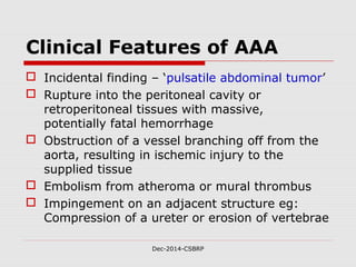
Dec-2014-CSBRP
Dec-2014-CSBRP
Aneurysms and DissectionAneurysms and Dissection
CSBR.Prasad, MD.,
Dec-2014-CSBRP
Aneurysm - Definition:
 Localized abnormal dilation of a blood
vessel or the heart
 It may be congenital or acquired
Common sites
Dec-2014-CSBRP
Aneurysm of left ventricle
Dec-2014-CSBRP
Aneurysm
of the left
ventricle
Dec-2014-CSBRP
The gross morphology of aneurysms
reveals several different pathological
features:
Dec-2014-CSBRP
Dec-2014-CSBRP
Dec-2014-CSBRP
Types:
 True aneurysms:
 Dilation of intact arterial wall or thinned
ventricular wall of the heart
 Atherosclerotic, syphilitic, and congenital
vascular aneurysms, ventricular aneurysms
following MI
 False aneurysms / Pseudoaneurysms:
 A defect in the vascular wall leading to an
extravascular hematoma that freely
communicates with the intravascular space
(“pulsating hematoma”)
 ventricular rupture after MI that is contained by
a pericardial adhesion
True & False
aneurysms
Dec-2014-CSBRP
Cardiac aneurysm
Dec-2014-CSBRP
Dec-2014-CSBRP
Complication:
 Both true and false aneurysms can
rupture, often with catastrophic
consequences
Dec-2014-CSBRP
Morphological types:
 Saccular aneurysms:
 Spherical outpouchings involving only a
portion of the vessel wall
 vary from 5 to 20 cm in diameter and
 often contain thrombus
 Fusiform aneurysms:
 Circumferential dilations of a long
vascular segment
 vary in diameter (up to 20 cm) and
 can involve extensive portions
These types are not specific for any disease
or clinical manifestations
Dec-2014-CSBRP
Aneurysms:
Dec-2014-CSBRP
Pathogenesis of Aneurysms:
 Aneurysms can occur when the
structure or function of the
connective tissue within the vascular
wall is compromised
 Inherited defects in connective tissues
 Acquired connective tissue defects
Dec-2014-CSBRP
Pathogenesis of Aneurysms:
 The intrinsic quality of the vascular wall
connective tissue is poor
 The balance of collagen degradation and
synthesis is altered by inflammation and
associated proteases
 The vascular wall is weakened through loss
of smooth muscle cells or the synthesis of
noncollagenous or nonelastic extracellular
matrix
Dec-2014-CSBRP
Pathogenesis of Aneurysms:
 The intrinsic quality of the vascular wall
connective tissue is poor
 Marfan syndrome:
 Defective protein fibrillin leads to aberrant TGF-
ß activity and weakening of elastic tissue
 Loeys-Dietz syndrome:
 Mutations in TGF-ß receptors lead to defective
synthesis of elastin and collagens I and III
 Ehlers-Danlos syndrome:
 Defective type III collagen synthesis
 Vitamin C deficiency:
 Altered collagen cross-linking
Dec-2014-CSBRP
Pathogenesis of Aneurysms:
 The intrinsic quality of the vascular wall
connective tissue is poor
 The balance of collagen degradation and
synthesis is altered by inflammation and
associated proteases
 The vascular wall is weakened through loss
of smooth muscle cells or the synthesis of
noncollagenous or nonelastic extracellular
matrix
Dec-2014-CSBRP
Pathogenesis of Aneurysms:
 The balance of collagen degradation and
synthesis is altered by inflammation and
associated proteases
 Increased matrix metalloprotease (MMP)
expression
 AS, Vasculitis
 Decreased expression of tissue inhibitors of
metalloproteases (TIMPs)
 Inflammatory lesions – AS
 Local Inflammation – production of IL-4, IL-10
Dec-2014-CSBRP
Pathogenesis of Aneurysms:
 The intrinsic quality of the vascular wall
connective tissue is poor
 The balance of collagen degradation and
synthesis is altered by inflammation and
associated proteases
 The vascular wall is weakened through loss
of smooth muscle cells or the synthesis of
noncollagenous or nonelastic extracellular
matrix
Dec-2014-CSBRP
Pathogenesis of Aneurysms:
 The vascular wall is weakened
through loss of smooth muscle cells
or the synthesis of noncollagenous or
nonelastic extracellular matrix
 Ischemia of the inner media
[Histo: Cystic medial degeneration]
 AS, Systemic hypertension
 Tertiary syphilis
Dec-2014-CSBRP
Cystic medial
degeneration
Alcian Blue-PAS
Marfan’s syndromeMarfan’s syndrome
NormalNormal
Dec-2014-CSBRP
Important causes of aortic
aneurysms
The two most important causes of aortic
aneurysms:
 Atherosclerosis [AAA]
 Hypertension [Thoracic aortic aneurysm]
 Others:
 Trauma
 Vasculitis
 Congenital defects &
 Infections
Dec-2014-CSBRP
Mycotic aneurysms:
Mycotic aneurysms can originate:
 Septic embolus [infective endocarditis]
 Extension of an adjacent suppurative
process
 Circulating organisms directly infecting
the arterial wall
Dec-2014-CSBRP
Abdominal Aortic Aneurysm: AAA
 Atherosclerosis
 Men
 >50yrs
 Smokers
Dec-2014-CSBRP
Abdominal Aortic Aneurysm:
MORPHOLOGY
 Usual position: below the renal
arteries and above the bifurcation of
the aorta
 Saccular or fusiform
 15 cm in diameter, and up to 25
cm in length
 Usually contains a bland, laminated,
mural thrombus
Abdominal Aortic Aneurysm
Usual position:
Dec-2014-CSBRP
Dec-2014-CSBRP
Dec-2014-CSBRP
Variants of AAA:
Three AAA variants merit special
mention because of their unusual
features
 Inflammatory AAA
 IgG4-related disease
 Mycotic AAA
Inflammatory AAA
 5% to 10% of all AAA
 Typically seen in younger patients
 Back pain and
 Elevated inflammatory markers – CRP
 Histology:
 Characterized by abundant lymphoplasmacytic
inflammation with many macrophages, giant cells
 Dense periaortic scarring that can extend into the
anterior retroperitoneum
 Localized immune response to the abdominal
aortic wall
Dec-2014-CSBRP
IgG4-related disease AAA
 High plasma levels of IgG4
 Fibrosis
 Infiltrated by IgG4-expressing plasma cells
 Other organ systems may also be involved
 Salivary, pancreas, biliary system
Dec-2014-CSBRP
Mycotic AAA
 Infected by the lodging of circulating
microorganisms in the wall
Dec-2014-CSBRP
Clinical Features of AAA
 Incidental finding – ‘pulsatile abdominal tumor’
 Rupture into the peritoneal cavity or
retroperitoneal tissues with massive,
potentially fatal hemorrhage
 Obstruction of a vessel branching off from the
aorta, resulting in ischemic injury to the
supplied tissue
 Embolism from atheroma or mural thrombus
 Impingement on an adjacent structure eg:
Compression of a ureter or erosion of vertebrae
Dec-2014-CSBRP
The risk of rupture - AAA
 Is directly related to the size
 NIL - <4 cm
 1% per year between 4 and 5 cm
 11% per year between 5 and 6 cm
 25% per year larger than 6 cm
 Most aneurysms expand at a rate of 0.2 to
0.3 cm/year, but 20% expand more
rapidly
Dec-2014-CSBRP
Aneurysms 5 cm or larger are managed aggressively
Thoracic Aortic Aneurysm - TAA
 Most commonly associated with
hypertension
 Other causes:
 Marfan syndrome
 Loeys-Dietz syndrome
Dec-2014-CSBRP
Thoracic Aortic Aneurysm – TAA
Presentation:
 Respiratory difficulties due to encroachment on
the lungs and airways
 Difficulty in swallowing due to compression of
the esophagus
 Persistent cough due to compression of the
recurrent laryngeal nerves
 Pain caused by erosion of bone
 Cardiac disease – IHD - as the aortic aneurysm
leads to AR
 Rupture
Dec-2014-CSBRP
Abdominal Aortic Aneurysm
Dec-2014-CSBRP
Thoracic Aortic Aneurysm – TAA
Presentation:
Most patients with syphilitic
aneurysms die of heart failure
secondary to aortic valvular
incompetence
Dec-2014-CSBRP
Thoracic Aortic Aneurysm –
TAA
Dec-2014-CSBRP
Thoracic Aortic Aneurysm –
TAA
Dec-2014-CSBRP
Thoracic Aortic Aneurysm – TAA
Dec-2014-CSBRP
Thoracic Aortic Aneurysm – TAA
Tree bark appearance - Syphilis
Dec-2014-CSBRP
Aortic Dissection
 Occurs when blood separates the
laminar planes of the media to form a
blood-filled channel within the aortic wall
 Occurs principally in two groups of
patients:
 Men, 40 to 60 years with antecedent
hypertension (>90%) and
 Younger adults with abnormalities of
connective tissue affecting the aorta
Dec-2014-CSBRP
Aortic Dissection
 Other causes:
 Arterial cannulization
 Pregnancy
Dec-2014-CSBRP
Aortic Dissection
Pathogenesis:
 Hypertension – Cystic medial degeneration
 Marfan syndrome
 Ehlers-Danlos syndrome
 Defects in copper metabolism
Dec-2014-CSBRP
Aortic Dissection -
MORPHOLOGY
Dec-2014-CSBRP
 Most frequent finding is cystic medial degeneration
 usually initiates with an intimal tear
 Spontaneous dissection:
 Within 10 cm of the aortic valve
 Tears are typically transverse with sharp, jagged edges up
to 1 to 5 cm in length
 Dissection can extend retrograde toward the heart as well
as distally
 Tear: Between the middle and outer thirds
 Rupture through the adventitia causing massive
hemorrhage
 Cardic tamponade
 “double-barreled aorta”
Aortic Dissection -
MORPHOLOGY
Dec-2014-CSBRP
Dec-2014-CSBRP
Aortic
dissection
Dec-2014-CSBRP
Dec-2014-CSBRP
Classification:
Dec-2014-CSBRP
Clinical Features:
 Proximal dissections are more serious and
dangerous
 (types I and II of the DeBakey)
 PAIN: sudden onset of excruciating pain,
usually beginning in the anterior chest,
radiating to the back between the scapulae,
and moving downward as the dissection
progresses
 Cardiac tamponade
 Aortic insufficiency
 Ischemic consequences
Dec-2014-CSBRP
Out come:
 In type A dissections: Mortality is
70%
 Most type B dissections can be
managed conservatively -75%
survival rate
Dec-2014-CSBRP
E N D
Dec-2014-CSBRP

Cvs aneurysms&amp;dissection-csbrp

  • 1.
  • 2.
    Dec-2014-CSBRP Aneurysms and DissectionAneurysmsand Dissection CSBR.Prasad, MD.,
  • 3.
    Dec-2014-CSBRP Aneurysm - Definition: Localized abnormal dilation of a blood vessel or the heart  It may be congenital or acquired
  • 4.
  • 5.
    Aneurysm of leftventricle Dec-2014-CSBRP
  • 6.
  • 7.
    The gross morphologyof aneurysms reveals several different pathological features: Dec-2014-CSBRP
  • 8.
  • 9.
    Dec-2014-CSBRP Types:  True aneurysms: Dilation of intact arterial wall or thinned ventricular wall of the heart  Atherosclerotic, syphilitic, and congenital vascular aneurysms, ventricular aneurysms following MI  False aneurysms / Pseudoaneurysms:  A defect in the vascular wall leading to an extravascular hematoma that freely communicates with the intravascular space (“pulsating hematoma”)  ventricular rupture after MI that is contained by a pericardial adhesion
  • 10.
  • 11.
  • 12.
    Dec-2014-CSBRP Complication:  Both trueand false aneurysms can rupture, often with catastrophic consequences
  • 13.
    Dec-2014-CSBRP Morphological types:  Saccularaneurysms:  Spherical outpouchings involving only a portion of the vessel wall  vary from 5 to 20 cm in diameter and  often contain thrombus  Fusiform aneurysms:  Circumferential dilations of a long vascular segment  vary in diameter (up to 20 cm) and  can involve extensive portions These types are not specific for any disease or clinical manifestations
  • 14.
  • 15.
    Dec-2014-CSBRP Pathogenesis of Aneurysms: Aneurysms can occur when the structure or function of the connective tissue within the vascular wall is compromised  Inherited defects in connective tissues  Acquired connective tissue defects
  • 16.
    Dec-2014-CSBRP Pathogenesis of Aneurysms: The intrinsic quality of the vascular wall connective tissue is poor  The balance of collagen degradation and synthesis is altered by inflammation and associated proteases  The vascular wall is weakened through loss of smooth muscle cells or the synthesis of noncollagenous or nonelastic extracellular matrix
  • 17.
    Dec-2014-CSBRP Pathogenesis of Aneurysms: The intrinsic quality of the vascular wall connective tissue is poor  Marfan syndrome:  Defective protein fibrillin leads to aberrant TGF- ß activity and weakening of elastic tissue  Loeys-Dietz syndrome:  Mutations in TGF-ß receptors lead to defective synthesis of elastin and collagens I and III  Ehlers-Danlos syndrome:  Defective type III collagen synthesis  Vitamin C deficiency:  Altered collagen cross-linking
  • 18.
    Dec-2014-CSBRP Pathogenesis of Aneurysms: The intrinsic quality of the vascular wall connective tissue is poor  The balance of collagen degradation and synthesis is altered by inflammation and associated proteases  The vascular wall is weakened through loss of smooth muscle cells or the synthesis of noncollagenous or nonelastic extracellular matrix
  • 19.
    Dec-2014-CSBRP Pathogenesis of Aneurysms: The balance of collagen degradation and synthesis is altered by inflammation and associated proteases  Increased matrix metalloprotease (MMP) expression  AS, Vasculitis  Decreased expression of tissue inhibitors of metalloproteases (TIMPs)  Inflammatory lesions – AS  Local Inflammation – production of IL-4, IL-10
  • 20.
    Dec-2014-CSBRP Pathogenesis of Aneurysms: The intrinsic quality of the vascular wall connective tissue is poor  The balance of collagen degradation and synthesis is altered by inflammation and associated proteases  The vascular wall is weakened through loss of smooth muscle cells or the synthesis of noncollagenous or nonelastic extracellular matrix
  • 21.
    Dec-2014-CSBRP Pathogenesis of Aneurysms: The vascular wall is weakened through loss of smooth muscle cells or the synthesis of noncollagenous or nonelastic extracellular matrix  Ischemia of the inner media [Histo: Cystic medial degeneration]  AS, Systemic hypertension  Tertiary syphilis
  • 22.
  • 23.
    Dec-2014-CSBRP Important causes ofaortic aneurysms The two most important causes of aortic aneurysms:  Atherosclerosis [AAA]  Hypertension [Thoracic aortic aneurysm]  Others:  Trauma  Vasculitis  Congenital defects &  Infections
  • 24.
    Dec-2014-CSBRP Mycotic aneurysms: Mycotic aneurysmscan originate:  Septic embolus [infective endocarditis]  Extension of an adjacent suppurative process  Circulating organisms directly infecting the arterial wall
  • 25.
    Dec-2014-CSBRP Abdominal Aortic Aneurysm:AAA  Atherosclerosis  Men  >50yrs  Smokers
  • 26.
    Dec-2014-CSBRP Abdominal Aortic Aneurysm: MORPHOLOGY Usual position: below the renal arteries and above the bifurcation of the aorta  Saccular or fusiform  15 cm in diameter, and up to 25 cm in length  Usually contains a bland, laminated, mural thrombus
  • 27.
    Abdominal Aortic Aneurysm Usualposition: Dec-2014-CSBRP
  • 28.
  • 29.
    Dec-2014-CSBRP Variants of AAA: ThreeAAA variants merit special mention because of their unusual features  Inflammatory AAA  IgG4-related disease  Mycotic AAA
  • 30.
    Inflammatory AAA  5%to 10% of all AAA  Typically seen in younger patients  Back pain and  Elevated inflammatory markers – CRP  Histology:  Characterized by abundant lymphoplasmacytic inflammation with many macrophages, giant cells  Dense periaortic scarring that can extend into the anterior retroperitoneum  Localized immune response to the abdominal aortic wall Dec-2014-CSBRP
  • 31.
    IgG4-related disease AAA High plasma levels of IgG4  Fibrosis  Infiltrated by IgG4-expressing plasma cells  Other organ systems may also be involved  Salivary, pancreas, biliary system Dec-2014-CSBRP
  • 32.
    Mycotic AAA  Infectedby the lodging of circulating microorganisms in the wall Dec-2014-CSBRP
  • 33.
    Clinical Features ofAAA  Incidental finding – ‘pulsatile abdominal tumor’  Rupture into the peritoneal cavity or retroperitoneal tissues with massive, potentially fatal hemorrhage  Obstruction of a vessel branching off from the aorta, resulting in ischemic injury to the supplied tissue  Embolism from atheroma or mural thrombus  Impingement on an adjacent structure eg: Compression of a ureter or erosion of vertebrae Dec-2014-CSBRP
  • 34.
    The risk ofrupture - AAA  Is directly related to the size  NIL - <4 cm  1% per year between 4 and 5 cm  11% per year between 5 and 6 cm  25% per year larger than 6 cm  Most aneurysms expand at a rate of 0.2 to 0.3 cm/year, but 20% expand more rapidly Dec-2014-CSBRP Aneurysms 5 cm or larger are managed aggressively
  • 35.
    Thoracic Aortic Aneurysm- TAA  Most commonly associated with hypertension  Other causes:  Marfan syndrome  Loeys-Dietz syndrome Dec-2014-CSBRP
  • 36.
    Thoracic Aortic Aneurysm– TAA Presentation:  Respiratory difficulties due to encroachment on the lungs and airways  Difficulty in swallowing due to compression of the esophagus  Persistent cough due to compression of the recurrent laryngeal nerves  Pain caused by erosion of bone  Cardiac disease – IHD - as the aortic aneurysm leads to AR  Rupture Dec-2014-CSBRP
  • 37.
  • 38.
    Thoracic Aortic Aneurysm– TAA Presentation: Most patients with syphilitic aneurysms die of heart failure secondary to aortic valvular incompetence Dec-2014-CSBRP
  • 39.
    Thoracic Aortic Aneurysm– TAA Dec-2014-CSBRP
  • 40.
    Thoracic Aortic Aneurysm– TAA Dec-2014-CSBRP
  • 41.
    Thoracic Aortic Aneurysm– TAA Dec-2014-CSBRP
  • 42.
    Thoracic Aortic Aneurysm– TAA Tree bark appearance - Syphilis Dec-2014-CSBRP
  • 43.
    Aortic Dissection  Occurswhen blood separates the laminar planes of the media to form a blood-filled channel within the aortic wall  Occurs principally in two groups of patients:  Men, 40 to 60 years with antecedent hypertension (>90%) and  Younger adults with abnormalities of connective tissue affecting the aorta Dec-2014-CSBRP
  • 44.
    Aortic Dissection  Othercauses:  Arterial cannulization  Pregnancy Dec-2014-CSBRP
  • 45.
    Aortic Dissection Pathogenesis:  Hypertension– Cystic medial degeneration  Marfan syndrome  Ehlers-Danlos syndrome  Defects in copper metabolism Dec-2014-CSBRP
  • 46.
    Aortic Dissection - MORPHOLOGY Dec-2014-CSBRP Most frequent finding is cystic medial degeneration  usually initiates with an intimal tear  Spontaneous dissection:  Within 10 cm of the aortic valve  Tears are typically transverse with sharp, jagged edges up to 1 to 5 cm in length  Dissection can extend retrograde toward the heart as well as distally  Tear: Between the middle and outer thirds  Rupture through the adventitia causing massive hemorrhage  Cardic tamponade  “double-barreled aorta”
  • 47.
  • 48.
  • 49.
  • 50.
  • 51.
  • 52.
    Clinical Features:  Proximaldissections are more serious and dangerous  (types I and II of the DeBakey)  PAIN: sudden onset of excruciating pain, usually beginning in the anterior chest, radiating to the back between the scapulae, and moving downward as the dissection progresses  Cardiac tamponade  Aortic insufficiency  Ischemic consequences Dec-2014-CSBRP
  • 53.
    Out come:  Intype A dissections: Mortality is 70%  Most type B dissections can be managed conservatively -75% survival rate Dec-2014-CSBRP
  • 54.

Editor's Notes

  • #7 Thorax x-ray showing the pacemaker, an enlarged heart, and an oval-shaped calcified structure related to a calcified anteroapical left ventricular aneurysm.
  • #10 When an aneurysm involves an attenuated but intact arterial wall or thinned ventricular wall of the heart, it is called a “true” aneurysm. Atherosclerotic, syphilitic, and congenital vascular aneurysms, as well as ventricular aneurysms that follow transmural myocardial infarctions are of this type. In contrast, a false aneurysm (also called pseudo-aneurysm) is a defect in the vascular wall leading to an extravascular hematoma that freely communicates with the intravascular space (“pulsating hematoma”). Examples include a ventricular rupture after myocardial infarction that is contained by a pericardial adhesion, or a leak at the sutured junction of a vascular graft with a natural artery. An arterial dissection arises when blood enters a defect in the arterial wall and tunnels between its layers. Dissections are often but not always aneurysmal (see later). Both true and false aneurysms as well as dissections can rupture, often with catastrophic consequences.
  • #14 Descriptively, aneurysms are classified by macroscopic shape and size (Fig. 11-18). Saccular aneurysms are spherical outpouchings involving only a portion of the vessel wall; they vary from 5 to 20 cm in diameter and often contain thrombus. Fusiform aneurysms are diffuse, circumferential dilations of a long vascular segment; they vary in diameter (up to 20 cm) and in length and can involve extensive portions of the aortic arch, abdominal aorta, or even the iliacs. These types are not specific for any disease or clinical manifestations.
  • #15 Figure 11-18 Aneurysms. A, Normal vessel. B, True aneurysm, saccular type. The wall focally bulges outward and may be attenuated but is otherwise intact. C, True aneurysm, fusiform type. There is circumferential dilation of the vessel, without rupture. D, False aneurysm. The wall is ruptured, and there is a collection of blood (hematoma) that is bounded externally by adherent extravascular tissues.
  • #18 The intrinsic quality of the vascular wall connective tissue is poor. In Marfan syndrome, for example (Chapter 5), defective Synthesis of the scaffolding protein fibrillin leads to aberrant TGF-ß activity and weakening of elastic tissue; in the aorta, this may result in progressive dilation. Loeys-Dietz syndrome is another cause of aneurysms; in this disorder, mutations in TGF-ß receptors lead to defective synthesis of elastin and collagens I and III. Aneurysms in such individuals can rupture fairly easily (even at small size) and are thus considered to follow an “aggressive” course. Weak vessel walls due to defective type III collagen synthesis are also a hallmark of the vascular forms of Ehlers-Danlos syndrome (Chapter 5), and altered collagen cross-linking associated with vitamin C deficiency (scurvy) is an example of a nutritional basis for aneurysm formation, that is thankfully rare these days.
  • #20 The balance of collagen degradation and synthesis is altered by inflammation and associated proteases. In particular, increased matrix metalloprotease (MMP) expression, especially by macrophages in atherosclerotic plaque or in vasculitis, likely contributes to aneurysm development; these enzymes have the capacity to degrade virtually all components of the extracellular matrix in the arterial wall (collagens, elastin, proteoglycans, laminin, fibronectin). Decreased expression of tissue inhibitors of metalloproteases (TIMPs) can also contribute to the extracellular matrix degradation. The risk of aneurysm formation in the setting of inflammatory lesions (e.g.,atherosclerosis) may be associated with MMP and/or TIMP polymorphisms, or altered by the nature of the local inflammatory response. For example, abdominal aortic aneurysms (AAA; see later) are associated with local production of cytokines (such as IL-4 and IL-10) that stimulate release of elastolytic MMP from macrophages.
  • #22 The vascular wall is weakened through loss of smooth muscle cells or the synthesis of noncollagenous or nonelastic extracellular matrix. Ischemia of the inner media occurs when there is atherosclerotic thickening of the intima, which increases the distance that oxygen and nutrients must diffuse. Systemic hypertension can also cause significant narrowing of arterioles of the vasa vasorum (e.g., in the aorta), which causes outer medial ischemia. Medial ischemia may lead to “degenerative changes” of the aorta, whereby smooth muscle cell loss—or change in synthetic phenotype—leads to scarring (and loss of elastic fibers), inadequate extracellular matrix synthesis, and production of increasing amounts of amorphous ground substance (glycos aminoglycan). Histologically, these changes are collectively recognized as cystic medial degeneration (Fig. 11-19), which can be seen in a variety of settings, including Marfan syndrome and scurvy. Tertiary syphilis is another rare cause of aortic aneurysms. The obliterative endarteritis characteristic of late stage syphilis shows a predilection for small vessels, including those of the vasa vasorum of the thoracic aorta. This leads to ischemic injury of the aortic media and aneurysmal dilation, which sometimes involves the aortic valve annulus.
  • #23 Figure 11-19 Cystic medial degeneration. A, Cross-section of aortic media from a patient with Marfan syndrome, showing elastin fragmentation and areas devoid of elastin that resemble cystic spaces but are actually filled with proteoglycans (asterisks). B, Normal media for comparison, showing the regular layered pattern of elastic tissue. In both A and B, elastin is stained black. Alcian Blue-PAS: Disruption and fragmentation of elastic lamellae of the aortic media. The cysts are filled with proteoglycan rich mucoid substance.
  • #24 AAA = Abdominal aortic aneurysm. TAA = Thoracic aortic aneurysm.
  • #29 Note the laminated thrombus
  • #32 There are four IgG subclasses (IgG1, 2, 3, and 4) in humans, named in order of their abundance in serum (IgG1 being the most abundant).
  • #35 Diameter of Aorta: The portion of the aorta in the chest is called the thoracic aorta; the portion in the abdomen is called the abdominal aorta. Normal diameter is about 3.0 cm (1.2 inches), tapering to a width of about 2.0 cm (0.8 inch) in the abdomen.
  • #36 Loeys-Dietz syndrome is a disorder that affects the connective tissue in many parts of the body. Connective tissue provides strength and flexibility to structures such as bones, ligaments, muscles, and blood vessels. There are four types of Loeys-Dietz syndrome, labelled types I through IV, which are distinguished by their genetic cause. Regardless of the type, signs and symptoms of Loeys-Dietz syndrome can become apparent anytime in childhood or adulthood, and the severity is variable. Loeys-Dietz syndrome is characterized by enlargement of the aorta, which is the large blood vessel that distributes blood from the heart to the rest of the body. The aorta can weaken and stretch, causing a bulge in the blood vessel wall (an aneurysm). Stretching of the aorta may also lead to a sudden tearing of the layers in the aorta wall (aortic dissection). People with Loeys-Dietz syndrome can also have aneurysms or dissections in arteries throughout the body and have arteries with abnormal twists and turns (arterial tortuosity). Individuals with Loeys-Dietz syndrome often have skeletal problems including premature fusion of the skull bones (craniosynostosis), an abnormal side-to-side curvature of the spine (scoliosis), either a sunken chest (pectus excavatum) or a protruding chest (pectus carinatum), an inward- and upward-turning foot (clubfoot), flat feet (pes planus), or elongated limbs with joint deformities called contractures that restrict the movement of certain joints. Degeneration of the discs that separate the bones of the spine (vertebrae), often affecting the neck, is a common finding. Some affected individuals have prominent joint inflammation (osteoarthritis) that commonly affects the knees and the joints of the hands, wrists, and spine. People with Loeys-Dietz syndrome may bruise easily and develop abnormal scars after wound healing. The skin is frequently described as translucent, often with stretch marks (striae) and visible underlying veins. Other characteristic features include widely spaced eyes (hypertelorism), a split in the soft flap of tissue that hangs from the back of the mouth (bifid uvula), and an opening in the roof of the mouth (cleft palate). Individuals with Loeys-Dietz syndrome frequently develop immune system-related problems such as food allergies, asthma, or inflammatory disorders such as eczema or inflammatory bowel disease. How common is Loeys-Dietz syndrome? The prevalence of Loeys-Dietz syndrome is unknown. Loeys-Dietz syndrome types I and II appear to be the most common forms. What genes are related to Loeys-Dietz syndrome? The four types of Loeys-Dietz syndrome are distinguished by their genetic cause: mutations in the TGFBR1 gene cause type I, mutations in the TGFBR2 gene cause type II, mutations in the SMAD3 gene cause type III, and mutations in the TGFB2 gene cause type IV. These four genes play a role in cell signaling that promotes growth and development of the body&amp;apos;s tissues. This signaling pathway also helps with bone and blood vessel development and plays a part in the formation of the extracellular matrix, an intricate lattice of proteins and other molecules that forms in the spaces between cells. Mutations in the TGFBR1, TGFBR2, TGFB2, and SMAD3 genes result in the production of proteins with little or no function. Even though these proteins have severely reduced function, cell signaling occurs at an even greater intensity than normal. Researchers speculate that the activity of proteins in this signaling pathway is increased to compensate for the protein whose function is reduced; however, the exact mechanism responsible for the increase in signaling is unclear. The overactive signaling pathway disrupts the development of connective tissue, the extracellular matrix, and various body systems, leading to the varied signs and symptoms of Loeys-Dietz syndrome. Read more about the SMAD3, TGFB2, TGFBR1, and TGFBR2 genes. How do people inherit Loeys-Dietz syndrome? Loeys-Dietz syndrome is considered to have an autosomal dominant pattern of inheritance, which means one copy of the altered gene in each cell is sufficient to cause the disorder. In about 75 percent of cases, this disorder results from a new gene mutation and occurs in people with no history of the disorder in their family. In other cases, an affected person inherits the mutation from one affected parent.
  • #38 Erosion of vertebra causing backpain, this may be the initial presentation.
  • #42 DDs: TAA and Gumma.