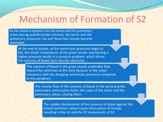
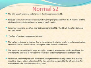
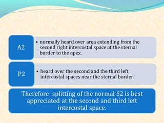
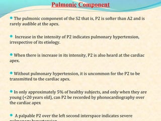
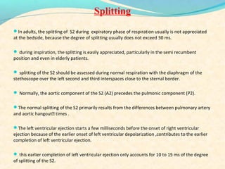
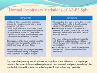
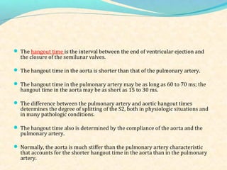
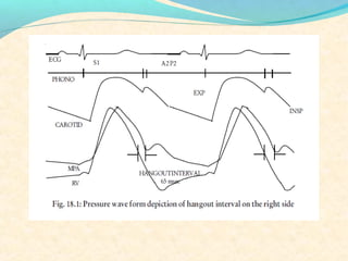
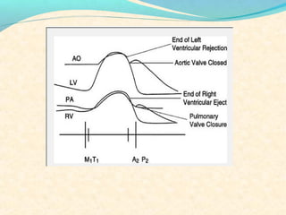
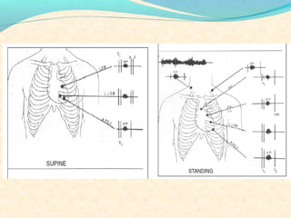
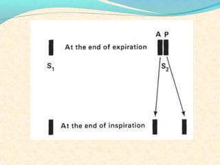
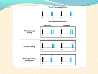
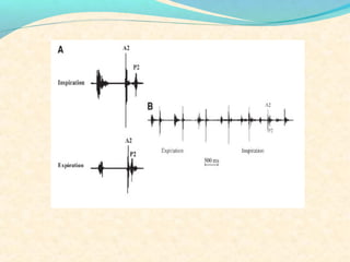
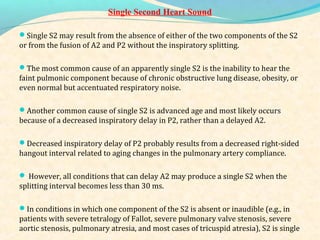
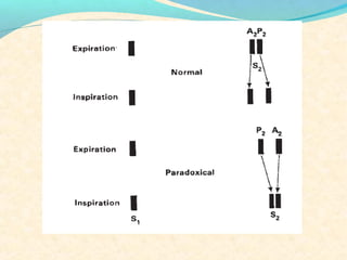
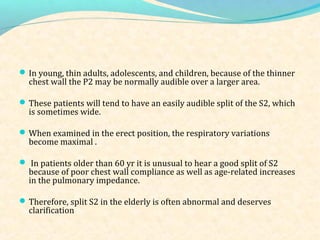
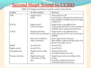
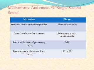
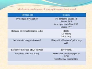
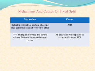
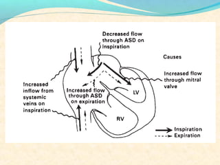
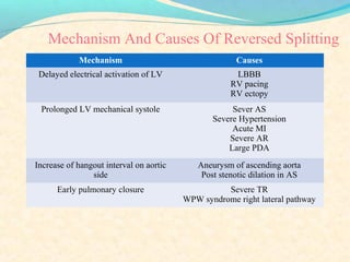
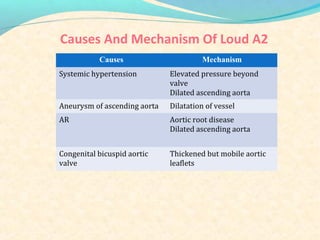
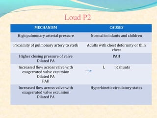
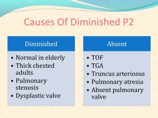
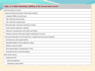
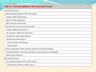
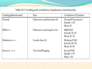
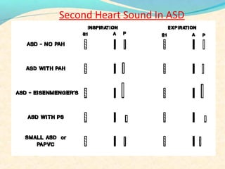
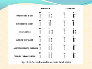
The second heart sound occurs at the end of systole due to closure of the semilunar valves. There are normally two components: A2 from aortic valve closure and P2 from pulmonary valve closure. A2 is typically louder due to higher pressures in the aorta. The components are normally split, with A2 occurring earlier due to differences in vascular resistance and compliance between the pulmonary and systemic circulations. Widening of the split may indicate conduction delays or pulmonary hypertension. Reversed or paradoxical splitting can occur in conditions that delay left ventricular ejection such as left bundle branch block. Single second heart sounds may result from fusion of the components or absence of one.
Wolff–Parkinson–White syndrome (WPW) is one of several disorders of the conduction system of the heart that are commonly referred to as pre-excitation syndromes. WPW is caused by the presence of an abnormal accessory electrical conduction pathway between the atria and the ventricles. Electrical signals traveling down this abnormal pathway (known as the bundle of Kent) may stimulate the ventricles to contract prematurely, resulting in a unique type of supra-ventricular tachycardia referred to as an atrio-ventricular reciprocating tachycardia.
Wolff–Parkinson–White syndrome (WPW) is one of several disorders of the conduction system of the heart that are commonly referred to as pre-excitation syndromes. WPW is caused by the presence of an abnormal accessory electrical conduction pathway between the atria and the ventricles. Electrical signals travelling down this abnormal pathway (known as the bundle of Kent) may stimulate the ventricles to contract prematurely, resulting in a unique type of supraventricular tachycardia referred to as an atrioventricular reciprocating tachycardia.The incidence of WPW is between 0.1% and 0.3% in the general population.Sudden cardiac death in people with WPW is rare (incidence of less than 0.6%), and is usually caused by the propagation of an atrial tachydysrhythmia (rapid and abnormal heart rate) to the ventricles by the abnormal accessory pathway.
The jugular venous pressure (JVP, sometimes referred to as jugular venous pulse) is the indirectly observed pressure over the venous system via visualization of the internal jugular vein. It can be useful in the differentiation of different forms of heart and lung disease.
Basics on heart murmurs, differentiate physiologic murmur from pathologic. Learn when to investigate further and when to monitor. Know the effect of different maneuvers on murmurs and physiology behind them. Listen to the heart sounds on the slides to appreciate the distinctive nature of each murmur.
Cardiac cycle refers to a complete heartbeat from its generation to the beginning of the next beat.
Cardiac events that occur from –
beginning of one heart beat to the beginning of the next are called the cardiac cycle.
Aortic insufficiency (AI), also known as aortic regurgitation (AR), is the leaking of the aortic valve of the heart that causes blood to flow in the reverse direction
Title: Sense of Smell
Presenter: Dr. Faiza, Assistant Professor of Physiology
Qualifications:
MBBS (Best Graduate, AIMC Lahore)
FCPS Physiology
ICMT, CHPE, DHPE (STMU)
MPH (GC University, Faisalabad)
MBA (Virtual University of Pakistan)
Learning Objectives:
Describe the primary categories of smells and the concept of odor blindness.
Explain the structure and location of the olfactory membrane and mucosa, including the types and roles of cells involved in olfaction.
Describe the pathway and mechanisms of olfactory signal transmission from the olfactory receptors to the brain.
Illustrate the biochemical cascade triggered by odorant binding to olfactory receptors, including the role of G-proteins and second messengers in generating an action potential.
Identify different types of olfactory disorders such as anosmia, hyposmia, hyperosmia, and dysosmia, including their potential causes.
Key Topics:
Olfactory Genes:
3% of the human genome accounts for olfactory genes.
400 genes for odorant receptors.
Olfactory Membrane:
Located in the superior part of the nasal cavity.
Medially: Folds downward along the superior septum.
Laterally: Folds over the superior turbinate and upper surface of the middle turbinate.
Total surface area: 5-10 square centimeters.
Olfactory Mucosa:
Olfactory Cells: Bipolar nerve cells derived from the CNS (100 million), with 4-25 olfactory cilia per cell.
Sustentacular Cells: Produce mucus and maintain ionic and molecular environment.
Basal Cells: Replace worn-out olfactory cells with an average lifespan of 1-2 months.
Bowman’s Gland: Secretes mucus.
Stimulation of Olfactory Cells:
Odorant dissolves in mucus and attaches to receptors on olfactory cilia.
Involves a cascade effect through G-proteins and second messengers, leading to depolarization and action potential generation in the olfactory nerve.
Quality of a Good Odorant:
Small (3-20 Carbon atoms), volatile, water-soluble, and lipid-soluble.
Facilitated by odorant-binding proteins in mucus.
Membrane Potential and Action Potential:
Resting membrane potential: -55mV.
Action potential frequency in the olfactory nerve increases with odorant strength.
Adaptation Towards the Sense of Smell:
Rapid adaptation within the first second, with further slow adaptation.
Psychological adaptation greater than receptor adaptation, involving feedback inhibition from the central nervous system.
Primary Sensations of Smell:
Camphoraceous, Musky, Floral, Pepperminty, Ethereal, Pungent, Putrid.
Odor Detection Threshold:
Examples: Hydrogen sulfide (0.0005 ppm), Methyl-mercaptan (0.002 ppm).
Some toxic substances are odorless at lethal concentrations.
Characteristics of Smell:
Odor blindness for single substances due to lack of appropriate receptor protein.
Behavioral and emotional influences of smell.
Transmission of Olfactory Signals:
From olfactory cells to glomeruli in the olfactory bulb, involving lateral inhibition.
Primitive, less old, and new olfactory systems with different path
Rasamanikya is a excellent preparation in the field of Rasashastra, it is used in various Kushtha Roga, Shwasa, Vicharchika, Bhagandara, Vatarakta, and Phiranga Roga. In this article Preparation& Comparative analytical profile for both Formulationon i.e Rasamanikya prepared by Kushmanda swarasa & Churnodhaka Shodita Haratala. The study aims to provide insights into the comparative efficacy and analytical aspects of these formulations for enhanced therapeutic outcomes.
263778731218 Abortion Clinic /Pills In Harare ,sisternakatoto
263778731218 Abortion Clinic /Pills In Harare ,ABORTION WOMEN’S CLINIC +27730423979 IN women clinic we believe that every woman should be able to make choices in her pregnancy. Our job is to provide compassionate care, safety,affordable and confidential services. That’s why we have won the trust from all generations of women all over the world. we use non surgical method(Abortion pills) to terminate…Dr.LISA +27730423979women Clinic is committed to providing the highest quality of obstetrical and gynecological care to women of all ages. Our dedicated staff aim to treat each patient and her health concerns with compassion and respect.Our dedicated group ABORTION WOMEN’S CLINIC +27730423979 IN women clinic we believe that every woman should be able to make choices in her pregnancy. Our job is to provide compassionate care, safety,affordable and confidential services. That’s why we have won the trust from all generations of women all over the world. we use non surgical method(Abortion pills) to terminate…Dr.LISA +27730423979women Clinic is committed to providing the highest quality of obstetrical and gynecological care to women of all ages. Our dedicated staff aim to treat each patient and her health concerns with compassion and respect.Our dedicated group of receptionists, nurses, and physicians have worked together as a teamof receptionists, nurses, and physicians have worked together as a team wwww.lisywomensclinic.co.za/
DISSERTATION on NEW DRUG DISCOVERY AND DEVELOPMENT STAGES OF DRUG DISCOVERYNEHA GUPTA
The process of drug discovery and development is a complex and multi-step endeavor aimed at bringing new pharmaceutical drugs to market. It begins with identifying and validating a biological target, such as a protein, gene, or RNA, that is associated with a disease. This step involves understanding the target's role in the disease and confirming that modulating it can have therapeutic effects. The next stage, hit identification, employs high-throughput screening (HTS) and other methods to find compounds that interact with the target. Computational techniques may also be used to identify potential hits from large compound libraries.
Following hit identification, the hits are optimized to improve their efficacy, selectivity, and pharmacokinetic properties, resulting in lead compounds. These leads undergo further refinement to enhance their potency, reduce toxicity, and improve drug-like characteristics, creating drug candidates suitable for preclinical testing. In the preclinical development phase, drug candidates are tested in vitro (in cell cultures) and in vivo (in animal models) to evaluate their safety, efficacy, pharmacokinetics, and pharmacodynamics. Toxicology studies are conducted to assess potential risks.
Before clinical trials can begin, an Investigational New Drug (IND) application must be submitted to regulatory authorities. This application includes data from preclinical studies and plans for clinical trials. Clinical development involves human trials in three phases: Phase I tests the drug's safety and dosage in a small group of healthy volunteers, Phase II assesses the drug's efficacy and side effects in a larger group of patients with the target disease, and Phase III confirms the drug's efficacy and monitors adverse reactions in a large population, often compared to existing treatments.
After successful clinical trials, a New Drug Application (NDA) is submitted to regulatory authorities for approval, including all data from preclinical and clinical studies, as well as proposed labeling and manufacturing information. Regulatory authorities then review the NDA to ensure the drug is safe, effective, and of high quality, potentially requiring additional studies. Finally, after a drug is approved and marketed, it undergoes post-marketing surveillance, which includes continuous monitoring for long-term safety and effectiveness, pharmacovigilance, and reporting of any adverse effects.
Basavarajeeyam is an important text for ayurvedic physician belonging to andhra pradehs. It is a popular compendium in various parts of our country as well as in andhra pradesh. The content of the text was presented in sanskrit and telugu language (Bilingual). One of the most famous book in ayurvedic pharmaceutics and therapeutics. This book contains 25 chapters called as prakaranas. Many rasaoushadis were explained, pioneer of dhatu druti, nadi pareeksha, mutra pareeksha etc. Belongs to the period of 15-16 century. New diseases like upadamsha, phiranga rogas are explained.
- Video recording of this lecture in English language: https://youtu.be/kqbnxVAZs-0
- Video recording of this lecture in Arabic language: https://youtu.be/SINlygW1Mpc
- Link to download the book free: https://nephrotube.blogspot.com/p/nephrotube-nephrology-books.html
- Link to NephroTube website: www.NephroTube.com
- Link to NephroTube social media accounts: https://nephrotube.blogspot.com/p/join-nephrotube-on-social-media.html
ABDOMINAL TRAUMA in pediatrics part one.drhasanrajab
Abdominal trauma in pediatrics refers to injuries or damage to the abdominal organs in children. It can occur due to various causes such as falls, motor vehicle accidents, sports-related injuries, and physical abuse. Children are more vulnerable to abdominal trauma due to their unique anatomical and physiological characteristics. Signs and symptoms include abdominal pain, tenderness, distension, vomiting, and signs of shock. Diagnosis involves physical examination, imaging studies, and laboratory tests. Management depends on the severity and may involve conservative treatment or surgical intervention. Prevention is crucial in reducing the incidence of abdominal trauma in children.
micro teaching on communication m.sc nursing.pdfAnurag Sharma
Microteaching is a unique model of practice teaching. It is a viable instrument for the. desired change in the teaching behavior or the behavior potential which, in specified types of real. classroom situations, tends to facilitate the achievement of specified types of objectives.
These simplified slides by Dr. Sidra Arshad present an overview of the non-respiratory functions of the respiratory tract.
Learning objectives:
1. Enlist the non-respiratory functions of the respiratory tract
2. Briefly explain how these functions are carried out
3. Discuss the significance of dead space
4. Differentiate between minute ventilation and alveolar ventilation
5. Describe the cough and sneeze reflexes
Study Resources:
1. Chapter 39, Guyton and Hall Textbook of Medical Physiology, 14th edition
2. Chapter 34, Ganong’s Review of Medical Physiology, 26th edition
3. Chapter 17, Human Physiology by Lauralee Sherwood, 9th edition
4. Non-respiratory functions of the lungs https://academic.oup.com/bjaed/article/13/3/98/278874
Muktapishti is a traditional Ayurvedic preparation made from Shoditha Mukta (Purified Pearl), is believed to help regulate thyroid function and reduce symptoms of hyperthyroidism due to its cooling and balancing properties. Clinical evidence on its efficacy remains limited, necessitating further research to validate its therapeutic benefits.
2. INTRODUCTION
The second heart sound occurs at the end of the ejection phase
of systole. It is related to the closure of the semilunar valves.
Since there are two semilunar valves, aortic and pulmonary,
there are also two components for the S2, namely the aortic
component (A2)and the pulmonary component (P2)
4. Normal s2
The S2 is usually sharper, and shorter in duration compared to S1.
because semilunar valve closures occur at much higher pressures than the A-V valves and the
dissipated energy in the columns of blood is much greater.
In normal young one can often hear both components of S2 . The S2 will therefore be heard
as a split sound.
The first of the two components is the A2.
The higher resistance to forward flow in the systemic circulation results in earlier acceleration
of reverse flow in the aortic root, causing the aortic valve to close earlier.
The pulmonary arterial bed is larger and offers markedly less resistance to forward flow. This
will make the tendency to reverse flow occur later and slower compared to the left side.
In addition, the lower pressures achieved by the right ventricle during systole may actually
result in a slower rate of relaxation of the right ventricle compared to the left ventricle. For
these reasons, the P2 component occurs later.
5.
6.
7. Intensity
Aortic Component
The amplitude and intensity of A2 and P2 are directly
proportional to the rate of change of the diastolic pressure
gradient that develops across the semilunar valves.
The rate of pressure decline in ventricle and level of the
diastolic pressure in the great vessels determine the pressure
gradient in the root of the great vessels.
Normally, the diastolic pressure gradient in the aorta is greater
than that in the pulmonary artery, which explains the normal
increased intensity of A2 compared with that of P2
8. The Increased Intensity Decreased Intensity
The most common cause of
the increased intensity of A2
is systemic hypertension.
Occasionally, in addition to
the increased intensity, a
tambour quality of A2 is
recognized in systemic
hypertension.
Such altered quality of A2
also is appreciated in some
patients with aneurysm of the
ascending aorta.
The decreased intensity of A2
most frequently occurs from
immobility of calcified,
sclerosed aortic valves in
calcific aortic stenosis.
In aortic regurgitation
resulting from fibrosed and
retracted aortic valve leaflets,
as in syphilitic aortic
regurgitation, the aortic
component of the S2 also is
decreased in intensity
9. Pulmonic Component
The pulmonic component of the S2 that is, P2 is softer than A2 and is
rarely audible at the apex.
Increase in the intensity of P2 indicates pulmonary hypertension,
irrespective of its etiology.
When there is increase in its intensity, P2 is also heard at the cardiac
apex.
Without pulmonary hypertension, it is uncommon for the P2 to be
transmitted to the cardiac apex.
In only approximately 5% of healthy subjects, and only when they are
young (<20 years old), can P2 be recorded by phonocardiography over
the cardiac apex
A palpable P2 over the left second interspace indicates severe
10. When the cardiac apex is occupied by the right ventricle as in patients
with large atrial septal defects P2 can be heard at the apex, even when
the pulmonary artery pressure is not increased.
Similarly, in patients with primary tricuspid regurgitation without
pulmonary hypertension, P2 occasionally is heard at the apex.
In patients with a widely split S2 secondary to right bundle branch
block, P2 rarely can be heard at the apex in the absence of pulmonary
hypertension.
Decreased intensity of P2 results from a reduction in the pulmonary
artery diastolic pressure, as in patients with pulmonary valve stenosis.
Decreased intensity of P2 or absence of P2 may also occur from the
loss of the pulmonary valve leaflets or from the congenital absence of
the pulmonary valves.
11. Splitting
In adults, the splitting of S2 during expiratory phase of respiration usually is not appreciated
at the bedside, because the degree of splitting usually does not exceed 30 ms.
during inspiration, the splitting is easily appreciated, particularly in the semi recumbent
position and even in elderly patients.
splitting of the S2 should be assessed during normal respiration with the diaphragm of the
stethoscope over the left second and third interspaces close to the sternal border.
Normally, the aortic component of the S2 (A2) precedes the pulmonic component (P2).
The normal splitting of the S2 primarily results from the differences between pulmonary artery
and aortic hangout times .
The left ventricular ejection starts a few milliseconds before the onset of right ventricular
ejection because of the earlier onset of left ventricular depolarization ,contributes to the earlier
completion of left ventricular ejection.
this earlier completion of left ventricular ejection only accounts for 10 to 15 ms of the degree
of splitting of the S2.
12. Normal Respiratory Variations of A2-P2 Split
The normal respiratory variation is not as prevalent in the elderly as it is in younger
patients. because of decreased compliance of the chest wall and great vessels and the
relatively increased impedances in both systemic and pulmonary circulation
13. The hangout time is the interval between the end of ventricular ejection and
the closure of the semilunar valves.
The hangout time in the aorta is shorter than that of the pulmonary artery.
The hangout time in the pulmonary artery may be as long as 60 to 70 ms; the
hangout time in the aorta may be as short as 15 to 30 ms.
The difference between the pulmonary artery and aortic hangout times
determines the degree of splitting of the S2, both in physiologic situations and
in many pathologic conditions.
The hangout time also is determined by the compliance of the aorta and the
pulmonary artery.
Normally, the aorta is much stiffer than the pulmonary artery characteristic
that accounts for the shorter hangout time in the aorta than in the pulmonary
artery.
14.
15.
16.
17.
18.
19.
20. The normal inspiratory splitting of the S2 is explained by an increase in the pulmonary
hangout time during inspiration that results from an increase in right ventricular stroke
volume.
An increase in the right ventricular ejection time after inspiration also contributes to the
inspiratory splitting of the S2.
More negative intrathoracic pressure during inspiration is associated with an increased
venous return to the right ventricle and an increased right ventricular stroke volume.
During inspiration, A2 occurs slightly earlier because of the slight reduction of left ventricular
ejection time associated with a transient, slight reduction of left ventricular stroke volume.
During normal respiration, prolongation of left ventricular ejection time and a delayed A2
usually occur during the expiratory phase, whereas lengthening of the right ventricular
ejection time and delay in P2 coincide with the inspiratory phase.
21. Wide Splitting
In adults, when splitting of the S2 is appreciated during expiration, abnormal
wide splitting of the S2 should be suspected.
The inspiratory increase in the degree of splitting of the S2 indicates the
presence of physiologic delay in the pulmonary valve closure sound.
The widely split S2 during expiration (with further increase in splitting during
inspiration) most frequently occurs in right bundle branch block.
A widely split S2 may be present in Wolff-Parkinson-White syndrome with left
ventricular preexcitation.
Left ventricular pacing also produces right bundle branch block types of
conduction disturbances and is associated with widely split S2.
The wide splitting of the S2 in conduction disturbances occurs from delayed
activation of the right ventricle and consequently delayed completion of right
ventricular ejection.
22. The wide splitting of the S2 may also result from increased resistance of right
ventricular ejection, as in patients with pulmonary valve stenosis, infundibular
stenosis, supravalvular stenosis, and pulmonary branch stenosis.
If the expiratory splitting of the S2 is approximately 40 to 50 ms, right
ventricular systolic pressure is also 40 to 50 mm Hg.
When the degree of splitting of the S2 exceeds 70 to 80 ms, the right ventricular
systolic pressure is extremely high and may exceed 80 mm Hg.
In patients with pulmonary branch stenosis, the intensity of P2 is increased,
and, frequently, unilateral or bilateral continuous murmurs are appreciated.
In adults, the most common cause of obvious expiratory splitting of the S2 with
increased intensity of P2 is precapillary or postcapillary pulmonary arterial
hypertension.
In pulmonary hypertension, although the expiratory splitting is obvious, the
degree of splitting is less than that expected from the degree of pulmonary
hypertension
23. Paradoxical Split
recognized when splitting of the S2 during expiration is appreciated. And, during
inspiration, the A2 P2 interval shortens, and the S2 may appear single .
The sequence is reversed, with P2 preceding A2 during expiration.
During inspiration, P2 moves toward A2, and the splitting of the interval narrows.
The reversed splitting of the S2 may occur because of a delay in the electrical activation of
the left ventricle, which results in a delay in the onset and completion of left ventricular
ejection.
The most common cause of reversed splitting of the S2 is left bundle branch block, which is
associated with a prolonged electromechanical interval.
Right ventricular ectopic beats and right ventricular pacing produce a delay in the onset of
left ventricular contraction and result in reversed splitting of the S2.
The Wolff-Parkinson-White syndrome with right ventricular preexcitation is associated
with reversed splitting of the S2.
24.
25. Reversed splitting of the S2 may occur owing to prolongation of the left ventricular
ejection time, resulting from selective increase in the left ventricular forward stroke
volume or a marked increase in resistance to left ventricular ejection.
A selective increase in left ventricular forward stroke volume can occur in patients with
significant aortic regurgitation or with patent ductus arteriosus with a large left-to-right
shunt.
Increased resistance to left ventricular ejection occurs in patients with significant aortic
stenosis and obstructive hypertrophic cardiomyopathy.
In patients with aortic stenosis, reversed splitting in the absence of left bundle branch
block indicates hemodynamically significant aortic stenosis.
Poststenotic aortic root dilatation is associated with a decrease in the impedance in the
systemic vascular bed; delayed A2 can occur, which may contribute to the reversed
splitting of the S2 .
26. Single Second Heart Sound
Single S2 may result from the absence of either of the two components of the S2
or from the fusion of A2 and P2 without the inspiratory splitting.
The most common cause of an apparently single S2 is the inability to hear the
faint pulmonic component because of chronic obstructive lung disease, obesity, or
even normal but accentuated respiratory noise.
Another common cause of single S2 is advanced age and most likely occurs
because of a decreased inspiratory delay in P2, rather than a delayed A2.
Decreased inspiratory delay of P2 probably results from a decreased right-sided
hangout interval related to aging changes in the pulmonary artery compliance.
However, all conditions that can delay A2 may produce a single S2 when the
splitting interval becomes less than 30 ms.
In conditions in which one component of the S2 is absent or inaudible (e.g., in
patients with severe tetralogy of Fallot, severe pulmonary valve stenosis, severe
aortic stenosis, pulmonary atresia, and most cases of tricuspid atresia), S2 is single
27. CLINICAL ASSESSMENT OF S2
S2 is a sharper, crisper sound and can be mimicked by the
syllable “dub.” It marks the end of systole and beginning of
diastole.
With normal heart rates, diastole is longer than systole.
If the jugular contour is normal and visible in the patient,
then the S2 can be noted to coincide with the systolic
descent or the x descent of the jugular pulse.′
The x descent is noted to fall onto the S2.′
28. When assessing the S2, one needs to pay attention to
the
-intensity.
-the nature of the individual components.
-variation with respiration.
Trying to pose a series of questions and answer them
in a systematic manner is a useful bedside method to
adapt:
29.
30.
31.
32.
33. The A2 is equally loud at the left ventricular apex as it is in the second right
intercostal space, and it may occasionally be loudest at the apex.
A2 is never palpably loud unless significant systemic hypertension is present.
The intensity of theA2 does not vary with respiration.
P2, is never heard normally beyond the second and third left interspace.
when heard over the lower sternal border region and/or to the xiphoid region
would indicate either a louder intensity P2 as in pulmonary hypertension or
that the right ventricle is enlarged because of a volume-overload state.
the P2 is not usually audible at the normal apex area, which is usually formed
by the left ventricle.
34. P2 often can be noted to increase in intensity with inspiration.
The increased volume in the right side presumably provides a greater right
ventricular stroke volume, distending the pulmonary root to a greater degree.
A palpable P2 in the second left intercostal space usually indicates pulmonary
hypertension, correlates to a pulmonary systolic pressure of at least 75mmHg.
Grade III A2 and grade III P2 fusing on expiration may occasionally become
palpable. If this happens, the S2 palpability will be restricted to expiration.
S2 were palpable throughout inspiration and expiration in the second left
intercostal space, it would definitely indicate pulmonary hypertension.
The exception,when an A2 may be actually palpable at the second left
interspace, is transposition of the great vessels (whether congenitally corrected
or not) where the aortic root is anterior, superior, and leftward
35.
36.
37.
38. In young, thin adults, adolescents, and children, because of the thinner
chest wall the P2 may be normally audible over a larger area.
These patients will tend to have an easily audible split of the S2, which
is sometimes wide.
When examined in the erect position, the respiratory variations
become maximal .
In patients older than 60 yr it is unusual to hear a good split of S2
because of poor chest wall compliance as well as age-related increases
in the pulmonary impedance.
Therefore, split S2 in the elderly is often abnormal and deserves
clarification
40. Sequence Identification
In the normal, A2 precedes P2.
While A2 is heard over the apex, P2 is usually not heard at the normal apex,
which is formed by the left ventricle.
If one auscultates over the second or third left intercostal space and hears a split
S2 and then quickly changes over to apex with the rhythm of the split S2 in mind,
one may be able to detect which of the two components is dropped or not heard at
the apex.
If the first of the two components is dropped at the apex, then the sequence will
have to be P2 A2.
If the second component of the split is dropped, then the sequence will be A2-
P2. These conclusions stem from the fact that the normal P2 is the one that is not
heard at the apex.
41.
42. Mechanism Disease
Only one semilunar valve is present Truncus arteriosus
One of semilnar valve is atretic Pulmonary atresia
Aortic atresia
Posterior location of pulmonary
valve
TGA
Severe stenosis of one semilunar
valve
AS or PS
Mechanisms And causes Of Single Second
Sound
43. Mechanism Causes
Prolonged RV ejection Moderate to severe PS
Severe PAH
Acute pul embolism ASD
Severe RVF
Delayed electrical impulse to RV RBBB
LV pacing
LV ectopy
Increase in hangout interval Idiopathic dilation of pul artery
ASD
Earlier completion of LV ejection Severe MR
Impaired diastolic filling Restrictive cardiomyopathy
HCM
Constrictive pericarditis
Mechanisms and causes of wide split second heart sound
44. Mechanism Causes
Defect in interatrial septum allowing
free communication between to atria
ASD
RVF failing to increase the stroke
volume from the increased venous
return
All causes of wide split with
associated severe RVF
Mehanisms And Causes Of Fixed Split
45.
46. Mechanism Causes
Delayed electrical activation of LV LBBB
RV pacing
RV ectopy
Prolonged LV mechanical systole Sever AS
Severe Hypertension
Acute MI
Severe AR
Large PDA
Increase of hangout interval on aortic
side
Aneurysm of ascending aorta
Post stenotic dilation in AS
Early pulmonary closure Severe TR
WPW syndrome right lateral pathway
Mechanism And Causes Of Reversed Splitting
47. Causes Mechanism
Systemic hypertension Elevated pressure beyond
valve
Dilated ascending aorta
Aneurysm of ascending aorta Dilatation of vessel
AR Aortic root disease
Dilated ascending aorta
Congenital bicuspid aortic
valve
Thickened but mobile aortic
leaflets
Causes And Mechanism Of Loud A2
48. MECHANISM CAUSES
High pulmonary arterial pressure Normal in infants and children
Proximity of pulmonary artery to steth Adults with chest deformity or thin
chest
Higher closing pressure of valve
Dilated PA
PAH
Increased flow across valve with
exagerrated valve excursion
Dilated PA
PAH
L R shunts
Increased flow across valve with
exagerrated valve excursion
Dilated PA
Hyperkinetic circulatory states
Loud P2