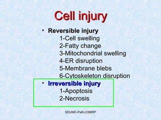
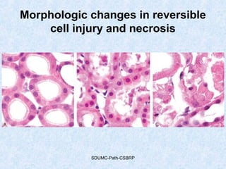
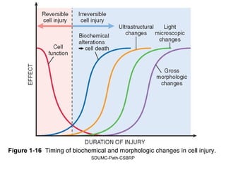
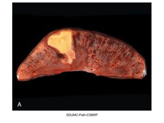
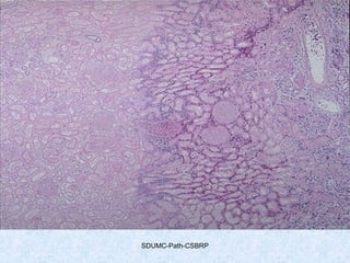
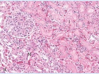
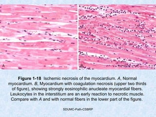
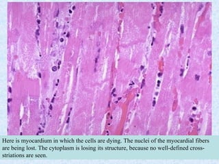
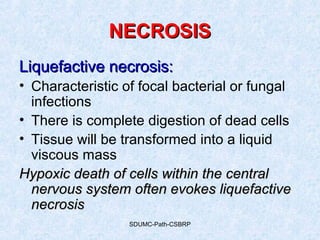
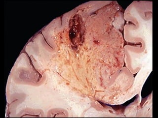
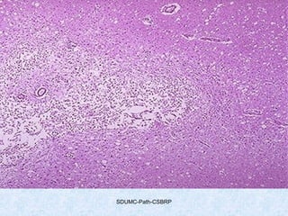
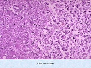
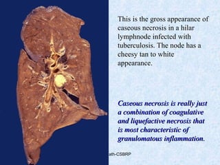
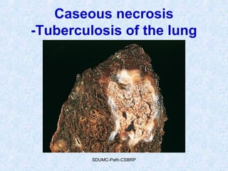
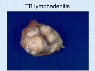
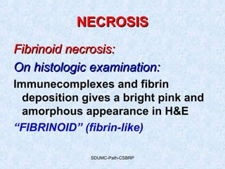
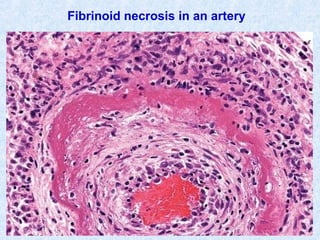
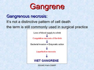
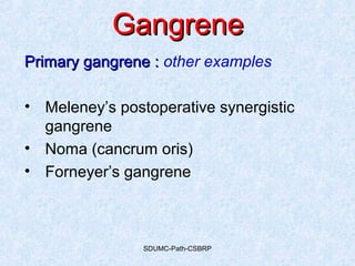
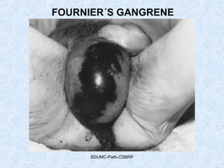
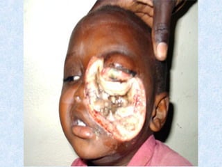
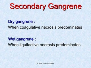
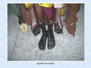
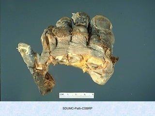
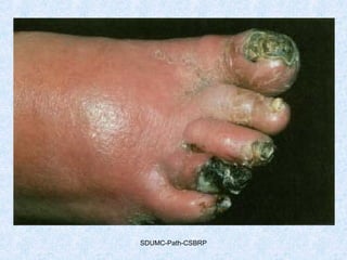
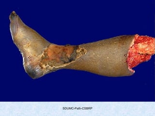
8. SDUMC-Path-CSBRP
• Is a manifestation of irreversible cell injury
• Loss of membrane integrity Leakage
of contents
• Leaked contents may elicit inflammation
Source of enzymes:Source of enzymes:
1. Lysosomes from the necrotic cell itself
(autolysis)
2. Enzymes from the immigrant leucocytes
NECROSISNECROSIS
14. SDUMC-Path-CSBRP
NECROSISNECROSIS
Coagulative necrosis:Coagulative necrosis:
• Basic outline of the cell is preserved
• The affected tissues has firm consistency
• Increasing intracellular acidosis denatures all
proteins including enzymes
Coagulative necrosis, is characteristic of hypoxicCoagulative necrosis, is characteristic of hypoxic
death of cells in all tissues except the braindeath of cells in all tissues except the brain
Eg: The myocardial infarct is an excellent example
in which acidophilic, coagulated, anucleate cells
may persist for weeks.
20. SDUMC-Path-CSBRP
Figure 1-18 Ischemic necrosis of the myocardium. A, Normal
myocardium. B, Myocardium with coagulation necrosis (upper two thirds
of figure), showing strongly eosinophilic anucleate myocardial fibers.
Leukocytes in the interstitium are an early reaction to necrotic muscle.
Compare with A and with normal fibers in the lower part of the figure.
21. SDUMC-Path-CSBRP
Here is myocardium in which the cells are dying. The nuclei of the myocardial fibers
are being lost. The cytoplasm is losing its structure, because no well-defined cross-
striations are seen.
23. SDUMC-Path-CSBRP
NECROSISNECROSIS
Liquefactive necrosis:Liquefactive necrosis:
• Characteristic of focal bacterial or fungal
infections
• There is complete digestion of dead cells
• Tissue will be transformed into a liquid
viscous mass
Hypoxic death of cells within the centralHypoxic death of cells within the central
nervous system often evokes liquefactivenervous system often evokes liquefactive
necrosisnecrosis
27. SDUMC-Path-CSBRP
Figure 1-19 Coagulative and liquefactive necrosis. A, Kidney infarct
exhibiting coagulative necrosis, with loss of nuclei and clumping of
cytoplasm but with preservation of basic outlines of glomerular and
tubular architecture. B, A focus of liquefactive necrosis in the kidney
caused by fungal infection. The focus is filled with white cells and cellular
debris, creating a renal abscess that obliterates the normal architecture.
28. SDUMC-Path-CSBRP
NECROSISNECROSIS
Caseous necrosis:Caseous necrosis:
• a distinctive form of coagulative necrosis
• Seen most often in foci of tuberculous infection
Caseous = cheesy whiteCaseous = cheesy white (gross appearance)
Microscopy:
amorphous granular debris, composed of
fragmented, coagulated cells
with a distinctive inflammatory border known as a
granulomatous reaction
Unlike coagulative necrosis, the tissue architectureUnlike coagulative necrosis, the tissue architecture
is completely obliterated.is completely obliterated.
29. SDUMC-Path-CSBRP
This is the gross appearance of
caseous necrosis in a hilar
lymphnode infected with
tuberculosis. The node has a
cheesy tan to white
appearance.
Caseous necrosis is really justCaseous necrosis is really just
a combination of coagulativea combination of coagulative
and liquefactive necrosis thatand liquefactive necrosis that
is most characteristic ofis most characteristic of
granulomatous inflammation.granulomatous inflammation.
36. SDUMC-Path-CSBRP
The edge of a pulmonary granuloma is shown here at medium power magnification.
Amorphous pink caseous material composed of the necrotic elements of the granuloma.
A Langhan’s giant cell is seen as well.
37. SDUMC-Path-CSBRP
At high magnification, the granuloma demonstrates that the epithelioid
macrophages are elongated with long, pale nuclei and pink cytoplasm.
The typical giant cell for infectious granulomas is called a Langhans
giant cell and has the nuclei lined up along one edge of the cell.
56. SDUMC-Path-CSBRP
GangreneGangrene
Types :Types : Based on the cause for tissue necrosis
1.1. PrimaryPrimary
2.2. SecondarySecondary
Criteria Primary. GPrimary. G Secondary. GSecondary. G
Necrosis Both caused by
same agent i.e.
bacteria
Both caused by
different agents
Putrefaction
57. SDUMC-Path-CSBRP
GangreneGangrene
Primary gangrene :Primary gangrene :
• Due to infection with specific bacteria
• Later, putrefaction is brought about by other
saprophytic bacteria
• Agent: Cl.perfringens, Cl.edematiens & Cl.septicum
• Source: intestines, soil
• Mode of entry: thru wounds
• Requirement: anerobic environment
58. SDUMC-Path-CSBRP
GangreneGangrene
Primary gangrene :Primary gangrene :
• Agent: Cl.perfringens, Cl.edematiens & Cl.septicum
• Toxins:
Lecithinase (lysis of cell membranes)
Hyaluronidase (digestion of intercellular cement)
Collagenases (digestion of connective tissue)
• Production of H2 & CO2 by fermentation of sugars
• Secondary invasion by saprophytes -- putrefaction
60. SDUMC-Path-CSBRP
GangreneGangrene
Primary gangrene :Primary gangrene :
Clinical course:
1. Rapidly spreading along tissue planes
2. Hemolysins may cause hemolysis
3. Severe toxaemia
4. Circulatory collapse
5. Distant spread is a terminal event
61. SDUMC-Path-CSBRP
GangreneGangrene
Primary gangrene :Primary gangrene :
• Wounds must be treated with early
excision of devitalized tissue
• Treated with antibiotics to prevent
infection
• With the clinical suspicion institution of
antigasgangrene serum
• Clostridia may also cause cellulitis without
affecting the underlying muscle
FIGURE 1-9 Morphologic changes in reversible cell injury and necrosis. A, Normal kidney tubules with viable epithelial cells. B, Early (reversible) ischemic injury showing surface blebs, increased eosinophilia of cytoplasm, and swelling of occasional cells. C, Necrosis (irreversible injury) of epithelial cells, with loss of nuclei, fragmentation of cells, and leakage of contents. (Courtesy of Drs. Neal Pinckard and M.A. Venkatachalam, University of Texas Health Sciences Center, San Antonio, TX.)
Common feature pertinent to all these terms is Dead Cells.
However, there are differences.
Necrosis is microscopic description
Infarction is grossly visible dead area in living tissue
Gangrene is grossly visible dead tissue with super added ‘infection
Tissue taken for Biopsy: Cells are dead but, they are fixed in a state as they were in the body before biopsy.
Necrosis is a manifestation of irreversible cell injury
Necrotic cells are unable to maintain membrane integrity and their contents often leak out.
Leaked contents may elicit inflammation in the surrounding tissue
Source of enzymes:
Lysosomes from the necrotic cell itself (autolysis)
Enzymes from the immigrant leucocytes
Increased eosinophilia attributable in part to loss of the normal basophilia imparted by the RNA in the cytoplasm and in part to the increased binding of eosin to denatured intracytoplasmic proteins.
The necrotic cell may have a more glassy homogeneous appearance than that of normal cells, mainly as a result of the loss of glycogen particles.
When enzymes have digested the cytoplasmic organelles, the cytoplasm becomes vacuolated and appears moth-eaten.
Dead cells may ultimately be replaced by large, whorled phospholipid masses called myelin figures. These phospholipid precipitates are then either phagocytosed by other cells or further degraded into fatty acids; calcification of such fatty acid residues results in the generation of calcium soaps.
Nuclear changes appear in one of three patterns, all due to nonspecific breakdown of DNA (see Fig. 1-9C ). The basophilia of the chromatin may fade (karyolysis), a change that presumably reflects loss of DNA because of enzymatic degradation by endonucleases. A second pattern (which is also seen in apoptotic cell death) is pyknosis, characterized by nuclear shrinkage and increased basophilia. Here the chromatin condenses into a solid, shrunken basophilic mass. In the third pattern, known as karyorrhexis, the pyknotic nucleus undergoes fragmentation. With the passage of time (a day or two), the nucleus in the necrotic cell totally disappears.
Although the terms are somewhat outmoded, they are routinely used and their meanings are understood by both pathologists and clinicians. When denaturation is the primary pattern, coagulative necrosis develops. In the instance of dominant enzyme digestion, the result is liquefactive necrosis; in special circumstances, caseous necrosis and fat necrosis may occur.
Coagulative necrosis implies preservation of the basic outline of the coagulated cell for a span of at least some days ( Fig. 1-19A ). The affected tissues exhibit a firm texture. Presumably, the injury or the subsequent increasing intracellular acidosis denatures not only structural proteins but also enzymes and so blocks the proteolysis of the cell. The myocardial infarct is an excellent example in which acidophilic, coagulated, anucleate cells may persist for weeks. Ultimately, the necrotic myocardial cells are removed by fragmentation and phagocytosis of the cellular debris by scavenger leukocytes and by the action of proteolytic lysosomal enzymes brought in by the immigrant white cells. The process of coagulative necrosis, with preservation of the general tissue architecture, is characteristic of hypoxic death of cells in all tissues except the brain.
The contrast between normal adrenal cortex and the small pale infarct is good. The area just under the capsule is spared because of blood supply from capsular arterial branches. This is an odd place for an infarct, but it illustrates the shape and appearance of an ischemic (pale) infarct well.
When many cells undergo necrosis at once, then definable patterns of necrosis are produced, depending upon the nature of the injury, the type of tissue, and the length of time. This is an example of coagulative necrosis. This is the typical pattern with ischemia and infarction (loss of blood supply and resultant tissue anoxia). Here, there is a wedge-shaped pale area of coagulative necrosis (infarction) in the renal cortex of the kidney.
Microscopically, the renal cortex has undergone anoxic injury at the left so that the cells appear pale and ghost-like. There is a hemorrhagic zone in the middle where the cells are dying or have not quite died, and then normal renal parenchyma at the far right. This is an example of coagulative necrosis.
FIGURE 1-11 Coagulative necrosis: B, Microscopic view of the edge of the infarct, with normal kidney (N) and necrotic cells in the infarct (I) showing preserved cellular outlines with loss of nuclei and an inflammatory infiltrate (which is difficult to discern at this magnification).
Two large infarctions (areas of coagulative necrosis) are seen in this sectioned spleen. Since the etiology of coagulative necrosis is usually vascular with loss of blood supply, the infarct occurs in a vascular distribution. Thus, infarcts are often wedge-shaped with a base on the organ capsule.
Here is myocardium in which the cells are dying. The nuclei of the myocardial fibers are being lost. The cytoplasm is losing its structure, because no well-defined cross-striations are seen.
Figure 1-19 Coagulative and liquefactive necrosis. A, Kidney infarct exhibiting coagulative necrosis, with loss of nuclei and clumping of cytoplasm but with preservation of basic outlines of glomerular and tubular architecture. B, A focus of liquefactive necrosis in the kidney caused by fungal infection. The focus is filled with white cells and cellular debris, creating a renal abscess that obliterates the normal architecture.
Can you guess this tissue?: Kidney. Though, cellular ddtails are lost, it’s possible to identify the tissue. This is what onemay see in coagulativenecrosis.
Liquefactive necrosis is characteristic of focal bacterial or, occasionally, fungal infections, because microbes stimulate the accumulation of inflammatory cells ( Fig. 1-19B ). For obscure reasons, hypoxic death of cells within the central nervous system often evokes liquefactive necrosis. Whatever the pathogenesis, liquefaction completely digests the dead cells. The end result is transformation of the tissue into a liquid viscous mass. If the process was initiated by acute inflammation ( Chapter 2 ), the material is frequently creamy yellow because of the presence of dead white cells and is called pus. Although gangrenous necrosis is not a distinctive pattern of cell death, the term is still commonly used in surgical clinical practice. It is usually applied to a limb, generally the lower leg, that has lost its blood supply and has undergone coagulation necrosis. When bacterial infection is superimposed, coagulative necrosis is modified by the liquefactive action of the bacteria and the attracted leukocytes (so-called wet gangrene).
Grossly, the cerebral infarction at the upper left here demonstrates liquefactive necrosis. Eventually, the removal of the dead tissue leaves behind a cavity.
This is liquefactive necrosis in the brain in a patient who suffered a "stroke" with focal loss of blood supply to a portion of cerebrum. This type of infarction is marked by loss of neurons and neuroglial cells and the formation of a clear space at the center left.
At high magnification, liquefactive necrosis of the brain demonstrates many macrophages at the right which are cleaning up the necrotic cellular debris. The job description of a macrophage includes janitorial services such as this, particularly when there is lipid.
Caseous necrosis, a distinctive form of coagulative necrosis, is encountered most often in foci of tuberculous infection ( Chapter 8 ). The term caseous is derived from the cheesy white gross appearance of the area of necrosis ( Fig. 1-20 ). On microscopic examination, the necrotic focus appears as amorphous granular debris seemingly composed of fragmented, coagulated cells and amorphous granular debris enclosed within a distinctive inflammatory border known as a granulomatous reaction ( Chapter 2 ). Unlike coagulative necrosis, the tissue architecture is completely obliterated.
This is the gross appearance of caseous necrosis in a hilar lymp node infected with tuberculosis. The node has a cheesy tan to white appearance. Caseous necrosis is really just a combination of coagulative and liquefactive necrosis that is most characteristic of granulomatous inflammation.
Multiple granulomas with areas of caseous necrosis -- most characteristic of secondary (reactivation) tuberculosis.
DD: Fungal granulomas (histoplasmosis, cryptococcosis, coccidioidomycosis).
Figure 1-20 A tuberculous lung with a large area of caseous necrosis. The caseous debris is yellow-white and cheesy.
Microscopically, caseous necrosis is characterized by acellular pink areas of necrosis, as seen here at the upper right, surrounded by a granulomatous inflammatory process.
Well-defined granulomas are seen here. They have rounded outlines. The one toward the center of the photograph contains several Langhans giant cells. Granulomas are composed of transformed macrophages called epithelioid cells along with lymphocytes, occasional PMN's, plasma cells, and fibroblasts. The localized, small appearance of these granulomas suggests that the immune response is fairly good.
The edge of a pulmonary granuloma is shown here at medium power magnification. Amorphous pink caseous material composed of the necrotic elements of the granuloma as well as the infectious organisms is present. This area is ringed by the inflammatory component with epithelioid cells, lymphocytes, and fibroblasts. A Langhans giant cell is seen as well.
At high magnification, the granuloma demonstrates that the epithelioid macrophages are elongated with long, pale nuclei and pink cytoplasm. The macrophages organize into committees called giant cells. The typical giant cell for infectious granulomas is called a Langhans giant cell and has the nuclei lined up along one edge of the cell. The process of granulomatous inflammation takes place over months to years (did you ever hear of a committee action that was completed in a short time?)
Fat necrosis is a term that is well fixed in medical parlance but does not in reality denote a specific pattern of necrosis. Rather, it is descriptive of focal areas of fat destruction, typically occurring as a result of release of activated pancreatic lipases into the substance of the pancreas and the peritoneal cavity. This occurs in the calamitous abdominal emergency known as acute pancreatitis ( Chapter 19 ). In this disorder, activated pancreatic enzymes escape from acinar cells and ducts, the activated enzymes liquefy fat cell membranes, and the activated lipases split the triglyceride esters contained within fat cells. The released fatty acids combine with calcium to produce grossly visible chalky white areas (fat saponification), which enable the surgeon and the pathologist to identify the lesions ( Fig. 1-21 ). On histologic examination, the necrosis takes the form of foci of shadowy outlines of necrotic fat cells, with basophilic calcium deposits, surrounded by an inflammatory reaction.
Figure 1-21 Foci of fat necrosis with saponification in the mesentery. The areas of white chalky deposits represent calcium soap formation at sites of lipid breakdown.
This is fat necrosis of the pancreas. Cellular injury to the pancreatic acini leads to release of powerful enzymes which damage fat by the production of soaps, and these appear grossly as the soft, chalky white areas seen here on the cut surfaces.
Microscopically, fat necrosis adjacent to pancreas is seen here. There are some remaining steatocytes at the left which are not necrotic. The necrotic fat cells at the right have vague cellular outlines, have lost their peripheral nuclei, and their cytoplasm has become a pink amorphous mass of necrotic material.
Fibrinoid necrosis is a special form of necrosis usually seen in immune reactions involving blood vessels. This pattern of necrosis typically occurs when complexes of antigens and antibodies are deposited in the walls of arteries. Deposits of these “immune complexes,” together with fibrin that has leaked out of vessels, result in a bright pink and amorphous appearance in H&E stains, called “fibrinoid” (fibrin-like) by pathologists ( Fig. 1-15 ). The immunologically mediated vasculitis syndromes in which this type of necrosis is seen are described in Chapter 6 .
FIGURE 1-15 Fibrinoid necrosis in an artery. The wall of the artery shows a circumferential bright pink area of necrosis with inflammation (neutrophils with dark nuclei).
Although gangrenous necrosis is not a distinctive pattern of cell death, the term is still commonly used in surgical clinical practice. It is usually applied to a limb, generally the lower leg, that has lost its blood supply and has undergone coagulation necrosis. When bacterial infection is superimposed, coagulative necrosis is modified by the liquefactive action of the bacteria and the attracted leukocytes (so-called wet gangrene).
The same changes are observed post mortem and in putrifaction of meat, et.c.
In primary gangrene: pathogenic bacteria both will kill the tissue by secreting potent exotoxins and then invade the tissue. i.e. necrosis and putrefaction are brought about by the same agent.
In secondary gangrene: Necrosis is due some other cause (ex: loss of blood supply) and then saprophytic bacteria then digests the dead tissue.
Clostridia present in the intestinal flora and in the perineum.
Gas gangrene
Findings
There is extensive gas in the soft tissues of the lower leg. There is arterial calcification visible, consistent with the known diabetes. The appearance are those of gas gangrene, which is more common in diabetics. Treatment includes antibiotics (penicillin) and prompt amputation.
NEJM-Volume 360:280 January 15, 2009 Number 3
A 55-year-old man with end-stage liver disease associated with alcohol abuse was evaluated for liver transplantation. He had a history of refractory ascites and hepatic encephalopathy. On physical examination, we noted muscle atrophy, jaundice, ascites, and numerous spider angiomas (Panel A). Eight months later, the patient underwent successful liver transplantation, when he had a score of 21 on the Model for End-Stage Liver Disease (MELD) scale (ranging from 6 to 40, with higher scores indicating greater severity of disease). Six months after transplantation, he was clinically well without evidence of organ dysfunction or infection, and most of the spider angiomas had disappeared (Panel B). Spider angiomas can be seen in healthy children and pregnant women. In such cases, angiomas are few in number and resolve with time or a normalization of estrogen levels. Numerous spider angiomas are more common in patients with chronic liver disease and consist of a central arteriole from which numerous small venules radiate, resembling a spider's legs. Possible mechanisms of formation include arteriolar vasodilatation, neovascularization from angiogenic factors such as vascular endothelial growth factor, direct effects of alcohol, and estrogen excess due to inadequate hepatic metabolism.
---
Olivier Detry, M.D., Ph.D. Arnaud De Roover, M.D., Ph.D. University of Liège B-4000 Liège, Belgium oli.detry@chu.ulg.ac.be
=======
Meleney’s postoperative synergistic gangrene: slow spreading infection of the skin and subcutaneous tissue of the chest or abdominal wall which is progresses and leaves the central zone gangrenous. The lesion may spread relentlessly to involve much of the trunk.
NOMA: gangrenous condition affecting the oral cavity. Usually secondary to malnourishment with debilitating infections. This condition is caused by Bacteroides spp togeather with Borrelia vincenti.
A patient with Fournier´s gangrene of the scrotum. Note the sharp demarcation of gangrenous changes and the marked edema of the scrotum and the penis.
FOURNIER´S GANGRENE
Fournier´s gangrene is a polymicrobial, synergistic infection of the subcutaneous tissues that originates from one of three sites: skin, urethra, or rectum. This infectious process typically begins as a benign infection or simple abscess that quickly becomes virulent, especially in an immunocompromised host, and leads to end-artery thrombosis in the subcutaneous tissue that promotes widespread necrosis of previously healthy tissue (see image).
In FG, suppurative bacterial infection results in microthrombosis of the small subcutaneous vessels leading to the development of gangrene of the overlying skin.
The diabetic male seems to be most at risk. Prompt recognition of Fournier´s gangrene in its early stages should prevent extensive tissue loss that accompanies delayed diagnosis. Aggressive fluid resuscitation; gram-positive, gram-negative, and anaerobic antibiotic coverage; and wide surgical debridement sometimes in conjunction with pre- and postoperative hyperbaric oxygen therapy are the mainstays of treatment. Urologic consultation is often required when periurethral abscess is the inciting event, or when other etiologies have secondarily invaded the urinary tract and supravesical urinary drainage is needed. It is imperative that emergency physicians maintain a very high index of suspicion for this entity in immunocompromised patients who present complaining of scrotal, rectal, or any genitalia pain out of proportion to their physical examination findings. Surgical consultation is strongly recommended in all such patients, rather than deciding on symptomatic treatment and discharge from the emergency department.
Meleney’s postoperative synergistic gangrene: slow spreading infection of the skin and subcutaneous tissue of the chest or abdominal wall which is progresses and leaves the central zone gangrenous. The lesion may spread relentlessly to involve much of the trunk.
This is gangrene, or necrosis of many tissues in a body part. In this case, the toes were involved in a frostbite injury. This is an example of "dry" gangrene in which there is mainly coagulative necrosis from the anoxic injury.
This is gangrene of the lower extremity. In this case the term "wet" gangrene is more applicable because of the liquefactive component from superimposed infection in addition to the coagulative necrosis from loss of blood supply. This patient had diabetes mellitus.