Downloaded 935 times


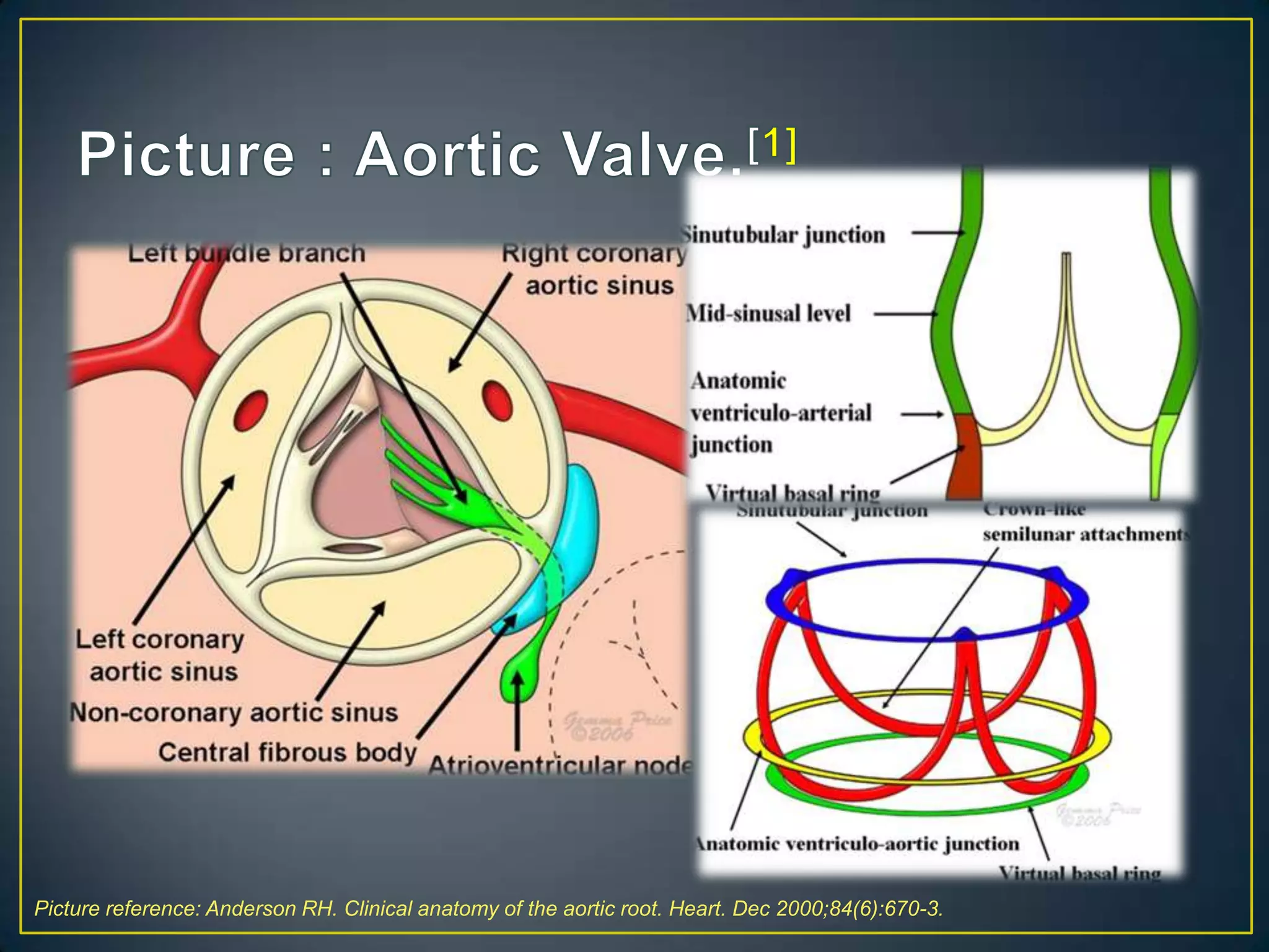
![The AV is located betwn the LVOT and the ascending aorta. It forms the centerpiece of t
heart and closely approximates many other important cardiac structures; specifically, th
PV anteriorly, MV posterolaterally, and TV posteromedially.[1]
Ref:
1. Anderson RH. Clinical anatomy of the aortic root. Heart. Dec 2000;84(6):670-3.](https://image.slidesharecdn.com/aorticregurgitation-131030042922-phpapp02/75/Aortic-regurgitation-3-2048.jpg)

![Bicuspid aortic valve
• Bicuspid aortic valves are the most common cardiac
valvular anomaly, occurring in 1- 2% of the general
population.
• It is twice as common in males as in females.[1]
Ref:
1. Tzemos N, Therrien J, Yip J et al (September 2008). "Outcomes in adults with bicuspid aortic valves". JAMA 300 (11): 1317–1325](https://image.slidesharecdn.com/aorticregurgitation-131030042922-phpapp02/75/Aortic-regurgitation-6-2048.jpg)
![• AR is a condition due to inadequate closure of the
aortic valve leaflets leading to abnormal retrograde
flow of blood through the aortic valve during cardiac
diastole.
• It can be induced either by damage to and dysfunction
of the aortic valve leaflets or by distortion or dilatation
of the aortic root and ascending aorta
• In the developing world, the most common cause of AR is
rheumatic heart disease. However, in developed
countries, AR is most often due to aortic root dilation or a
congenital bicuspid aortic valve .[1]
Ref:
1. Maurer G. Aortic regurgitation. Heart. Jul 2006;92(7):994-1000.](https://image.slidesharecdn.com/aorticregurgitation-131030042922-phpapp02/75/Aortic-regurgitation-7-2048.jpg)
![Leaflet abnormalities
Aortic root or ascending aorta
Rheumatic fever
Systemic hypertension
Endocarditis
Aortitis (eg, syphilis)
Trauma
Reactive arthritis
Bicuspid aortic valve
Ankylosing spondylitis
Rheumatoid arthritis
Trauma/ Dissecting aneurysm
Myxomatous degeneration
Osteogenesis imperfecta
Ankylosing spondylitis
Marfan syndrome/ EDS
Acromegaly
Inflammatory bowel disease
AR is seen more commonly in men than in women. As in the Framingham study, AR was
13% of men versus 8.5% of women.[1] The greater prevalence of AR in men may reflect, in
the male preponderance of underlying conditions such as Marfan syndrome[2] or bicusp
valve[3] .
Ref:
1. Singh JP, Evans JC, et al. Prevalence and clinical determinants of mitral, tricuspid, and AR (the Framingham Heart Study). Am J Cardiol. Mar 15 1999;83(6
2. Keane MG, Pyeritz RE. Medical management of Marfan syndrome. Circulation. May 27 2008;117(21):2802-13.
3. Ortiz JT, Shin DD, Rajamannan NM. Approach to the patient with bicuspid AVand ascending aorta aneurysm. Curr Treat Options Cardiovasc Med. Dec 200](https://image.slidesharecdn.com/aorticregurgitation-131030042922-phpapp02/75/Aortic-regurgitation-8-2048.jpg)


![• In a review of the literature, the presence of an early
diastolic murmur, as heard by a cardiologist, was the
most useful finding for establishing the presence of AR
(positive likelihood ratio 8.8 [ie, the odds of AR are
increased 8.8 fold]) and its absence the most useful
finding for eliminating the presence of AR (negative
likelihood ratio 0.2 to 0.3 [ie, the odds of disease are
reduced by a factor of 0.2 to 0.3]) .[1]
Ref:
1. Choudhry NK, Etchells EE. The rational clinical examination. Does this patient have aortic regurgitation? JAMA 1999; 281:2231](https://image.slidesharecdn.com/aorticregurgitation-131030042922-phpapp02/75/Aortic-regurgitation-15-2048.jpg)



![Class I indications for CC under current ACC/AHA
guidelines include the following:[1]
• Assessment of coronary anatomy prior to aortic valve
surgery in patients with risk factors for coronary artery
disease.
Ref:
1. Bonow RO, Carabello BA, Chatterjee K, de Leon AC Jr, Faxon DP, Freed MD. 2008 focused update incorporated into the
ACC/AHA 2006 guidelines for the management of patients with valvular heart disease.
.](https://image.slidesharecdn.com/aorticregurgitation-131030042922-phpapp02/75/Aortic-regurgitation-21-2048.jpg)

![• In acute severe AR, surgical intervention is usually
indicated, but the patient may be supported medically
with dobutamine to augment cardiac output and shorten
diastole and sodium nitroprusside to reduce afterload
in hypertensive patients.
• In chronic severe AR, vasodilator therapy may be used
in select conditions to reduce afterload in patients with
systolic hypertension to minimize wall stress and
optimize LV function; in normotensive patients,
vasodilator therapy is not likely to reduce regurgitant
volume (preload) significantly and thus may not be of
Ref:
1. Bekeredjian R, Grayburn PA. Valvular heart disease: aortic regurgitation. Circulation. Jul 5 2005;112(1):125-34.
clinical benefit.[1]](https://image.slidesharecdn.com/aorticregurgitation-131030042922-phpapp02/75/Aortic-regurgitation-23-2048.jpg)

![• Reduced EDV and ↑EF have also been observed in
small number of pts receiving long term oral therapy with
hydralazine and nifedipine for a period of 1-2 yrs. With
nifedipine these effects are A/W ↓ in LV mass.[1,2]
• Reduced BP with enalapril and quinapril has been A/W
↓ in EDV and mass but no change in EF.[3,4]
References:
1. Scognamiglio R, Fasoli G. Long-term nifedipine unloading therapy in asymptomatic patients with chronic severe AR. J Am Coll Cardiol 1990; 16:424–9.
2. Greenberg B, Massie B, et al. Long-term vasodilator therapy of chronic AI: a randomized double-blinded, placebo-controlled clinical trial. Circulation 1988;7
3. Lin M, Chiang HT,etal.Vasodilator therapy in chronic asymptomatic AR: enalapril versus hydralazine therapy. J Am Coll Cardiol 1994;24:1046–53.
4. Schon HR, Dorn R. Effects of 12 months quinapril therapy in asymptomatic patients with chronic AR. J Heart Valve Dis 1994;3:500–9.](https://image.slidesharecdn.com/aorticregurgitation-131030042922-phpapp02/75/Aortic-regurgitation-25-2048.jpg)

![• Antibiotic prophylaxis prior to dental procedures is no
longer routinely recommended for all patients with AR
under current ACC/AHA guidelines.[1]
Ref:
1. Bonow RO, Carabello BA, Chatterjee K, de Leon AC Jr, Faxon DP, Freed MD. 2008 focused update incorporated into the ACC/A
2006 guidelines for the management of patients with valvular heart disease](https://image.slidesharecdn.com/aorticregurgitation-131030042922-phpapp02/75/Aortic-regurgitation-27-2048.jpg)


![• For patients undergoing AV replacement, careful
consideration should be given to the relative risks and
benefits of mechanical vs bioprosthetic valves.
• Mechanical valves : more durable but require long-term
anticoagulation with warfarin due to increased risk of
thrombosis.
• Bioprosthetic valves carry a greater risk of long-term
deterioration and risk of reoperation but avoid the need
for long-term warfarin.[1]
Ref:
1. Bonow RO, Carabello BA, Chatterjee K, de Leon AC Jr, Faxon DP, Freed MD. 2008 focused update incorporated into the ACC/A
2006 guidelines for the management of patients with valvular heart disease.
.](https://image.slidesharecdn.com/aorticregurgitation-131030042922-phpapp02/75/Aortic-regurgitation-30-2048.jpg)


![The prognosis for patients with severe AR depends on the
presence or absence of LV dysfunction and symptoms, as
follows:[1]
• In asymptomatic patients with normal EF
1. Rate of progression to symptoms &/or LVD = < 6% per yr
2. Rate of progression to asymptomatic LVD = < 3.5% per yr
3. Rate of sudden death = less than 0.2% per yr
• In asymptomatic patients with decreased EF, rate of
progression to symptoms = >25% per year.
• In symptomatic patients, mortality rate = >10% per yr.
The strongest predictors of outcome are echocardiographic
parameters (EF and LV end-systolic dimension).
Ref:
1. Bonow RO, Carabello BA, Chatterjee K, de Leon AC Jr, Faxon DP, Freed MD. 2008 focused update
incorporated into the ACC/AHA 2006 guidelines for the management of patients with valvular heart disease.](https://image.slidesharecdn.com/aorticregurgitation-131030042922-phpapp02/75/Aortic-regurgitation-34-2048.jpg)


The document discusses the anatomy, causes, diagnosis, and management of aortic regurgitation (AR). It provides details on the location of the aortic valve, variants such as bicuspid aortic valve, and common causes of AR including rheumatic heart disease. Physical exam findings, echocardiography parameters, and indications for surgery to replace the aortic valve are summarized. Medical management including vasodilator therapy to reduce afterload is also reviewed.


