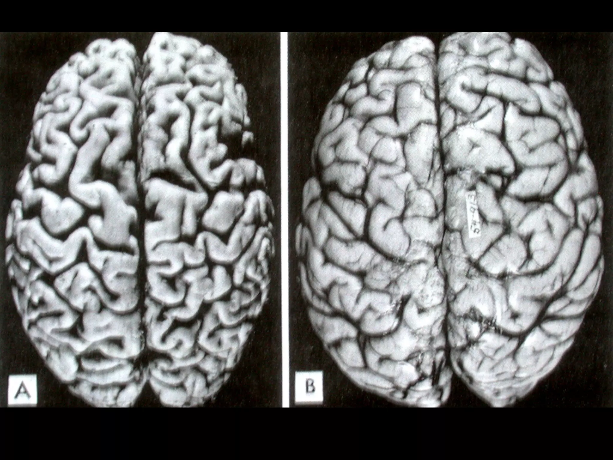
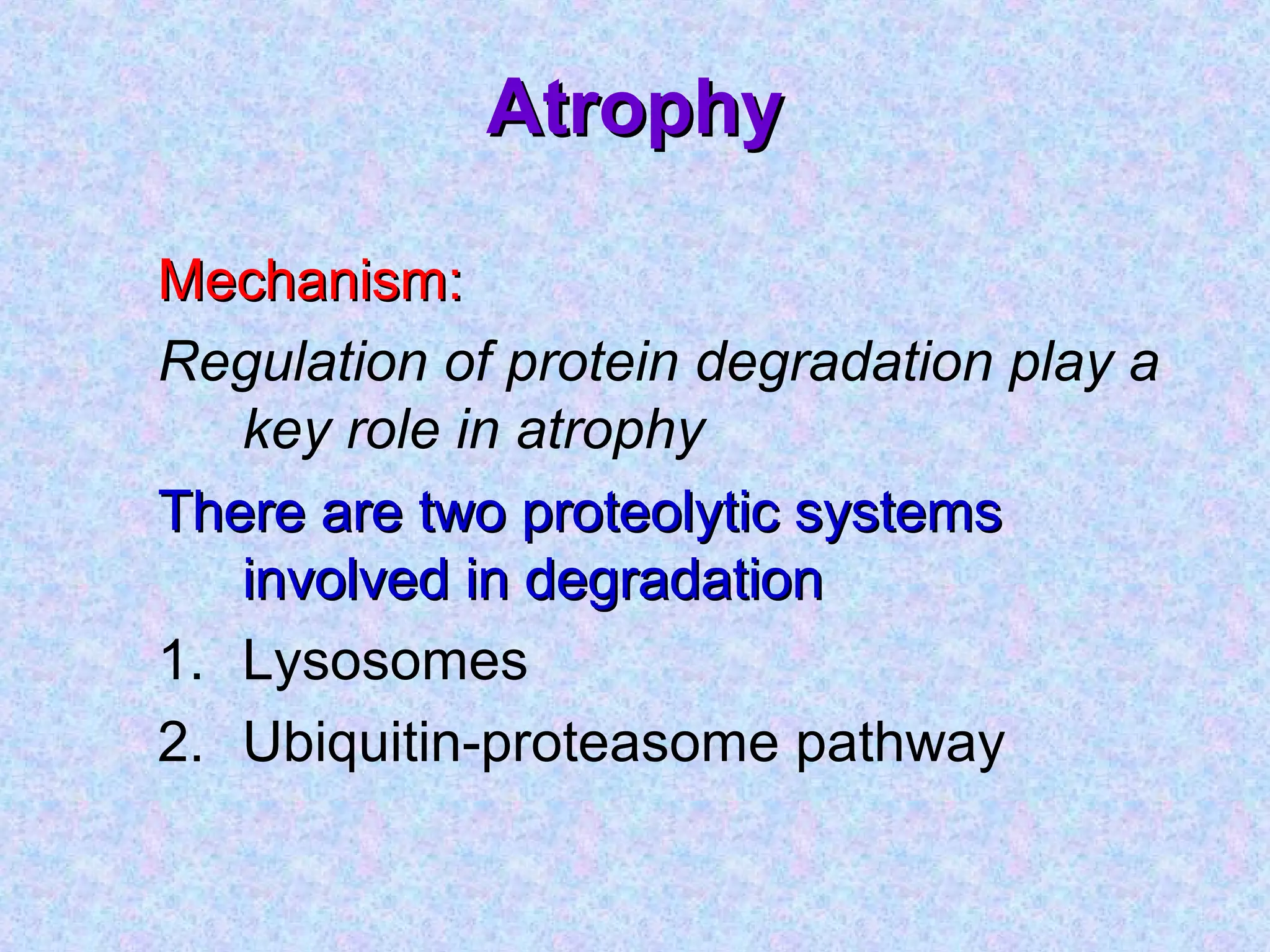
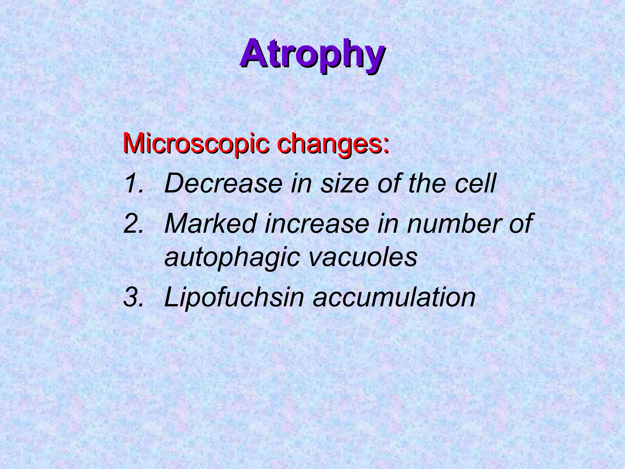
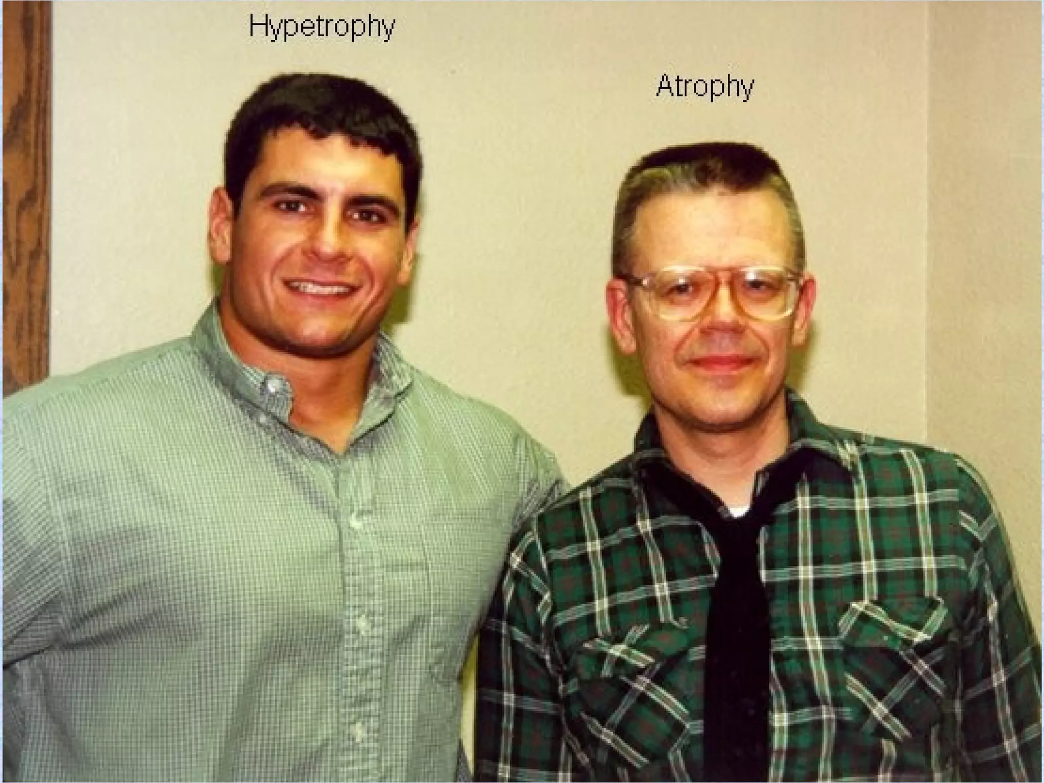
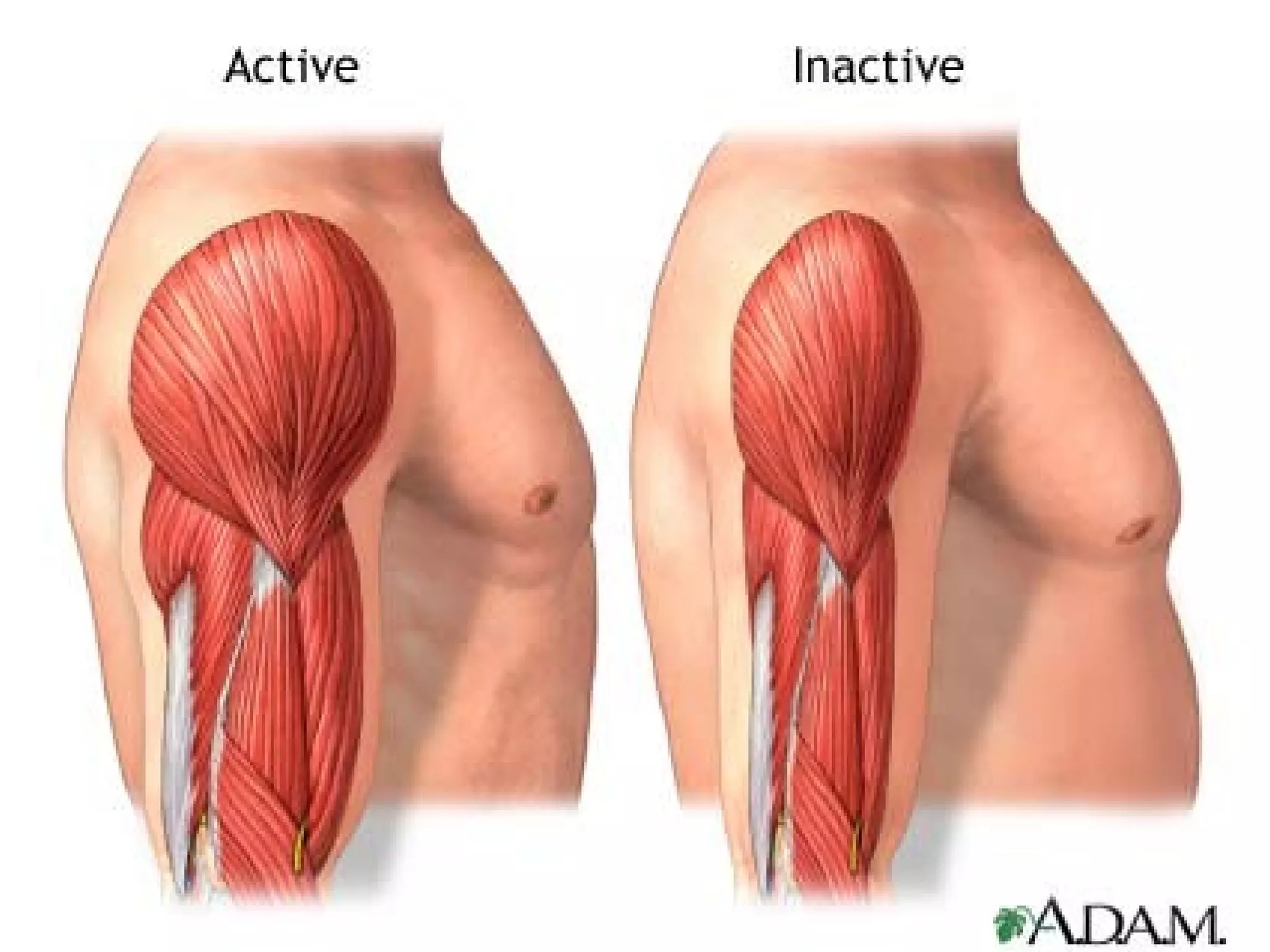
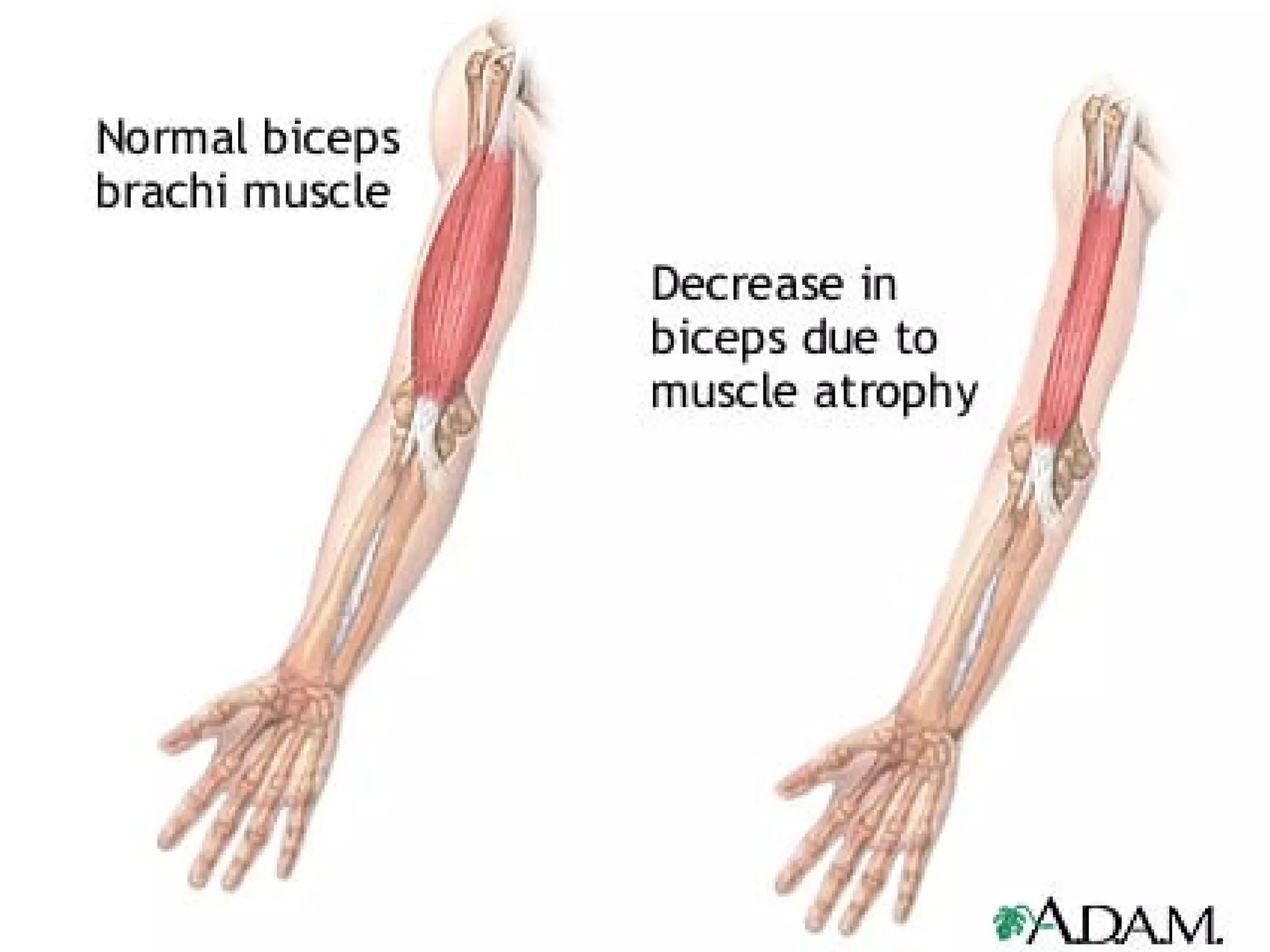
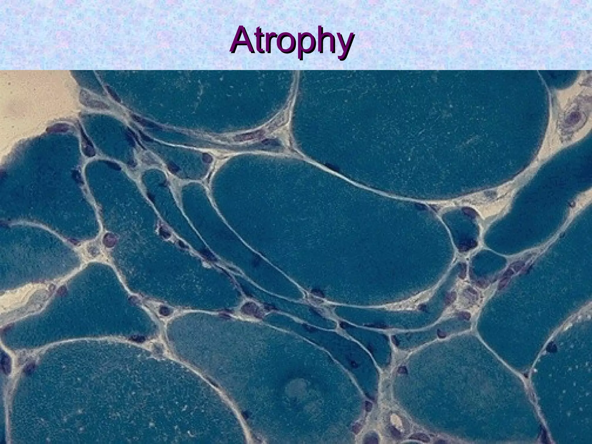
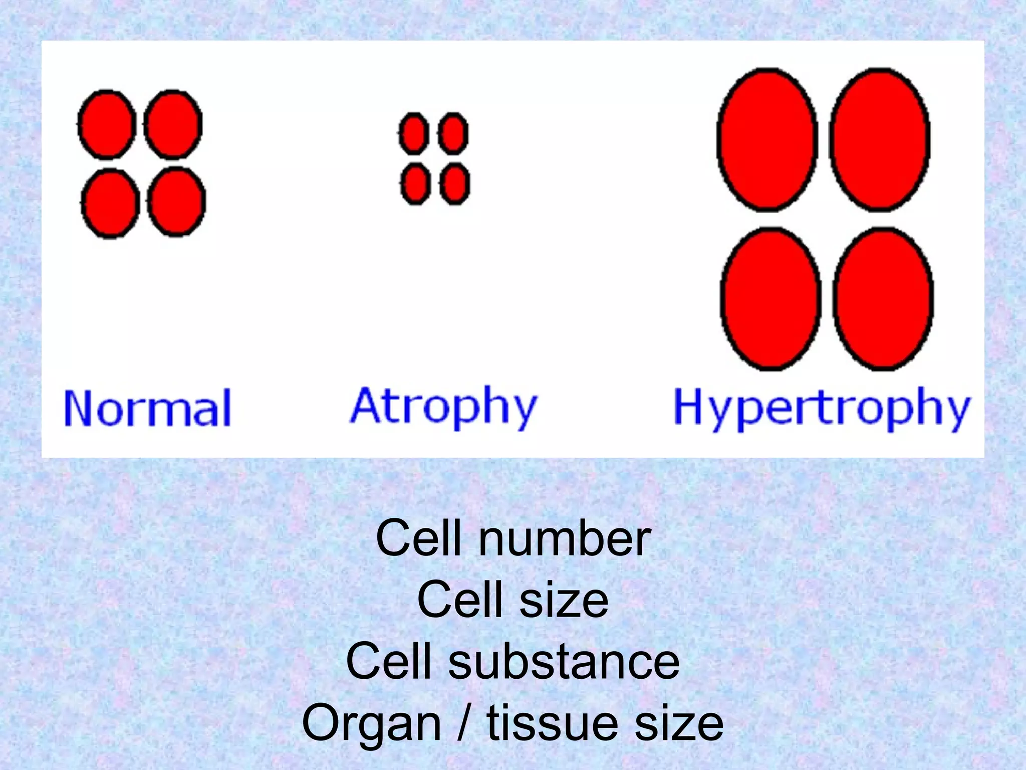
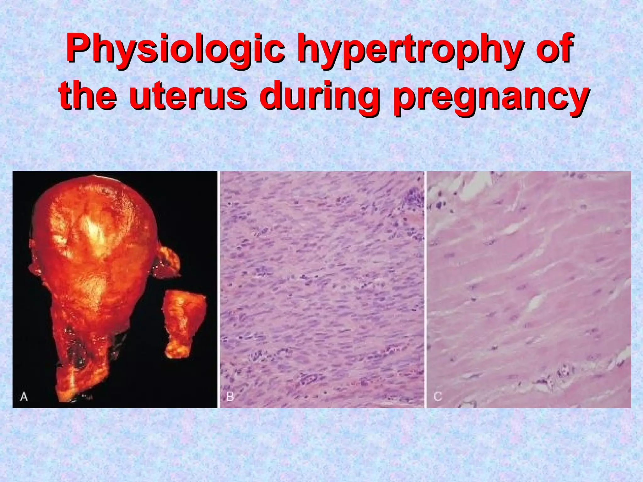
Atrophy is the shrinkage of cells and tissues due to loss of cellular components. It can result from decreased workload, blood supply, nutrition, endocrine stimulation or aging. At the microscopic level, atrophied cells appear smaller with more autophagic vacuoles and lipofuscin accumulation. Cancer cachexia involves activation of the proteasome pathway leading to muscle wasting.
Hypertrophy is the enlargement of cells and tissues due to an increase in cellular components but without an increase in cell number. It can be physiological such as in pregnancy or pathological such as in response to hypertension. The mechanisms involve increased protein synthesis, DNA content or cellular phenotype. Over time, degenerative changes can occur.
orbital cellulitis / DENTAL COURSES certified fixed orthodontic courses,oral...Indian dental academy
The Indian Dental Academy is the Leader in continuing dental education , training dentists in all aspects of dentistry and offering a wide range of dental certified courses in different formats.
Indian dental academy provides dental crown & Bridge,rotary endodontics,fixed orthodontics,
Dental implants courses.for details pls visit www.indiandentalacademy.com ,or call
00919248678078
- Coagulative Necrosis / summarized
- Description of the GROSS appearance of Coagulative necrosis.
- Description of the MICROSCOPIC appearance of Coagulative necrosis.
- Coagulative Necrosis / summarized
- Description of the GROSS appearance of Coagulative necrosis.
- Description of the MICROSCOPIC appearance of Coagulative necrosis.
Cellular Adaptation
as cells encounter stresses they undergo functional or structural adaptations to maintain viability / homeostasis.
Injury - altered homeostasis
if limits of the adaptive response are exceeded or if adaptation not possible, a sequence of events called cell injury occurs.
Reversible Cell Injury
removal of stress results in complete restoration of structural & functional integrity.
b) Irreversible Cell Injury / Cell Death
if stimulus persists or is severe enough from the start, the cell suffers irreversible cell injury and death.
2 main morphologic patterns: necrosis & apoptosis.
Adaptations are reversible changes in the size, number, phenotype, metabolic activity, or functions of cells in response to changes in their environment.
Physiologic adaptations are responses of cells to normal stimulation by hormones or endogenous chemical mediators
Pathologic adaptations are responses to stress that allow cells to modulate their structure and function and thus escape injury.
Hypertrophy refers to an increase in the size of cells, that results in an increase in the size of the affected organ
The hypertrophied organ has no new cells, just larger cells.
Types:
a) physiologic b) pathologic
Causes:
a) increased functional demand b) hormonal stimulation
Information about how cell get injured from different stimuli. Mechanism of cellular injury. Different types of cellular injury. Different examples of cellular injury with images which makes it easy to understand.
Couples presenting to the infertility clinic- Do they really have infertility...Sujoy Dasgupta
Dr Sujoy Dasgupta presented the study on "Couples presenting to the infertility clinic- Do they really have infertility? – The unexplored stories of non-consummation" in the 13th Congress of the Asia Pacific Initiative on Reproduction (ASPIRE 2024) at Manila on 24 May, 2024.
Title: Sense of Smell
Presenter: Dr. Faiza, Assistant Professor of Physiology
Qualifications:
MBBS (Best Graduate, AIMC Lahore)
FCPS Physiology
ICMT, CHPE, DHPE (STMU)
MPH (GC University, Faisalabad)
MBA (Virtual University of Pakistan)
Learning Objectives:
Describe the primary categories of smells and the concept of odor blindness.
Explain the structure and location of the olfactory membrane and mucosa, including the types and roles of cells involved in olfaction.
Describe the pathway and mechanisms of olfactory signal transmission from the olfactory receptors to the brain.
Illustrate the biochemical cascade triggered by odorant binding to olfactory receptors, including the role of G-proteins and second messengers in generating an action potential.
Identify different types of olfactory disorders such as anosmia, hyposmia, hyperosmia, and dysosmia, including their potential causes.
Key Topics:
Olfactory Genes:
3% of the human genome accounts for olfactory genes.
400 genes for odorant receptors.
Olfactory Membrane:
Located in the superior part of the nasal cavity.
Medially: Folds downward along the superior septum.
Laterally: Folds over the superior turbinate and upper surface of the middle turbinate.
Total surface area: 5-10 square centimeters.
Olfactory Mucosa:
Olfactory Cells: Bipolar nerve cells derived from the CNS (100 million), with 4-25 olfactory cilia per cell.
Sustentacular Cells: Produce mucus and maintain ionic and molecular environment.
Basal Cells: Replace worn-out olfactory cells with an average lifespan of 1-2 months.
Bowman’s Gland: Secretes mucus.
Stimulation of Olfactory Cells:
Odorant dissolves in mucus and attaches to receptors on olfactory cilia.
Involves a cascade effect through G-proteins and second messengers, leading to depolarization and action potential generation in the olfactory nerve.
Quality of a Good Odorant:
Small (3-20 Carbon atoms), volatile, water-soluble, and lipid-soluble.
Facilitated by odorant-binding proteins in mucus.
Membrane Potential and Action Potential:
Resting membrane potential: -55mV.
Action potential frequency in the olfactory nerve increases with odorant strength.
Adaptation Towards the Sense of Smell:
Rapid adaptation within the first second, with further slow adaptation.
Psychological adaptation greater than receptor adaptation, involving feedback inhibition from the central nervous system.
Primary Sensations of Smell:
Camphoraceous, Musky, Floral, Pepperminty, Ethereal, Pungent, Putrid.
Odor Detection Threshold:
Examples: Hydrogen sulfide (0.0005 ppm), Methyl-mercaptan (0.002 ppm).
Some toxic substances are odorless at lethal concentrations.
Characteristics of Smell:
Odor blindness for single substances due to lack of appropriate receptor protein.
Behavioral and emotional influences of smell.
Transmission of Olfactory Signals:
From olfactory cells to glomeruli in the olfactory bulb, involving lateral inhibition.
Primitive, less old, and new olfactory systems with different path
The prostate is an exocrine gland of the male mammalian reproductive system
It is a walnut-sized gland that forms part of the male reproductive system and is located in front of the rectum and just below the urinary bladder
Function is to store and secrete a clear, slightly alkaline fluid that constitutes 10-30% of the volume of the seminal fluid that along with the spermatozoa, constitutes semen
A healthy human prostate measures (4cm-vertical, by 3cm-horizontal, 2cm ant-post ).
It surrounds the urethra just below the urinary bladder. It has anterior, median, posterior and two lateral lobes
It’s work is regulated by androgens which are responsible for male sex characteristics
Generalised disease of the prostate due to hormonal derangement which leads to non malignant enlargement of the gland (increase in the number of epithelial cells and stromal tissue)to cause compression of the urethra leading to symptoms (LUTS
Anti ulcer drugs and their Advance pharmacology ||
Anti-ulcer drugs are medications used to prevent and treat ulcers in the stomach and upper part of the small intestine (duodenal ulcers). These ulcers are often caused by an imbalance between stomach acid and the mucosal lining, which protects the stomach lining.
||Scope: Overview of various classes of anti-ulcer drugs, their mechanisms of action, indications, side effects, and clinical considerations.
Lung Cancer: Artificial Intelligence, Synergetics, Complex System Analysis, S...Oleg Kshivets
RESULTS: Overall life span (LS) was 2252.1±1742.5 days and cumulative 5-year survival (5YS) reached 73.2%, 10 years – 64.8%, 20 years – 42.5%. 513 LCP lived more than 5 years (LS=3124.6±1525.6 days), 148 LCP – more than 10 years (LS=5054.4±1504.1 days).199 LCP died because of LC (LS=562.7±374.5 days). 5YS of LCP after bi/lobectomies was significantly superior in comparison with LCP after pneumonectomies (78.1% vs.63.7%, P=0.00001 by log-rank test). AT significantly improved 5YS (66.3% vs. 34.8%) (P=0.00000 by log-rank test) only for LCP with N1-2. Cox modeling displayed that 5YS of LCP significantly depended on: phase transition (PT) early-invasive LC in terms of synergetics, PT N0—N12, cell ratio factors (ratio between cancer cells- CC and blood cells subpopulations), G1-3, histology, glucose, AT, blood cell circuit, prothrombin index, heparin tolerance, recalcification time (P=0.000-0.038). Neural networks, genetic algorithm selection and bootstrap simulation revealed relationships between 5YS and PT early-invasive LC (rank=1), PT N0—N12 (rank=2), thrombocytes/CC (3), erythrocytes/CC (4), eosinophils/CC (5), healthy cells/CC (6), lymphocytes/CC (7), segmented neutrophils/CC (8), stick neutrophils/CC (9), monocytes/CC (10); leucocytes/CC (11). Correct prediction of 5YS was 100% by neural networks computing (area under ROC curve=1.0; error=0.0).
CONCLUSIONS: 5YS of LCP after radical procedures significantly depended on: 1) PT early-invasive cancer; 2) PT N0--N12; 3) cell ratio factors; 4) blood cell circuit; 5) biochemical factors; 6) hemostasis system; 7) AT; 8) LC characteristics; 9) LC cell dynamics; 10) surgery type: lobectomy/pneumonectomy; 11) anthropometric data. Optimal diagnosis and treatment strategies for LC are: 1) screening and early detection of LC; 2) availability of experienced thoracic surgeons because of complexity of radical procedures; 3) aggressive en block surgery and adequate lymph node dissection for completeness; 4) precise prediction; 5) adjuvant chemoimmunoradiotherapy for LCP with unfavorable prognosis.
Title: Sense of Taste
Presenter: Dr. Faiza, Assistant Professor of Physiology
Qualifications:
MBBS (Best Graduate, AIMC Lahore)
FCPS Physiology
ICMT, CHPE, DHPE (STMU)
MPH (GC University, Faisalabad)
MBA (Virtual University of Pakistan)
Learning Objectives:
Describe the structure and function of taste buds.
Describe the relationship between the taste threshold and taste index of common substances.
Explain the chemical basis and signal transduction of taste perception for each type of primary taste sensation.
Recognize different abnormalities of taste perception and their causes.
Key Topics:
Significance of Taste Sensation:
Differentiation between pleasant and harmful food
Influence on behavior
Selection of food based on metabolic needs
Receptors of Taste:
Taste buds on the tongue
Influence of sense of smell, texture of food, and pain stimulation (e.g., by pepper)
Primary and Secondary Taste Sensations:
Primary taste sensations: Sweet, Sour, Salty, Bitter, Umami
Chemical basis and signal transduction mechanisms for each taste
Taste Threshold and Index:
Taste threshold values for Sweet (sucrose), Salty (NaCl), Sour (HCl), and Bitter (Quinine)
Taste index relationship: Inversely proportional to taste threshold
Taste Blindness:
Inability to taste certain substances, particularly thiourea compounds
Example: Phenylthiocarbamide
Structure and Function of Taste Buds:
Composition: Epithelial cells, Sustentacular/Supporting cells, Taste cells, Basal cells
Features: Taste pores, Taste hairs/microvilli, and Taste nerve fibers
Location of Taste Buds:
Found in papillae of the tongue (Fungiform, Circumvallate, Foliate)
Also present on the palate, tonsillar pillars, epiglottis, and proximal esophagus
Mechanism of Taste Stimulation:
Interaction of taste substances with receptors on microvilli
Signal transduction pathways for Umami, Sweet, Bitter, Sour, and Salty tastes
Taste Sensitivity and Adaptation:
Decrease in sensitivity with age
Rapid adaptation of taste sensation
Role of Saliva in Taste:
Dissolution of tastants to reach receptors
Washing away the stimulus
Taste Preferences and Aversions:
Mechanisms behind taste preference and aversion
Influence of receptors and neural pathways
Impact of Sensory Nerve Damage:
Degeneration of taste buds if the sensory nerve fiber is cut
Abnormalities of Taste Detection:
Conditions: Ageusia, Hypogeusia, Dysgeusia (parageusia)
Causes: Nerve damage, neurological disorders, infections, poor oral hygiene, adverse drug effects, deficiencies, aging, tobacco use, altered neurotransmitter levels
Neurotransmitters and Taste Threshold:
Effects of serotonin (5-HT) and norepinephrine (NE) on taste sensitivity
Supertasters:
25% of the population with heightened sensitivity to taste, especially bitterness
Increased number of fungiform papillae
New Directions in Targeted Therapeutic Approaches for Older Adults With Mantl...i3 Health
i3 Health is pleased to make the speaker slides from this activity available for use as a non-accredited self-study or teaching resource.
This slide deck presented by Dr. Kami Maddocks, Professor-Clinical in the Division of Hematology and
Associate Division Director for Ambulatory Operations
The Ohio State University Comprehensive Cancer Center, will provide insight into new directions in targeted therapeutic approaches for older adults with mantle cell lymphoma.
STATEMENT OF NEED
Mantle cell lymphoma (MCL) is a rare, aggressive B-cell non-Hodgkin lymphoma (NHL) accounting for 5% to 7% of all lymphomas. Its prognosis ranges from indolent disease that does not require treatment for years to very aggressive disease, which is associated with poor survival (Silkenstedt et al, 2021). Typically, MCL is diagnosed at advanced stage and in older patients who cannot tolerate intensive therapy (NCCN, 2022). Although recent advances have slightly increased remission rates, recurrence and relapse remain very common, leading to a median overall survival between 3 and 6 years (LLS, 2021). Though there are several effective options, progress is still needed towards establishing an accepted frontline approach for MCL (Castellino et al, 2022). Treatment selection and management of MCL are complicated by the heterogeneity of prognosis, advanced age and comorbidities of patients, and lack of an established standard approach for treatment, making it vital that clinicians be familiar with the latest research and advances in this area. In this activity chaired by Michael Wang, MD, Professor in the Department of Lymphoma & Myeloma at MD Anderson Cancer Center, expert faculty will discuss prognostic factors informing treatment, the promising results of recent trials in new therapeutic approaches, and the implications of treatment resistance in therapeutic selection for MCL.
Target Audience
Hematology/oncology fellows, attending faculty, and other health care professionals involved in the treatment of patients with mantle cell lymphoma (MCL).
Learning Objectives
1.) Identify clinical and biological prognostic factors that can guide treatment decision making for older adults with MCL
2.) Evaluate emerging data on targeted therapeutic approaches for treatment-naive and relapsed/refractory MCL and their applicability to older adults
3.) Assess mechanisms of resistance to targeted therapies for MCL and their implications for treatment selection
Ethanol (CH3CH2OH), or beverage alcohol, is a two-carbon alcohol
that is rapidly distributed in the body and brain. Ethanol alters many
neurochemical systems and has rewarding and addictive properties. It
is the oldest recreational drug and likely contributes to more morbidity,
mortality, and public health costs than all illicit drugs combined. The
5th edition of the Diagnostic and Statistical Manual of Mental Disorders
(DSM-5) integrates alcohol abuse and alcohol dependence into a single
disorder called alcohol use disorder (AUD), with mild, moderate,
and severe subclassifications (American Psychiatric Association, 2013).
In the DSM-5, all types of substance abuse and dependence have been
combined into a single substance use disorder (SUD) on a continuum
from mild to severe. A diagnosis of AUD requires that at least two of
the 11 DSM-5 behaviors be present within a 12-month period (mild
AUD: 2–3 criteria; moderate AUD: 4–5 criteria; severe AUD: 6–11 criteria).
The four main behavioral effects of AUD are impaired control over
drinking, negative social consequences, risky use, and altered physiological
effects (tolerance, withdrawal). This chapter presents an overview
of the prevalence and harmful consequences of AUD in the U.S.,
the systemic nature of the disease, neurocircuitry and stages of AUD,
comorbidities, fetal alcohol spectrum disorders, genetic risk factors, and
pharmacotherapies for AUD.
Explore natural remedies for syphilis treatment in Singapore. Discover alternative therapies, herbal remedies, and lifestyle changes that may complement conventional treatments. Learn about holistic approaches to managing syphilis symptoms and supporting overall health.
Pulmonary Thromboembolism - etilogy, types, medical- Surgical and nursing man...VarunMahajani
Disruption of blood supply to lung alveoli due to blockage of one or more pulmonary blood vessels is called as Pulmonary thromboembolism. In this presentation we will discuss its causes, types and its management in depth.
Report Back from SGO 2024: What’s the Latest in Cervical Cancer?bkling
Are you curious about what’s new in cervical cancer research or unsure what the findings mean? Join Dr. Emily Ko, a gynecologic oncologist at Penn Medicine, to learn about the latest updates from the Society of Gynecologic Oncology (SGO) 2024 Annual Meeting on Women’s Cancer. Dr. Ko will discuss what the research presented at the conference means for you and answer your questions about the new developments.
Flu Vaccine Alert in Bangalore Karnatakaaddon Scans
As flu season approaches, health officials in Bangalore, Karnataka, are urging residents to get their flu vaccinations. The seasonal flu, while common, can lead to severe health complications, particularly for vulnerable populations such as young children, the elderly, and those with underlying health conditions.
Dr. Vidisha Kumari, a leading epidemiologist in Bangalore, emphasizes the importance of getting vaccinated. "The flu vaccine is our best defense against the influenza virus. It not only protects individuals but also helps prevent the spread of the virus in our communities," he says.
This year, the flu season is expected to coincide with a potential increase in other respiratory illnesses. The Karnataka Health Department has launched an awareness campaign highlighting the significance of flu vaccinations. They have set up multiple vaccination centers across Bangalore, making it convenient for residents to receive their shots.
To encourage widespread vaccination, the government is also collaborating with local schools, workplaces, and community centers to facilitate vaccination drives. Special attention is being given to ensuring that the vaccine is accessible to all, including marginalized communities who may have limited access to healthcare.
Residents are reminded that the flu vaccine is safe and effective. Common side effects are mild and may include soreness at the injection site, mild fever, or muscle aches. These side effects are generally short-lived and far less severe than the flu itself.
Healthcare providers are also stressing the importance of continuing COVID-19 precautions. Wearing masks, practicing good hand hygiene, and maintaining social distancing are still crucial, especially in crowded places.
Protect yourself and your loved ones by getting vaccinated. Together, we can help keep Bangalore healthy and safe this flu season. For more information on vaccination centers and schedules, residents can visit the Karnataka Health Department’s official website or follow their social media pages.
Stay informed, stay safe, and get your flu shot today!
13. AtrophyAtrophy
Def: Shrinkage in the size of the cell by the
loss of cell substance
As a result organ / tissue size diminishes
Ex:
Skeletal muscle in disuse
Ischemia causing reduction in size of a limb
14. AtrophyAtrophy
Because of atrophy of cells, organ /
tissue size diminishes
Atrophic cells have diminished function
but they are not dead
There may be over all loss of number of
cells in an organ
It’s a retreat for the cells to a smaller
size at which survival is still possible
16. AtrophyAtrophy
Mechanism:Mechanism:
Regulation of protein degradation play a
key role in atrophy
There are two proteolytic systemsThere are two proteolytic systems
involved in degradationinvolved in degradation
1. Lysosomes
2. Ubiquitin-proteasome pathway
40. HypertrophyHypertrophy
Basic Mechanisms:
1.1. Increased synthesis of structural proteinsIncreased synthesis of structural proteins
/ organelle/ organelle
2.2. Increased DNA contentIncreased DNA content
3.3. Rarely - a change in cellular phenotypeRarely - a change in cellular phenotype
43. HypertrophyHypertrophy
Types:Types:
1-Physiological1-Physiological Eg:Eg:
- Uterus during pregnancy- Uterus during pregnancy
- Exercise induced increase in muscle bulk- Exercise induced increase in muscle bulk
2-Pathological2-Pathological Eg:Eg:
- Concentric hypertrophy of LV in HTN / AS / AR- Concentric hypertrophy of LV in HTN / AS / AR
- Hypertrophy of residual cardiac myocytes after MI- Hypertrophy of residual cardiac myocytes after MI
- Hypertrophy of smooth muscle in the intestinal wall proximal- Hypertrophy of smooth muscle in the intestinal wall proximal
to obstructionto obstruction
Note:Note: some times hypertrophy and hyperplasia may occursome times hypertrophy and hyperplasia may occur
togethertogether
eg: uterus in pregnancyeg: uterus in pregnancy
44. HypertrophyHypertrophy
Whatever the mechanism, after a stage,Whatever the mechanism, after a stage,
degenerative changes take placedegenerative changes take place
Eg: fragmentation and loss of
myofilamentary contractile protein
This may be due to increased demand for
blood supply which is finite
54. HyperplasiaHyperplasia
• Increase in number of cells
• Increase in organ size and tissue size
• NEW CELLS will formNEW CELLS will form
• New cell form from stem cells / resting
cell
55. HyperplasiaHyperplasia
Types:Types:
• Physiological
1.1. HormonalHormonal eg: EM, Breast, Uterus in pregnancy
2.2. CompensatoryCompensatory eg: Partial hepatectomy, wound
healing
• Pathological
EM hyperplasias, adrenal cortical hyperplasia due to
pituitary tumor; TSH secreting adenoma of
pituitary, Stimulatory Ig against TSH receptor--->
thyroid hyperplasia; Androgens & prostate
56. HyperplasiaHyperplasia
Pathological
- Mostly due to excessive hormone stimulation or
growth factor stimulation
eg: EM proliferation
- Increased sensitivity to growth factors
eg: HPV infection of skin
Note: pathological hyperplasias constitute a fertile
soil for possible future cancer
Eg: EM hyperplasia
HPV infection and cervical cancer
57. HyperplasiaHyperplasia
Main difference between hyperplasia andMain difference between hyperplasia and
cancers:cancers:
In Hyperplasia : proliferation is controlled
In Cancers : proliferation is uncontrolled
59. 1 - In a patient with bleeding PV, US1 - In a patient with bleeding PV, US
showed endometrial hyperplasia.showed endometrial hyperplasia.
• What do you elicit in history?
• Assume that this patient had an ovarian tumor.
Can you guess what is the nature of tumor?
Collaterals:
• What do you call such tumors? Can you give
some more examples?
• Unfortunately, endometrial biopsy turned out to
be endometrial carcinoma. Surgeon considered
oophorectomy as a part of treatment. What is
your comment?
60. 2 - Earlier breast carcinomas were2 - Earlier breast carcinomas were
treated by mastectomy andtreated by mastectomy and
oophorectomy and adrenalectomy.oophorectomy and adrenalectomy.
• How oophorectomy will benefit the
patient?
62. MetaplasiaMetaplasia
Def: one adult cell type is replaced by
another adult cell type
It’s a reversiblereversible change
It may involve epitheliumepithelium or
mesenchymalmesenchymal tissue
65. MetaplasiaMetaplasia
Examples:Examples:
Smoking : squamous metaplasia of respiratory
epithelium
Vit-A deficiency: squamous metaplasia of respiratory
epithelium
Chronic cervical infections: squamous metaplasia of
endocervical epithelium
Urinary stones: squamous metaplasia of urothelium
GERD: gatric / intestinal metaplasia in squamous
epithelium of esophagus
Gastritis: Mucous metaplasia in gastric epithelium
66. MetaplasiaMetaplasia
Also occur in mesenchymal tissue
BUT - It’s not an adaptive response
Examples:Examples:
- bone & cartilage formation in soft
tissues after an injury
- tumor metaplasias
78. Intracellular accumulationsIntracellular accumulations
Processes that result in intracellular accumulations:Processes that result in intracellular accumulations:
1. Normal endogenous product at the normal rate /
induced rate but with reduced rate of removal
2. Normal / abnormal endogenous products accumulate
bec’ of defective metabolism
(packge/transport/secretion). It’s usually a gentic
defect
3. Accumulation of abnormal exogenous substance
bec’ cell has no machinary to degrade or export to
other sites
80. Intracellular accumulationsIntracellular accumulations
Accumulation of LIPIDSAccumulation of LIPIDS:
Any class of lipids may get accumulatedAny class of lipids may get accumulated
TriglyceridesTriglycerides
Cholesterol / cholesterol esterCholesterol / cholesterol ester
PhospholipidsPhospholipids
Complex of lipids & carbohydratesComplex of lipids & carbohydrates
(Eg: Lysosomal storage diseases)(Eg: Lysosomal storage diseases)
81. Intracellular accumulationsIntracellular accumulations
FATTY CHANGE: (STEATOSIS)FATTY CHANGE: (STEATOSIS)
Accumulation of triglyceridesAccumulation of triglycerides
Usually seen inUsually seen in liverliver
(can also occur in kidney, heart, skeletal(can also occur in kidney, heart, skeletal
muscle)muscle)
84. Intracellular accumulationsIntracellular accumulations
FATTY CHANGE:FATTY CHANGE: LiverLiver
Significance:Significance: depends on thedepends on the
CauseCause
Severity of accumulationSeverity of accumulation
NASHNASH (may lead to cirrhosis, HCC)(may lead to cirrhosis, HCC)
85. Intracellular accumulationsIntracellular accumulations
FATTY CHANGE:FATTY CHANGE: LiverLiver
Morphology:Morphology:
Uniform enlargementUniform enlargement
Yellowish with greater accumulation of fatYellowish with greater accumulation of fat
Borders are sharpBorders are sharp
Capsule is stretchedCapsule is stretched
c/s greasyc/s greasy
86. Intracellular accumulationsIntracellular accumulations
FATTY CHANGE:FATTY CHANGE: LiverLiver
Microscopy:Microscopy:
Minute membrane bound vesiclesMinute membrane bound vesicles
First seen around the nucleusFirst seen around the nucleus
With progressive accumulation cellsWith progressive accumulation cells
resembles adipocyteresembles adipocyte
Fatty cystsFatty cysts
87.
88. Intracellular accumulationsIntracellular accumulations
FATTY CHANGE:FATTY CHANGE: HeartHeart
Gross:Gross:
Two patterns of accumulationsTwo patterns of accumulations
Tigroid effect (hypoxia)Tigroid effect (hypoxia)
alternating layers of brown and yellowalternating layers of brown and yellow
Yellow heart (Diphtheria, severe hypoxia)Yellow heart (Diphtheria, severe hypoxia)
Diffuse accumulation in almost all cellsDiffuse accumulation in almost all cells
93. Intracellular accumulationsIntracellular accumulations
FATTY CHANGE:FATTY CHANGE: DDDD
Intracellular accumulations ofIntracellular accumulations of fatfat,, glycogenglycogen
andand waterwater cannot be differentiated bycannot be differentiated by
routine histologyroutine histology
Special stains are needed forSpecial stains are needed for
differentiationdifferentiation
94. Intracellular accumulationsIntracellular accumulations
FATTY CHANGE:FATTY CHANGE: DDDD
Special stains that will help to differentiate fat andSpecial stains that will help to differentiate fat and
glycogenglycogen
Sudan III / IVSudan III / IV fat – red / orangefat – red / orange
Sudan blackSudan black fat – blackfat – black
Oil Red – OOil Red – O fat – red / orangefat – red / orange
Nile blue ANile blue A fat – redfat – red
PASPAS stains glycogen (pink)stains glycogen (pink)
NoteNote:: for the demonstration of fatfor the demonstration of fat
frozen sections are used.frozen sections are used.
97. Intracellular accumulationsIntracellular accumulations
Cholesterol / cholestryl ester:Cholesterol / cholestryl ester:
Every cell uses choleterol for the synthesis of itsEvery cell uses choleterol for the synthesis of its
cell membranecell membrane
Accumulation of cholesterol is alwaysAccumulation of cholesterol is always
pathologicalpathological
Conditions:Conditions:
CholesterolosisCholesterolosis
ASAS
XanthomasXanthomas
Inflammation / necrosisInflammation / necrosis
Niemann – Pick disease type-CNiemann – Pick disease type-C
98. Intracellular accumulationsIntracellular accumulations
Atherosclerosis:Atherosclerosis:
Seen in large and medium sized arteriesSeen in large and medium sized arteries
MØ smooth muscle cells accumulate withMØ smooth muscle cells accumulate with
in the intimal layerin the intimal layer
They are filled with lipid vacuoles – FoamThey are filled with lipid vacuoles – Foam
cellscells
GrossGross: produces yellow plaques: produces yellow plaques
Cholesterol clefts may be seenCholesterol clefts may be seen
105. Intracellular accumulationsIntracellular accumulations
Xanthomas:Xanthomas:
Intracellular accumulation of fat in MØIntracellular accumulation of fat in MØ
Some hereditary hyperlipidemiasSome hereditary hyperlipidemias
Foam cells are seen in the subepithelialFoam cells are seen in the subepithelial
connective tissueconnective tissue
Clinically they produce tumorsClinically they produce tumors
106. Intracellular accumulationsIntracellular accumulations
Inflammation / necrosis:Inflammation / necrosis:
foamy MØ are seen at these sitesfoamy MØ are seen at these sites
When they are in excessive number theyWhen they are in excessive number they
produce yellowish colour to the siteproduce yellowish colour to the site
Eg:Eg:
Xanthogranulomatous pyelonphritisXanthogranulomatous pyelonphritis
xanthogranulomasxanthogranulomas
It’s a retreat for the cells to a smaller size at which survival is still possible. A new equilibrium is achieved between size and diminished blood supply / nutrition / trophic stimulation.
Decreased synthesis and increased catabolism or both may cause atrophy.
Synthesis and degradation of cellular constituents are influenced by hormones like: insulin, TSH, glucocorticoids.
Lysosomes: Contain proteases and other enzymes that degrade molecules endocytosed from the extracellular environment as well as catabolize subcellular components such as Senescent organelle.
Ubiquitin-proteasome pathway: Many cytosolic and nuclear proteins are degraded in this fashion. First the protein to be degraded is bound to Ubiquitin. Then it enters ‘Proteasome’, a cytoplasmic proteolytic complex, where it’s degraded. (this pathway is responsible for the accelerated proteolysis in hypercatabolic states ex: cancer cachexia and for regulation of intracellular activation molecules).
Autophagic vacuole: a fusion of lysosomes with intracellular organelle and cytosol that allows the catabolism and turnover of self-components in a given cell.
Lipofuschsin: Some of the cell debris within the autophagic vacuole may resist digestion and persist as membrane bound residual bodies.
There are some muscle fibers here that show atrophy. The number of cells is the same as before the atrophy occurred, but the size of some fibers is reduced. This is a response to injury by "downsizing" to conserve the cell. In this case, innervation of the small fibers in the center was lost. This is a trichrome stain.
The testis at the right has undergone atrophy and is much smaller than the normal testis at the left.
FIGURE 1-33 Lipofuscin granules in a cardiac myocyte shown by (A) light microscopy (deposits indicated by arrows), and (B) electron microscopy (note the perinuclear, intralysosomal location).
Increased functional demand : Skeletal muscle hypertrophy in athletes, ER hypertrophy in liver cells with long term phenytoin consumption, Nesidioblastosis.
Specific hormonal stimulus: Increased ACTH secretion and Adrenal cortical hyperplasia, Increased TSH secretion and hyperplastic goitre.
Increased synthesis of structural proteins / organelle: Many of the cells in adults are fixed cells, unable to divide. Hence to meet the demand there is increased synthesis of proteins and myofilaments (in skeletal muscle) satisfies the demand for increased activity. It permits an increased work load with a a level of metabolic activity per unit volume of cell not different from that borne by the normal cell.
Change in cellular phenotype: in cardiac volume overload some genes which are normally active in neonatal heart are reactivated. Hence contractile protein switched to fetal isoform which contracts slowly.
FIGURE 1-4 Biochemical mechanisms of myocardial hypertrophy. The major known signaling pathways and their functional effects are shown. Mechanical sensors appear to be the major triggers for physiologic hypertrophy, and agonists and growth factors may be more important in pathologic states. ANF, atrial natriuretic factor; IGF-1, insulin-like growth factor.
Increased synthesis of structural proteins / organelle: Many of the cells in adults are fixed cells, unable to divide. Hence to meet the demand there is increased synthesis of proteins and myofilaments (in skeletal muscle) satisfies the demand for increased activity. It permits an increased work load with a a level of metabolic activity per unit volume of cell not different from that borne by the normal cell.
Change in cellular phenotype: in cardiac volume overload some genes whixh are normally active in neonatal heart are reactivated. Hence contractile protein switched to fetal isoform which contracts slowly.
This is cardiac hypertrophy involving the left ventricle. The number of myocardial fibers does not increase, but their size can increase in response to an increased workload, leading to the marked thickening of the left ventricle in this patient with systemic hypertension.
FIGURE 1-3 Physiologic hypertrophy of the uterus during pregnancy. A, Gross appearance of a normal uterus (right) and a gravid uterus (removed for postpartum bleeding) (left). B, Small spindle-shaped uterine smooth muscle cells from a normal uterus, compared with C, large plump cells from the gravid uterus, at the same magnification.
In wound healing there will be proliferation of fibroblasts and endothelial cells.
HPV infection: type-6 & 11: low risk for cervical cancer; types-16 & 18 high risk for cervical cancer.
In wound healing there will be proliferation of fibroblasts and endothelial cells.
FIGURE 1-3 Physiologic hypertrophy of the uterus during pregnancy. A, Gross appearance of a normal uterus (right) and a gravid uterus (removed for postpartum bleeding) (left). B, Small spindle-shaped uterine smooth muscle cells from a normal uterus, compared with C, large plump cells from the gravid uterus, at the same magnification.
-- What you elicit in history? (Hormonal intake ex: HRT)
-- Assume that this patient had an ovarian tumor. Can you guess what is the nature of tumor? (Granulosa cell tumor)
-- What do you call such tumors? Can you give some more examples? (Functional tumors, Insulinoma, glucagonoma, VIPoma, Acromegaly)
-- Unfortunately, endometrial biopsy turned out to be endometrial carcinoma. Surgeon considered oophorectomy as a part of treatment. What is your comment? (Endometrial carcinomas are estrogen dependent, hence surgeon wanted to remove the source of stimulus)
How oophorectomy will benefit the patient? (Removal of source for estrogen)
In wound healing there will be proliferation of fibroblasts and endothelial cells.
Gastric epithelium for stratified squamous epithelium.
Squmous for columnar in cervix.
Squamous for transitional in bladder.
Squamous for ciliated columnar in respiratory system.
Loss of protective mechanism: although adaptive metaplastic wpithelium has survival advantage, important protective mechanisms are lost. Ex: mucus secretion, ciliary action in the case respiratory epithelium.
Fertile soil for cancers: insults that induce metaplastic transformation if persist may induce cancer.
Squamous metaplasia in cervix.
Metaplasia of esophageal squamous mucosa has occurred here, with gastric type columnar mucosa at the left.
This is dysplasia. The normal squamous epithelium at the left transforms to a disorderly growth pattern at the right. This is farther down the road toward neoplasia.
Examples:
1- fatty change in liver; protein resorbtion by renal tubular epithelium in NS
2- storage disorder (lipid / carbohydrate); A1AT deificiency (A1AT enzyme because of it has abnormal structure which prevents it ti form foldings-hence it’s useless and get accumulated)
3- accumulation of carbon particles, silica particles.
Accumulation of LIPIDS:
Any class of lipids may get accumulated
Triglycerides
Cholesterol / cholesterol ester
Phospholipids
Complex of lipids & carbohydrates (lysosomal storage diseases)
Examples:
1- fatty change in liver; protein resorbtion by renal tubular epithelium in NS
2- storage disorder (lipid / carbohydrate); A1AT deificiency (A1AT enzyme because of it has abnormal structure which prevents it ti form foldings-hence it’s useless and get accumulated)
3- accumulation of carbon particles, silica particles.
4-fatty change in heart in diphtheria / ischemic injury.
Examples:
1- fatty change in liver; protein resorbtion by renal tubular epithelium in NS
2- storage disorder (lipid / carbohydrate); A1AT deificiency (A1AT enzyme because of it has abnormal structure which prevents it ti form foldings-hence it’s useless and get accumulated)
3- accumulation of carbon particles, silica particles.
With progressive accumulation vacuoles coalesce to form a single large vacuole with peripherally pushed nucleus (resembles adipocyte).
Some times neighbouring cells rupture to produce cystic spaces fiolled with lipid – fatty cysts.
Oil Red O & Sudan Black:
Lipids in tissues present something of a challenge to histologists and pathologists. Lipids are relatively unreactive, chemically speaking. Even unsaturated lipids have few available sites to which stain molecules can bind. We've seen that osmium tetroxide will covalently add to lipids, but OsO4 is pretty nasty stuff to handle and it's extremely expensive. As a routine light microscopic stain for lipids it has little real practical application.
Another problem is that lipids don't remain in the tissues in routine processing: they get leached out by the numerous solvents used in preparing the typical wax sections for staining (i.e., xylene and alcohol). Hence, by the time the stains get applied, the presence of lipids is inferred by their absence in the section: large areas without any staining and without, in fact, any tissue at all. This isn't a problem in plastic-embedded tissues, which are processed without the prolonged solvent immersions needed for wax; but 99% of all clinical labs still use wax embedment because of its low cost, its suitability to automatic tissue processors, and the long-established and well-tested staining routines that they use. Plastic sections are superior in definition and detail, but wax is faster and cheaper, and for diagnostic purposes, almost as good.
Most lipid "stains" aren't truly stains at all. They don't form covalent bonds with lipid components, and they can't for ionic bonds or hydrogen bonds, either. Instead, lipid "stains" depend on preferential solubility of some chemicals. Twp examples are Oil Red O and Sudan Black. Both of these use preferential partitioning into the lipid compartment to achieve their results.
Arch Dis Child 1999;81:483-486 doi:10.1136/adc.81.6.483
Original article
Xanthogranulomatous pyelonephritis in childhood
F M J Quinna, A C Dicka, M T Corballya, M B McDermottb, E J Guineya
+ Author Affiliations
aDepartment of Surgery, Our Lady's Hospital for Sick Children, Crumlin, Dublin 12, Republic of Ireland, bDepartment of Pathology, Our Lady's Hospital for Sick Children
Dr Quinn.
Accepted 25 June 1999
Abstract
BACKGROUND Xanthogranulomatous pyelonephritis is a severe, atypical form of chronic renal parenchymal infection accounting for 6/1000 surgically proved cases of chronic pyelonephritis. Its manifestations mimic those of neoplastic and other inflammatory renal parenchymal diseases and, consequently, it is often misdiagnosed preoperatively.
AIM To examine the relation between clinical history and the results of renal investigations performed in children with xanthogranulomatous pyelonephritis.
METHOD A retrospective review of 31 cases presenting with the histopathological diagnosis of xanthogranulomatous pyelonephritis between 1963 and 1999.
RESULTS The mean follow up was 8.2 years. The male:female ratio was 1:1.1. The left kidney was affected in 26 of the 31 patients. The positive findings on examination and investigation at presentation were: fever, 16 children; pyuria, 26 children; positive urine culture, 16 children. A haemoglobin of < 100 g/l was measured in 27 of 31 patients and 15 of 18 patients tested had a raised erythrocyte sedimentation rate of > 20 mm in the first hour. Twenty six children had renal calculi, with a large reduction in the function of the affected kidney on isotope scintigraphy in 27 of the 29 patients tested. Hypertrophy of the contralateral kidney, shown on imaging, was present in 17 of 31 patients.
CONCLUSIONS Increasing awareness of this condition should lead to the diagnosis being suspected preoperatively.
Cholesterolosis of Gall Bladder •Gallbladder has been opened displaying the mucosa •Innumerable tiny (1mm or less) bright yellow dots represent foci of cholesterol filled macrophages in the mucosa •No stones are present in this case although it is common to find evidence of chronic cholecystitis and cholelithiasis in these cases (Description By:Melinda Sanders,M.D. ) (Image Contrib. by:Melinda Sanders,M.D. UCHC )
Familial hypercholesterolemia.
Hypertriglyceridemia.
Xanthelasma:
Xanthelasma (or xanthelasma palpebrarum) is a sharply demarcated yellowish collection of cholesterol underneath the skin, usually on or around the eyelids. Although not harmful or painful, these minor growths may be disfiguring and can be removed. The plural is "xanthelasmata". They are common in people of Asian origin and those from the Mediterranean region. Xanthelasmata can be removed with trichloroacetic acid peel, surgery, lasers or cryotherapy. Removal can cause scarring and pigment changes, but it is unusual after treatment with trichloroacetic acid. Because of the hereditary component, they may or may not indicate high blood levels of cholesterol. Where there is no family history of xanthelasmata they usually indicate high cholesterol and may correlate with a risk of atheromatous disease (cholesterol building up in arteries). The root of the word is from Greek xanthos,"yellow".
MPS I is a progressive, multi-systemic disease and presenting features vary depending on disease severity. Early diagnosis is critical. However, the earliest signs may be common in non-affected children and infants, which may complicate diagnosis. Still, the signs and symptoms below may lead to clinical suspicion of MPS I and warrant more definitive testing. Because the disease affects many organ systems, diagnosis may require collaboration with several specialists.
Coarse features
On the severe end of the disease spectrum, patients may have coarse facial features that develop slowly in the first year. By 2 years of age coarse features may be fairly obvious. This coarseness, which leads to the loss of fine detail in the infant’s facial features, is caused by storage of glycosaminoglycans (GAGs) in the orofacial region, as well as by underlying facial bone dysostosis. Thickened nostrils, lips, and ear lobules and enlargement of the tongue are all characteristics that become progressively more evident. Facial and body hypertrichosis is often seen by 24 months of age, at which time the facial and scalp hair may be coarse, straight and thatch-like.
The appearance of individuals with attenuated MPS I is extremely variable. They may have short stiff necks, broad mouths, square jaws and receding chins (micrognathia). Some with less severe MPS I can have almost normal appearance.
Developmental delay
Patients with MPS I manifest a wide range of intellectual involvement. MPS I patients will suffer progressive and profound mental retardation, while patients on the attenuated end of the disease spectrum will exhibit little or no intellectual dysfunction.[6] In severe patients historically known as Hurler patients, early development may be normal but developmental delay is usually suspected by 12 months.[6] Thereafter, there is usually progressive deterioration, and by 18 months, developmental delay is usually apparent. From this point on, patients generally do not progress in development but plateau for a number of years followed by a slow decline in intellectual capabilities.
Gibbus deformity
Gibbus deformity refers to a bump in the lower back due to an abnormal curvature of the spine. This forward bend, or thoracolumbar kyphosis, in the lower spine occurs in about 90% of children with severe MPS I. It develops from poor bone growth in the upper front part of the vertebrae, which results in a wedging of the vertebrae. Occasionally, children may suffer from both kyphosis and scoliosis, making surgical intervention more likely and more complex. Gibbus deformity is usually observed within 10-14 months.
Hepatosplenomegaly
In severe patients and attenuated patients with moderate-to-severe symptoms, glycosaminoglycan (GAG) accumulation causes enlargement of both the liver and spleen (hepatosplenomegaly). The liver may also be enlarged in less severely affected individuals. The large liver does not usually cause liver problems, but it can interfere with eating and breathing.
Joint restriction
Joint manifestations are among the most significant disability and discomfort for patients with MPS I.[2] Almost all patients experience progressive arthropathy affecting all joints, and eventually leading to the loss of (or severe restriction of) range of motion. This can be the first disease manifestation to be noticed in MPS I patients
Macrocephaly
Children with severe MPS I generally have large heads, in part a consequence of the thickened calvaria that also produces a characteristic cranial appearance.[2] The head tends to be longer than normal from front to back (scaphocephaly) and the forehead is often particularly prominent, or prow-shaped, as a consequence of cranio-synostosis. Thickened nostrils, lips, and ear lobules and enlargement of the tongue are all characteristics that become progressively more evident.
Recurrent infections
In patients on the severe end of the disease spectrum, glycosaminoglycan (GAG) storage within the oro-pharynx with associated enlargement of the tongue, tonsils, and adenoids can lead to significant upper airway complications.[6] Lung volumes are often reduced because of the small thorax and hepatosplenomegaly limiting excursion of the diaphragm. Recurrent respiratory infections are common, and respiratory insufficiency is a major cause of mortality. Mildly affected patients with attenuated disease may not be affected by ear, nose, throat, and chest problems.
Otitis Media
What is otitis media? Otitis media (OM) is an infection or inflammation of the middle ear. This inflammation often begins when infections that cause sore throats, colds, or other respiratory or breathing problems spread to the middle ear. In MPS I, OM is caused, in part, by the buildup of glycosaminoglycans (GAGs) in the middle ear, nose, mouth, and throat.
Acute Otitis Media
Acute OM occurs when fluid is present in the middle ear, along with signs or symptoms of ear infection such as bulging eardrum often with pain, ear tugging, fever, irritability, decreased appetite, vomiting, and diarrhea. OM with effusion occurs when fluid is present without signs of infection. Although rare, complications can include tympanic membrane perforation, acute mastoiditis, cholesteatoma, meningitis, and epidural abscess.[1] Some degree of deafness is common for both severe and attenuated patients, and can be made worse by frequent ear infections. Mildly affected individuals with attenuated MPS I may be unaffected by ear, nose, throat, and chest problems.
Recurrent Otitis Media
Recurrent otitis media, infection of the middle ear, is one of the more stubborn problems for children with severe MPS I and to a lesser extent, individuals with attenuated MPS I who have moderate-to-severe symptoms.[1] Recurrent OM is caused, in part, by the buildup of GAGs in the middle ear, nose, mouth, and throat.
Acid Sphingomyelinase Deficiency (Niemann-Pick Disease), Types A and B:
Etiology and Pathogenesis[1,2,3]
Acid sphingomyelinase breaks down a lipid called sphingomyelin. With both types A and B, the enzyme is deficient, with the more severe deficiency associated with the A phenotype. Sphingomyelin and cholesterol accumulate in lysosomes to form fatty deposits with a foamy, swollen appearance. Acid sphingomyelinase (ASM) deficiency is distinguished clinically from Niemann-Pick type C (which results from cholesterol transport defects) by the following:
Absence of vertical supranuclear ophthalmoplegia in type C
Early and rapid neurological decline in neuronopathic ASM deficiency
Apparent lack of progression or total absence of neurologic symptoms in non-neuronopathic ASM deficiency
Clinical Description[3,4]
Patients with acid sphingomyelinase (ASM) deficiency show evidence of fatty deposits in one or more organs. Those with neuronopathic ASM deficiency generally present within the first few months and were historically classified as having Niemann-Pick disease type A. Patients with non-neuronopathic ASM deficiency can present at any age and were historically classified as having Niemann-Pick disease type B. However, these clinical descriptions represent the extremes of a heterogeneous disease continuum and should not be used to predict disease course.
In patients with partial enzyme deficiencies, the accumulation of sphingomyelin and Niemann-Pick cells leads to hepatosplenomegaly, pulmonary infiltration and insufficiency, significant growth and pubertal delay, and elevated tissue cholesterol levels. The age and cause of death are variable. More severe enzyme deficiencies also lead to clinically significant accumulation of sphingomyelin in the brain with progressive neurological impairment and death, usually by age 3.
Inheritance pattern: autosomal recessive
Incidence: pan-ethnic, type A higher frequency among Ashkenazi Jews[5]; 1 in 248,000 live births[5]
Diagnosis: enzyme assay
Conditions with similar presentations: Gaucher disease; GM1 Gangliodosis
Management: no disease-specific treatment available; clinical trials for enzyme replacement therapy under investigation
Other medical care:[2] symptom management
Wegener’s granulomatosis: “The name adds insult to injury,” a patient with the condition says.
A Nazi Past Casts a Pall on Name of a Disease
By BARNABY J. FEDER
Published: January 22, 2008, New York Times.
What’s in a disease’s name? Long-hidden shame, perhaps.
In most cases, as with lung cancer or heart failure, names are not much more than thumbnail descriptions. But for a few common illnesses like Alzheimer’s disease and for hundreds of rarer ones, doctors and patients often use names honoring the first doctor to publicize the illness or its symptoms.
Some doctors have criticized that as a source of confusion. Different names are used in different countries in some cases.
And because some names became widely used before the disease was fully understood, the names may obscure ties to related problems.
What’s in a disease’s name? Long-hidden shame, perhaps.
Skip to next paragraph Enlarge This Image
James Cavallini/Photo Researchers
Wegener’s granulomatosis. “The name adds insult to injury,” a patient with the condition says.
Related
Times Health Guide: Wegener’s granulomatosis
Web Links
An Editorial in a Medical Journal With Links to Other Sources on the Issue of Diseases Named After Doctors
National Institutes of Health on Wegener's disease
Non-profit Clearinghouse for Information on Wegener's and Related Diseases
In most cases, as with lung cancer or heart failure, names are not much more than thumbnail descriptions. But for a few common illnesses like Alzheimer’s disease and for hundreds of rarer ones, doctors and patients often use names honoring the first doctor to publicize the illness or its symptoms.
Some doctors have criticized that as a source of confusion. Different names are used in different countries in some cases.
And because some names became widely used before the disease was fully understood, the names may obscure ties to related problems.
Another shortcoming is drawing new attention. The controversy centers on Dr. Friedrich Wegener, a German pathologist who in 1936 identified a rare blood vessel inflammation that since the 1950s has been called Wegener’s granulomatosis. The American College of Chest Physicians awarded Wegener a “master clinician” prize in 1989, a year before he died.
In 2000, Dr. Eric Matteson, a rheumatologist at the Mayo Clinic, and Dr. Alexander Woywodt, a kidney specialist now living in England, set out to write a column celebrating Wegener for The Lancet, the British medical journal. They uncovered a Nazi past that Wegener had kept secret after World War II.
Unlike doctors who joined the Nazi Party to be allowed to practice, Wegener joined the movement in 1932, before Hitler took power. He rose to a relatively high military rank and spent some of the war in a medical office three blocks from the Jewish ghetto in Lodz, Poland. Sketchy records suggest that he might have participated in experiments on concentration camp inmates.
“I can’t directly link him to a specific experiment,” Dr. Matteson said. Records show that other members of the Lodz health office did such research and that Wegener autopsied a prisoner with oxygen injected in his bloodstream in an embolism study.
“He was a true believer, and it’s inconceivable that he didn’t know about what was going on there,” Dr. Matteson said.
Wegener’s granulomatosis can be fatal without continuous drug therapy. Because it is often mistaken for more common diseases, patients feel grateful when doctors finally identify it, said Dianne G. Shaw of Chapel Hill, N.C., who received a diagnosis of the disorder in 1995.
But she said she and many others were distressed to learn of the name’s Nazi associations. “The name adds insult to injury,” said Ms. Shaw, who prefers calling her condition vasculitis, the broader term for the family of diseases that includes Wegener’s.
The case recalls that of Dr. Hans Conrad Reiter, a senior Nazi official who left a much clearer trail of war crimes. In the early 1940s, Reiter’s name became linked to a form of arthritis that he had written about decades earlier. His war record was publicized in the 1990s. Although many doctors still refer to Reiter’s syndrome, some journals and textbooks now call it reactive arthritis.
The movement to discredit the Wegener name is beginning to gather momentum. The chest physicians’ group has rescinded its prize. Dr. Matteson and others, including some of the tens of thousands of American patients with Wegener’s granulomatosis, seek a more descriptive term.
At the moment, the most precise candidate is ANCA-associated granulomatous vasculitis, which does not exactly trip off the tongue.
==========================
Friedrich Wegener (1907-1990) was a German pathologist who is notable for his description of a rare disease. Although this disease was known before Wegener's description, since the 1950s it has been called by the name Wegener’s granulomatosis.Wegener joined the Nazi Party in 1932. As a relatively high ranking military doctor he spent some of the war in a medical office three blocks from the Lodz Ghetto, a Jewish ghetto in Lodz, Poland. There is speculation that he participated in experiments on concentration camp inmates.The American College of Chest Physicians (ACCP) awarded Wegener a “master clinician” prize in 1989. After his Nazi past was discovered in 2000, the ACCP rescinded the prize and, separately, a campaign was begun to rename Wegener’s granulomatosis to ANCA-associated granulomatous vasculitis.
READ MORE:
http://www.nytimes.com/2008/01/22/health/22dise.htmlhttp://www.chestjournal.org/content/132/6/2065.full.pdf+html
HEINRICH RUDOLF HERTZ AND WEGENER'S GRANULOMATOSIS:
The hertz standard unit was named in honor of Heinrich R. Hertz, a German physicist who was a pioneer in the study of electromagnetic radiation.Heinrich Hertz is credited with the discovery of electromagnetic waves. Hertz performed numerous experiments and provided a wealth of data concerning reflection, refraction, and interference with regards to electric waves.Heinrich Hertz died in 1892 of Wegener's granulomatosis at the age of 36.