
Recommended
More Related Content
What's hot
What's hot (20)
Similar to Aortic regurgitation
Similar to Aortic regurgitation (20)
More from Vitrag Shah
More from Vitrag Shah (18)
Recently uploaded
Recently uploaded (20)
Aortic regurgitation
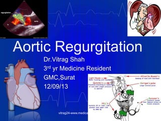
- 1. Aortic Regurgitation Dr.Vitrag Shah 3rd yr Medicine Resident GMC,Surat 12/09/13 vitrag24-www.medicalgeek.com
- 2. Aortic Regurgitation • Valve Anatomy • Etiology • Pathophysiology • Epidemiology • History & Physical Examination • Natural History • Differential diagnosis • Investigations • Assessing severity • Medical Management • Surgical Management • Long term monitoring • Prognosis vitrag24-www.medicalgeek.com
- 3. Anatomy • Located between the left ventricular outflow tract and the ascending aorta • The aortic valve functions to prevent the regurgitation of blood from the aorta into the left ventricle during ventricular diastole and to allow the appropriate flow of blood—the cardiac output —from the left ventricle into the aorta during ventricular systole. • The aortic valve has 3 principle components: the annulus, cusps, and commissures. • The normal aortic valve area in adults is 3.0 to 4.0 cm2 vitrag24-www.medicalgeek.com
- 4. Annulus • The aortic valve annulus is a collagenous structure lying at the level of the junction of the aortic valve and the ventricular septum, which is the nadir of the aortic valve complex. This area is also referred to as the aortic ring and serves to provide structural support to the aortic valve complex. Cusps • There are 3 aortic valve cusps - left, right, and posterior (or noncoronary), each half-moon shaped or semilunar in appearance. Commissures • Each cusp is attached to the wall of the aorta by the outward edges of its semicircular border. The small spaces between each cusp's attachment point are called the aortic valve commissuresvitrag24-www.medicalgeek.com
- 5. Etiology Acute AR Chronic AR Infective Endocarditis Aortic Dissection Trauma Bicuspid Aortic Valve Rheumatic SLE Degenerative Hypertension Anorectic drugs Syphilitic aortitis Takayasu Aortitis Giant cell arteritis Rheumatoid arthritis Whipple’s disease Ankylosing spodylitis Connective tissue disorders • Marfan syndrome • Ehlers-Danlos syndrome • Floppy aortic valve • Aortic valve prolapse • Sinus of Valsalva aneurysm • Aortic annular fistulavitrag24-www.medicalgeek.com
- 6. Primary Valve disease Primary Root disease Congenital (bicuspid aortic valve) Rheumatic fever Infective endocarditis Collagen vascular diseases Degenerative aortic valve disease Myxomatous (prolapse) Traumatic Certain weight loss medications, such as fenfluramine and dexfenfluramine Longstanding, uncontrolled hypertension Marfan syndrome Idiopathic aortic dilation Cystic medial necrosis Senile aortic ectasia and dilation Aortic dissection Marfan's syndrome Syphilitic aortitis Giant cell arteritis Takayasu arteritis Ankylosing spondylitis Whipple disease Other spondyloarthropathies vitrag24-www.medicalgeek.com
- 8. Valvular Abnormalities Nodular Rheumatic Disease Aortic Root Dilation Endocarditis vitrag24-www.medicalgeek.com
- 9. Pathophysiology • In contrast to MR, in which a fraction of the LV stroke volume is ejected into the low-pressure left atrium, in AR the entire LV stroke volume is ejected into a high- pressure chamber (i.e., the aorta), although the low aortic diastolic pressure does facilitate ventricular emptying during early systole • In MR, especially acute MR, the reduction of wall tension (i.e., reduced afterload) allows more complete systolic emptying; in AR the increase in LV end-diastolic volume (i.e., increased preload) provides hemodynamic compensation. vitrag24-www.medicalgeek.com
- 12. Pressure Volume Relationships in Chronic AR CO at rest may approach 25 L/min in severe AI with little increase in EDP very large EDV (Cor Bovinum)vitrag24-www.medicalgeek.com
- 14. Pathophysiology – Acute AR • Acute AR of significant severity leads to increased blood volume in the LV during diastole. The LV does not have sufficient time to dilate in response to the sudden increase in volume. As a result, LV end-diastolic pressure increases rapidly, causing an increase in pulmonary venous pressure and altering coronary flow dynamics. As pressure increases throughout the pulmonary circuit, the patient develops dyspnea and pulmonary edema. • In severe cases, heart failure may develop and potentially deteriorate to cardiogenic shock. Decreased myocardial perfusion may lead to myocardial ischemia. vitrag24-www.medicalgeek.com
- 15. Pathophysiology – Chronic AR • Severe AR may occur with a normal effective forward stroke volume and a normal ejection fraction ([forward plus regurgitant stroke volume]/[end-diastolic volume]), together with an elevated LV end-diastolic volume, pressure, and stress. • According to Laplace's law, which indicates that wall tension is related to the product of the intraventricular pressure and radius divided by wall thickness, LV dilation also increases the LV systolic tension required to develop any level of systolic pressure. Thus, in AR, there is an increase in preload and afterload. LV systolic function is maintained through the combination of chamber dilation and hypertrophy. This leads to eccentric hypertrophy. In compensated AR, there is sufficient wall thickening so that the ratio of ventricular wall thickness to cavity radius remains normal. vitrag24-www.medicalgeek.com
- 16. Pathophysiology – Chronic AR …Cont’d • During the early phases of chronic AR, the LV ejection fraction (EF) is normal or even increased (due to the increased preload and the Frank-Starling mechanism). Patients may remain asymptomatic during this period. • As AR progresses, LV enlargement surpasses preload reserve on the Frank-Starling curve, with the EF falling to normal and then subnormal levels. • The LV end-systolic volume rises and is a sensitive indicator of progressive myocardial dysfunction. • The severity of AR is dependent on the diastolic valve area, the diastolic pressure gradient between the aorta and LV, and the duration of diastole. vitrag24-www.medicalgeek.com
- 17. Pathophysiology – Chronic AR …Cont’d • Eventually, the LV reaches its maximal diameter and diastolic pressure begins to rise, resulting in symptoms (dyspnea) that may worsen during exercise. Increasing LV end-diastolic pressure may also lower coronary perfusion gradients, causing subendocardial and myocardial ischemia, necrosis, and apoptosis. • Severe chronic AR pts have the largest EDV of those with any form of heart disease, resulting in cor bovinum. • Grossly, the LV gradually transforms from an elliptical to a spherical configuration. vitrag24-www.medicalgeek.com
- 18. Myocardial ischemia • Increase in wall tension – Increase in 02 demand • Decrease in diastolic pressure – coronary perfusion pressure is reduced • So increased O2 demand & decreased O2 supply sets mmyocardial ischemia vitrag24-www.medicalgeek.com
- 19. Epidemiology • Approximately three-fourths of patients with pure or predominant valvular AR are men; women predominate among patients with primary valvular AR who have associated rheumatic mitral valve disease. • Chronic AR often begins in patients when they are in their late 50s. In general, the prevalence and severity of AR increase with age. • However, there are many exceptions to this observation. Patients with bicuspid aortic valve and, especially, those with Marfan syndrome tend to present much earlier vitrag24-www.medicalgeek.com
- 20. History – Acute AR • Sudden, severe shortness of breath • Chest pain if myocardial perfusion pressure is decreased or an aortic dissection is present • Rapidly developing heart failure, Pulmonary edema & Cardiogenic shock vitrag24-www.medicalgeek.com
- 21. History – Chronic AR • Relatively asymptomatic for as long as 10–15 years. • Palpitations, especially on lying down. • Sinus tachycardia, during exertion or with emotion, or VPCs may produce uncomfortable palpitations as well as head pounding. These complaints may persist for many years before the development of exertional dyspnea, usually the first symptom of diminished cardiac reserve. • The dyspnea is followed by orthopnea, paroxysmal nocturnal dyspnea, and excessive diaphoresis. vitrag24-www.medicalgeek.com
- 22. History – Chronic AR • Anginal chest pain even in the absence of CAD - at rest as well as during exertion. • Nocturnal angina may be a particularly troublesome symptom, and it may be accompanied by marked diaphoresis. • The anginal episodes can be prolonged and often do not respond satisfactorily to sublingual nitroglycerin. • Congestive heart failure • Sudden cardiac death vitrag24-www.medicalgeek.com
- 23. Aortic Regurgitation Clinical Manifestations • Physical Examination • Chronic AR Acute AR • Systolic BP Systolic BP = • Diastolic BP Diastolic BP = • Aortic Pulse Pressure Aortic Pulse Pressure = • Heart Rate = Heart Rate • S1 = (in CHF ) S1 • S2 = S2 • S3 absent(except CHF) S3 present • S4 usually not present S4 never present vitrag24-www.medicalgeek.com
- 24. Physical Examination – Acute AR • Tachycardia • Peripheral vasoconstriction • Cyanosis • Pulmonary edema • Arterial pulsus alternans; normal LV impulse • Early mitral valve closure • Early diastolic murmur (lower pitched and shorter than in chronic AR) may be present. • A murmur at the right sternal border is associated more often with dissection than it is with any other cause of aortic regurgitation. vitrag24-www.medicalgeek.com
- 25. Physical Examination – Chronic AR • Manifestations of severe chronic AR are often the result of widened pulse pressure because (1) elevated stroke volume exists during systole and (2) the incompetent aortic valve allows the diastolic pressure within the aorta to fall significantly. • On palpation, the point of maximal impulse may be diffuse or hyperdynamic but is often displaced inferiorly and toward the axilla. Peripheral pulses are prominent or bounding. Auscultation may reveal an S3 gallop if LV dysfunction is present. • A diastolic thrill may be palpable along the left sternal border in thin-chested individuals, and a prominent systolic thrill may be palpable in the suprasternal notch and transmitted upward along the carotid arteries.vitrag24-www.medicalgeek.com
- 26. Auscultation • Early-Holo diastolic murmur, immediately after A2, usually as a high-pitched blowing sound that is loudest at the left sternal border, decrescendo, best heard in at end-expiration & in sitting & leaning forward position. The duration of the murmur correlates better with the severity of AR than does the loudness of the murmur. • When the murmur is musical (cooing dove murmur), it usually signifies eversion or perforation of an aortic cusp. • The diastolic murmur of AR when well or predominantly heard in the left axilla – Cole-Cecil murmur • Harvey sign - When regurgitation is caused by primary valvular disease, the diastolic murmur is heard best along the left sternal border in the 3rd and 4th ICS. When it is caused mainly by dilation of the ascending aorta, the murmur is often more readily audible along the right sternal border.vitrag24-www.medicalgeek.com
- 27. Auscultation • A functional systolic flow murmur may also be present because of increased stroke volume, although concurrent AS may also be present. • An Austin-Flint murmur may be present at the cardiac apex in severe AR; it is a low-pitched, mid-diastolic rumbling murmur due to blood jets from the AR striking the anterior leaflet of the mitral valve, which results in premature closure of the mitral leaflets. • A2 may be normal or accentuated when AR is caused by disease of the aortic root but is soft or absent when the valve is causing AR. P2 may be obscured by the early diastolic murmur. Thus, S2 may be absent or single or exhibit narrow or paradoxical splitting. vitrag24-www.medicalgeek.com
- 28. Auscultation • A systolic ejection sound, presumably related to abrupt distention of the aorta by the augmented stroke volume, is frequently audible. • A third heart sound (S3) correlates with an increased LV end-diastolic volume. Its development may be a sign of impaired LV function, which is useful in identifying patients with severe AR who are candidates for surgical treatment. • The auscultatory features of AR are intensified by strenuous and sustained handgrip, which augments systemic vascular resistance. vitrag24-www.medicalgeek.com
- 29. Peripheral Signs • Becker sign - Visible systolic pulsations of the retinal arterioles • Corrigan pulse ("water-hammer" pulse) - Abrupt distention and quick collapse on palpation of the peripheral arterial pulse • de Musset sign - Bobbing motion of the patient's head with each heartbeat • Hill sign – (Popliteal cuff systolic blood pressure 20 mm Hg higher than brachial cuff systolic blood pressure) is an artifact of sphygmomanometric measurements and is no longer considered a sign of severe AR. • Duroziez sign - Systolic murmur over the femoral artery with proximal compression of the artery, and diastolic murmur (Duroziez murmur) over the femoral artery with distal compression of the arteryvitrag24-www.medicalgeek.com
- 30. • Müller sign - Visible systolic pulsations of the uvula • Light-house sign – Alternate flushing & blanching of forehead • Landolfi’s sign - Change in pupil size with each systole • Gerhardt’s sign - Visible systolic pulsations of spleen • Rosenbach’s sign - Visible systolic pulsations of liver • Corrigan’s sign – Dancing carotid in neck • Quincke sign - Visible pulsations of the fingernail bed with light compression of the fingernail • Traube sign ("pistol-shot" pulse) - Booming systolic and diastolic sounds auscultated over the femoral artery • A bisferiens pulse in AS + AR – readily recognized in the brachial and femoral arteries than in the carotid arteries.(Two systolic waves separated by a trough ) vitrag24-www.medicalgeek.com
- 31. Physical Examination …Cont’d As heart failure develops, peripheral vasoconstriction may occur and arterial diastolic pressure may rise, even though severe AR is present, so pulse pressure can be normal or narrow in severe AR. Causes of AR with normal/low pulse pressure: • Acute AR • AR with AS or severe MS • AR with CHF • AR with systemic hypertension vitrag24-www.medicalgeek.com
- 32. vitrag24-www.medicalgeek.com Assessing Severity of AR • Assess severity by impact on peripheral signs and LV • peripheral signs = severity • LV = severity • S3 • Long duration of diastlic murmur • Austin –Flint murmur • LVH • radiological cardiomegaly
- 33. Features AS > AR AR > AS Symptoms: Anginal pain, blackout Palpitation +++ + + +++ Pulse Low volume Bisferians pulse High volume Corrigan’s pulse Pulse pressure Normal to low Wide Peripheral signs +/- + Apex Heaving Hyperdynamic Thril Always systolic Rarely S3 Absent May be present S4 May be present Absent Ejection click Present Rare Diastolic murmur Very short Classic murmur of AR Systolic murmur Classic murmur of AS Function systolic murmur +/- Chest X-Ray Calcification + Cardiomegaly ECG Pressure overload Volume overloadvitrag24-www.medicalgeek.com
- 35. Differential Diagnosis Hyperdynamic circulation (wide pulse pressure), including the following: • Thyrotoxicosis • Severe anemia • Pregnancy • Thiamine deficiency (wet beriberi) • Arteriovenous fistula - Such as patent ductus arteriosus or peripheral arteriovenous malformations • Volume depletion • Sympathetic overdrive vitrag24-www.medicalgeek.com
- 36. ECG • LV hypertrophy • Left axis deviation • Left atrial enlargement • LV volume overload pattern - Prominent Q waves in leads I, aVL, and V3 to V6 and relatively small r waves in V1 • LV conduction defects - Typically late in the disease process • Left-axis deviation and/or QRS prolongation denote diffuse myocardial disease, generally associated with patchy fibrosis, and usually signify a poor prognosis. vitrag24-www.medicalgeek.com
- 37. The patient’s 12-lead electrocardiogram shows normal sinus rhythm and a rate of 55 beats per minute. The frontal-plane QRS complex vector is deviated leftward. Left atrial abnormality is present, given the terminally negative P wave in lead V1 and the bifid P wave in lead II. Left ventricular volume overload is supported by the following findings: increased QRS complex voltage, best seen in the precordial (chest) leads, indicative of increased left ventricular mass; prominent septal depolarization, as reflected by Q waves in leads V4 to V6; the absence of an ST-segment or T-wave abnormality; and negative U waves in leads V4 to V6 (arrows). vitrag24-www.medicalgeek.com
- 38. Chronic Acute Normal size LV with pulmonary vascular congestion LVE with normal pulmonary vasculature Aortic Regurgitation CxR vitrag24-www.medicalgeek.com
- 39. Chest X-Ray • In acute AR, there may be minimal cardiac enlargement, but marked enlargement is a common finding in chronic AR. • The apex is displaced downward and to the left in the frontal projection. In the left anterior oblique and lateral projections, the LV is displaced posteriorly and encroaches on the spine. • Distinct left atrial enlargement in the absence of heart failure suggests associated mitral valve disease. • When AR is caused by primary disease of the aortic root, aneurysmal dilation of the aorta may be noted, and the aorta may fill the retrosternal space in the lateral view. vitrag24-www.medicalgeek.com
- 40. Echocardiography • Aortic valve structure and morphology - Bileaflet versus trileaflet, flail, thickening • Presence of vegetations or nodules - May require transesophageal echocardiography in selected cases • Severity of AR • Color Doppler central jet width & Vena contracta (narrowest portion of the jet located at or just distal to its orifice) width - In severe AR, the vena contracta width is usually more than 65% of the width of the LV outflow tract • Regurgitant volume, fraction, and orifice area • Premature closure of the mitral valve (seen in severe AR) and opening of the aortic valve (with severely elevated LV end-diastolic pressure) • Holodiastolic flow reversal in the descending thoracic or abdominal aorta vitrag24-www.medicalgeek.com
- 41. • Pressure half-time - Usually less than 300-350 ms with significant AR • Associated lesions of the aorta - Including dilation, aneurysm, dissection, or ectasia • A rapid, high-frequency diastolic fluttering of the anterior mitral leaflet produced by the impact of the regurgitant jet is a characteristic finding - it does not develop when the mitral valve is rigid, as occurs with rheumatic involvement. & unlike the Austin Flint murmur, occurs even in mild AR • LV structure and function, LV hypertrophy and dilation • Ejection fraction (EF) and end-systolic dimension - These are key determinants of outcome; surgery is recommended if the EF is 50% or less or if the end- systolic dimension is more than 55 mm[4] vitrag24-www.medicalgeek.com
- 42. AMVL fluttering Color Flow – top mild, bottom moderate vitrag24-www.medicalgeek.com
- 43. Chronic AI Acute AI Continuous Wave Doppler vitrag24-www.medicalgeek.com
- 44. Regurgitant jet width/LVOT diameter ratio greater than or equal to 60 percent vitrag24-www.medicalgeek.com
- 45. Vena contracta greater than 6 mm vitrag24-www.medicalgeek.com
- 46. Regurgitant jet area/LVOT area ratio greater than or equal to 60 percent vitrag24-www.medicalgeek.com
- 48. Holodiastolic flow reversal in the descending thoracic or abdominal aorta vitrag24-www.medicalgeek.com
- 52. Cardiac Catheterization Class I indications for cardiac catheterization under current ACC/AHA guidelines: • Assessment of coronary anatomy prior to aortic valve surgery in patients with risk factors for coronary artery disease • Assessment of severity of AR, LV function, or aortic root size when noninvasive tests are inconclusive or are discordant with clinical findings vitrag24-www.medicalgeek.com
- 53. Aortic angiography • Mild (1+) - A small amount of contrast enters the LV during diastole and clears with each systole • Moderate AR (2+) - Contrast enters the LV with each diastole, but the LV chamber is less dense than the aorta • Moderately severe AR (3+) - The LV chamber is equal in density to the ascending aorta. • Severe AR (4+) - Complete, dense opacification of the LV chamber occurs on the first beat, and the LV is more densely opacified than the ascending aorta vitrag24-www.medicalgeek.com
- 54. Cardiac MRI • CMR provides accurate measurements of regurgitant volumes and the regurgitant orifice in AR. It is the most accurate noninvasive technique for assessing LV end- systolic volume, diastolic volume, and mass vitrag24-www.medicalgeek.com
- 55. Radionuclide imaging • Provides AR regurgitant fraction and the LV/right ventricular (RV) stroke volume ratio • In the absence of mitral regurgitation and tricuspid regurgitation, an LV/RV stroke volume ratio of 2.5 or more denotes severe aortic regurgitation. vitrag24-www.medicalgeek.com
- 56. Management – Acute AR • Prompt surgical intervention is indicated • Administer a positive inotrope (eg, dopamine, dobutamine) and a vasodilator (eg, nitroprusside). • Administration of vasodilators may be appropriate to improve systolic function and to decrease afterload. • The administration of cardiac glycosides (eg, digoxin) for rate control may in rare cases be necessary. • Beta-blocking agents and intra-aortic balloon counterpulsation are contraindicated, because either lowering the heart rate or augmenting peripheral resistance during diastole can lead to rapid hemodynamic decompensation. vitrag24-www.medicalgeek.com
- 58. Management – Chronic MR The current ACC/AHA guidelines say the following about vasodilator therapy (Nifedipine, ACE Inhibitors): • Vasodilator therapy is indicated for long-term treatment in patients who have severe chronic AR and symptoms of LV dysfunction but who are not candidates for surgery. • Vasodilator therapy is reasonable for short-term therapy in patients with severe LV dysfunction and heart failure symptoms, in order to improve their hemodynamic profile before surgery • Vasodilator therapy is acceptable for long-term therapy in asymptomatic patients with severe AR and LV dilation with normal EF (To control diastolic BP) vitrag24-www.medicalgeek.com
- 59. • Chronic medical therapy for some patients who refuse surgery or are considered to be inoperable because of comorbid conditions - These patients should receive an aggressive heart failure regimen with ACE inhibitors (and perhaps other vasodilators), digoxin, diuretics, and salt restriction; beta blockers may also be beneficial • Although nitroglycerin and long-acting nitrates are not as helpful in relieving anginal pain as they are in patients with ischemic heart disease, they are worth a trial. • Beta blockers (contraindicated in Valvular AR) and the angiotensin receptor blocker, losartan, may be useful to retard the rate of aortic root enlargement in young patients with Marfan's syndrome and aortic root dilation. • Avoid isometric exercises • Avoidance of atrial fibrillation and bradycardia are important as these are poorly tolerated vitrag24-www.medicalgeek.com
- 60. Antibiotic Prophylaxis The prophylactic use of antibiotics prior to dental procedures is no longer routinely recommended for all patients with AR. Select patient groups for whom prophylactic antibiotic therapy prior to dental procedures may be reasonable include the following: • Patients with prosthetic material in their heart - Such as an artificial valve or a valve repaired with prosthetic material • Patients with prior infective endocarditis • Patients who, following cardiac transplantation, have valve regurgitation due to a structurally abnormal valve vitrag24-www.medicalgeek.com
- 61. Antibiotic Prophylaxis • Patients with congenital heart disease (CHD) who meet any of the following criteria: (1) Cyanotic CHD that has not been repaired or has been incompletely repaired (including patients with palliative shunts and conduits); (2) repaired CHD using prosthetic material, for the first 6 months postprocedurally (ie, prior to endothelialization of the material); or (3) repaired CHD but the patient is at risk for inhibited endothelialization (ie, with residual defects at or adjacent to the site of the prosthetic material) vitrag24-www.medicalgeek.com
- 62. Surgical treatment – ACC/AHA guideline summary Class I - There is evidence and/or general agreement that aortic valve replacement or repair (AVR) is indicated in patients with chronic AR in the following settings • Symptomatic patients with severe chronic AR, irrespective of left ventricular ejection fraction (LVEF). • If the presence of symptoms in patients with severe chronic AR is equivocal, the development of symptoms during an exercise test. • Asymptomatic patients with severe chronic AR and an LVEF ≤50 percent at rest. • Patients with severe chronic AR who undergo coronary artery bypass graft surgery (CABG) or surgery on the aorta or other heart valves. The indications for surgery for patients with severe AR secondary to aortic root disease are similar to those with primary valvular disease. However, progressive expansion of the aortic root and/or a diameter more than 50 mm by echocardiography with any degree of regurgitation in patients with a bicuspid valve (or other connective tissue disorder) or with a diameter more than 55 mm in other patients is also an indication for aortic root replacement surgery. vitrag24-www.medicalgeek.com
- 63. Class IIa - The weight of evidence or opinion is in favor of the usefulness of AVR in patients with chronic AR in the following setting • Asymptomatic patients with severe chronic AR and a normal LVEF (LVEF >50 percent) who have severe left ventricular dilatation (end- diastolic dimension >75 mm or end-systolic dimension >55 mm). Lower threshold values can be considered for patients of small stature. Class IIb - The weight of evidence or opinion is less well established for the usefulness of AVR in patients with chronic AR in the following settings • Patients with moderate chronic AR who undergo CABG or surgery on the ascending aorta. • Asymptomatic patients with severe chronic AR and an LVEF >50 percent in whom the end-diastolic dimension is >70 mm or the end- systolic dimension is >50 mm, and there is evidence of progressive left ventricular dilatation, declining exercise tolerance, or an abnormal hemodynamic response to exercise. vitrag24-www.medicalgeek.com
- 64. ACC/AHA guideline summary: Criteria for selection of an aortic valve in patients undergoing aortic valve replacement (AVR) Class I • A mechanical valve in patients who already have a mechanical valve in the mitral or tricuspid position. • A bioprosthetic valve in patients who will not take or are incapable of taking warfarin or have a major contraindication to warfarin therapy. Class IIa • A bioprosthesis in patients ≥65 years of age who do not have risk factors for thromboembolism. • Patient preference can be considered in patients less than 65 years of age: 1. A mechanical valve is reasonable in patients who do not have a contraindication to warfarin therapy. 2. A bioprosthetic valve may be chosen after a detailed discussion of the risks of warfarin therapy compared to the likelihood of repeat valve replacement in the future. • A homograft when aortic valve re-replacement is performed for active prosthetic valve endocarditis. Class IIb • A bioprosthesis in women of child-bearing age to avoid the problems associated anticoagulation during pregnancy. vitrag24-www.medicalgeek.com
- 65. Figure 29-15 A. Björk-Shiley Monostrut mechanical prosthesis. B. Sorin Allcarbon monoleaflet mechanical prosthesis. C. Medtronic-Hall mechanical prosthesis. D. Omnicarbon mechanical prosthesis. Figure 29-16 A. Carpentier-Edwards Supra- annular porcine bioprosthesis. B. Hancock II porcine bioprosthesis. C. Hancock modified orifice porcine bioprosthesis. D. St. Jude Medical Bioimplant porcine bioprosthesis.vitrag24-www.medicalgeek.com
- 66. Surgery – Primary Root disease • Annuloplasty or other valve sparing surgery vitrag24-www.medicalgeek.com
- 67. Transcatheter aortic valve replacement • TAVR involves the implantation of a bioprosthetic aortic valve using a catheter that is inserted peripherally, typically through the femoral artery, and implanted without requiring a median sternotomy (ie, without ―open heart surgery‖). Initial reports are promising but further studies are needed before TAVR becomes clinically available • If MS + AR, first go for AR as correcting MS will increase LV load LVF. vitrag24-www.medicalgeek.com
- 68. Long term monitoring • All patients with an artificial heart valve should receive antibiotic prophylaxis prior to dental procedures. For antithrombotic therapy, all patients with an artificial heart valve should receive daily aspirin, and many should also receive oral anticoagulation therapy with warfarin according to the ACC/AHA guidelines. • After the initial study, clinical evaluation and a repeat echocardiogram are recommended in 3 months. The recommended frequency of subsequent follow-up evaluations is based on the stability of the LVESD and LVEDD – every 3 to 12 months. vitrag24-www.medicalgeek.com
- 69. Prognosis • Death usually occurs within 4 yrs after onset of angina pectoris and within 3 yrs of onset of heart failure • It is imperative to intervene before irreversible LV dysfunction occurs • After AVR, Long term results reveal 80-90% three year survival in patients with preserved LV function, and 40- 60% three year survival in patients with poor LV function • Mortality rate of AVR ranges from 3-8% vitrag24-www.medicalgeek.com
- 70. References: American Heart Association & American College of Cardiology Guidelines (http://www.cardiosource.org) Harrison’s PRINCIPLES OF INTERNAL MEDICINE : Eighteenth Edition Braunwald's Heart Disease : A Textbook of Cardiovascular Medicine : Ninth Edition Hurst's the Heart Manual of Cardiology, Thirteenth Edition UpToDate (http://www.uptodate.com) eMedicine (http://www.emedicine.com) vitrag24-www.medicalgeek.com
Editor's Notes
- The normal human heart contains 4 valves that regulate blood flow into and out of the heart. The aortic and pulmonic valves are known as the semilunar valves, whereas the tricuspid and mitral valves are referred to as the atrioventricular valves. All the valves are trileaflet, with the exception of the mitral valve, which has 2 leaflets. All 4 cardiac valves are surrounded by fibrous tissue forming partial or complete valvular rings, or annuli. These annuli join the fibrous skeleton of the heart to anchor and support the valvular structures.
- Bicuspid aortic valve is the most common congenital lesion of the human heart.Connective tissue disorders that can cause significant AR include the following:Marfan syndromeEhlers-Danlos syndromeFloppy aortic valveAortic valve prolapseSinus of Valsalva aneurysmAortic annular fistula
- Hemodynamics of aortic regurgitation. A, Normal conditions. B, The hemodynamic changes that occur in severe acute aortic regurgitation. Although total stroke volume is increased, forward stroke volume is reduced. Left ventricular end-diastolic pressure (LVEDP) rises dramatically. C, Hemodynamic changes occurring in chronic compensated aortic regurgitation are shown. Eccentric hypertrophy produces increased end-diastolic volume (EDV), which permits an increase in total, as well as forward, stroke volume. The volume overload is accommodated, and left ventricular filling pressure is normalized. Ventricular emptying and end-systolic volume (ESV) remain normal. D, In chronic decompensated aortic regurgitation, impaired left ventricular emptying produces an increase in end-systolic volume and a fall in ejection fraction (EF), total stroke volume, and forward stroke volume. There is further cardiac dilation and reelevation of left ventricular filling pressure. E, Immediately following valve replacement, preload estimated by EDV decreases, as does filling pressure. ESV also is decreased, but to a lesser extent. The result is an initial fall in EF. Despite these changes, elimination of regurgitation leads to an increase in forward stroke volume, and with time ejection fraction increases. Aop = aortic pressure; RF = regurgitant fraction.(From Carabello BA: Aortic regurgitation: Hemodynamic determinants of prognosis. In Cohn LH, DiSesa VJ [eds]: Aortic Regurgitation: Medical and Surgical Management. New York, Marcel Dekker, 1986, p 99-101.)
- Patients with a projected long life-span generally receive a mechanical valve because of far greater durability and improved patient survival at 15 years .The main indications for a bioprosthesis are patients who cannot or will not tolerate warfarin or for whom compliance is uncertain, and patients ≥65 years of age who do not have risk factors for thromboembolism since valve durability is less of an issue